TECHNOLOGY EXPLAINER
Drug Delivery Systems and Targeting
— How well does "aim and deliver" really work? Starting from 0.7%
A drug delivery system (DDS) is a technology for getting a drug to the place it is needed, for only as long as it is needed. Yet a ten-year pooled analysis of animal studies of cancer nanoparticles found that the median amount reaching a solid tumor was just 0.7% of the injected dose. Even so, products have been approved, and when their contents are weighed, most of the mass is the carrier materials (lipids, polymers and albumin).
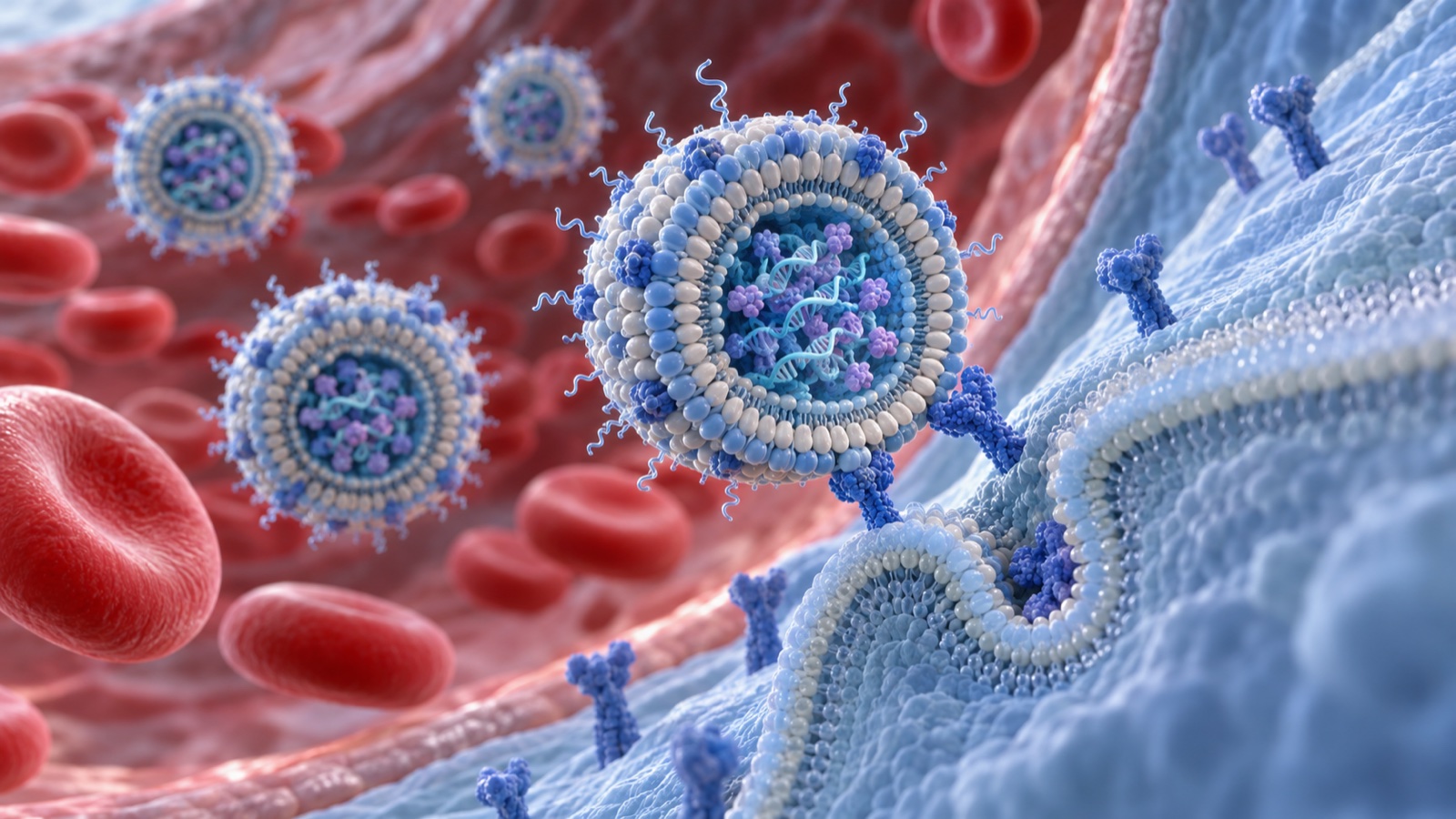
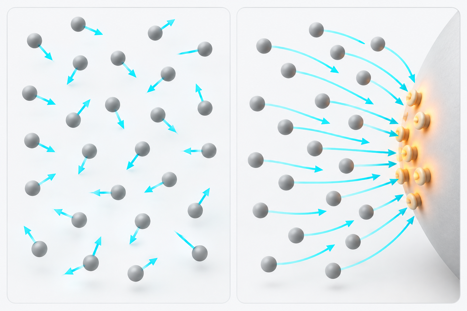
- What a DDS is (the short version)
- Four types of nanocarrier — liposomes, albumin particles, polymeric micelles and polymer conjugates
- Passive targeting and the EPR effect — the 1986 report
- The EPR debate — does it "not work in humans"?
- Our calculation: putting 0.7% into amounts
- A materials engineer's view 1: the other 99% is decided by the surface
- Active targeting and ligands — what changes is not how much arrives but how it gets in
- For small molecules, active targeting has already become a product
- Controlled release — how a drug is held decides how it is released
- Our calculation: how many times the drug's mass is the carrier?
- A materials engineer's view 2: why generics took 17 years, read from FDA guidance
- Challenges, and what could not be confirmed
- Glossary / References / Claim-to-source audit
Sourced = stated in published material (link given)
Our calculation = a figure this article derived, with the assumptions spelled out
Not yet confirmed = a plan, target or research-stage item whose results have not been confirmed
Structural readings and interpretations of materials or process design are marked separately as Commentary. Inference is shown in bold.
This article is an explanation of materials and formulation technology. It does not evaluate or compare the efficacy or safety of individual drugs, and it is not medical advice. Product compositions and approval dates are quoted only where they appear in the FDA labels and Drugs@FDA. Results in animals do not necessarily apply to humans.
1. What a DDS is (the short version)
- What it does: without changing the drug molecule itself, a DDS changes where the drug goes in the body and how fast it is released, by wrapping, attaching or embedding it (this article's framing). It is a separate axis from the "types of drug" covered in our explainer on drug discovery modalities: it is a "how to carry it" technology that can be combined with any type of drug
- What materials are used: Duncan calls nano-sized (5–100 nm) polymeric drugs "polymer therapeutics" and groups under that name polymer–drug conjugates, polymer–protein conjugates, polymeric micelles with covalently bound drug, and polyplexes for DNA deliverySourced[Ref. 12]. To these can be added liposomes made of lipids and albumin particles
- What is hard: only a small fraction reaches the intended place. Wilhelm and co-authors pooled ten years of literature and reported that the median share of the administered nanoparticles reaching a solid tumor was only 0.7%Sourced[Ref. 2]
Nanoparticles gather at the intended place far less than the word "targeting" suggests. There is an animal-study report that even with active targeting (attaching a ligand) the amount accumulating in the tumor hardly changed, and what changed was whether the particles got inside the cellsSourced. Meanwhile, when the contents of approved products are weighed, the carrier materials (lipids, polymers and albumin) account for 2 to 9 times the mass of the drugOur calculation. A large part of DDS performance is therefore set not by the drug but by the design and manufacture of the carrier (this article's commentary).
2. Four types of nanocarrier — liposomes, albumin particles, polymeric micelles and polymer conjugates
The "containers" that carry a drug (nanocarriers) come in several structural types. As passive-targeting nanocarriers used in the clinic, Rosenblum and co-authors list Doxil, Abraxane, Marqibo, DaunoXome and Onivyde in the United States, Myocet and Mepact in Europe, Genexol-PM in South Korea and SMANCS in JapanSourced[Ref. 3].
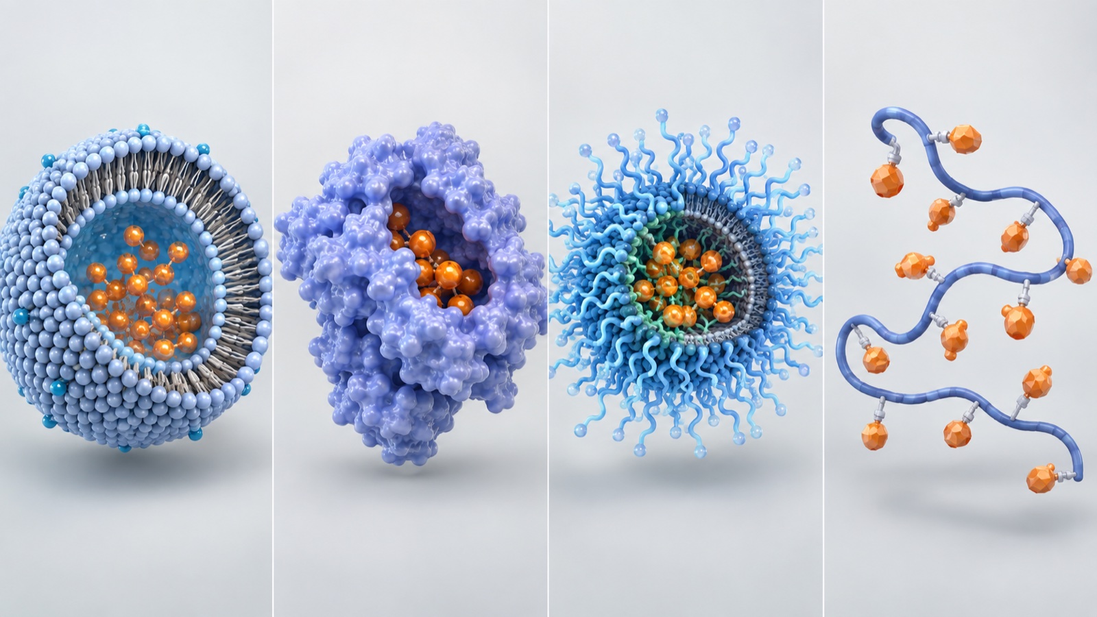
Show the original schematic with sources and numbers
Fig. 1 Conceptual diagram (vector drawing). Product examples are from Rosenblum et al. (2018) [Ref. 3], compositions from the FDA labels [Refs. 16–20], and the type grouping from Duncan (2003) [Ref. 12] and Cabral et al. (2018) [Ref. 13]. The split into four types is this article's own, and the drawing is schematic. It does not show real molecular structures or relative particle sizes.
| Type | Approved product (FDA), example | Composition as written in the label | US approval |
|---|---|---|---|
| PEG-modified liposome | DOXIL (doxorubicin hydrochloride) | STEALTH liposome. Cholesterol 3.19 mg/mL, hydrogenated soy phosphatidylcholine (HSPC) 9.58 mg/mL, MPEG-DSPE 3.19 mg/mL. More than 90% of the drug is encapsulated inside the liposome | 17 November 1995 |
| PEG-modified liposome | ONIVYDE (irinotecan) | Unilamellar liposome with a diameter of about 110 nm. DSPC 6.81 mg/mL, cholesterol 2.22 mg/mL, MPEG-2000-DSPE 0.12 mg/mL. Inside, irinotecan is gelled or precipitated as the sucrose octasulfate salt | 22 October 2015 |
| Liposome (two drugs) | VYXEOS (daunorubicin and cytarabine) | The two drugs are encapsulated at a 1:5 molar ratio. The membrane is DSPC, DSPG and cholesterol at a 7:2:1 molar ratio. One vial holds 618 mg of lipid in total, 100 mg of copper gluconate and 2,054 mg of sucrose | 2017 |
| Albumin-bound particle | ABRAXANE (paclitaxel) | Average particle diameter about 130 nm. The paclitaxel is amorphous. One vial holds 100 mg of paclitaxel and about 900 mg of human albumin. It contains no solvent | 2005 |
| Polymer–protein conjugate | ONCASPAR (pegaspargase) | An enzyme made of four 34.5 kDa subunits, with 69 to 82 mPEG chains of about 5 kDa attached | 1994 |
All Sourced (FDA labels [Refs. 16–20]; approval dates from Drugs@FDA [Refs. 24 and 26]; years from "Initial U.S. Approval" in each label). For the particle size and layer structure of VYXEOS, Anselmo and Mitragotri describe it as a 100 nm bilamellar liposomeSourced[Ref. 15]. This article does not cover indications or clinical results.
For polymeric micelles, the review by Cabral and co-authors addresses micelles assembled through multimolecular interactions between block copolymers and the drugs, proteins or nucleic acids they carry, and says that the type of interaction between polymer and cargo and the chemistry and engineering of the block copolymer are at the center of the designSourced[Ref. 13]. The idea is to use the self-assembly of amphiphilic molecules to load a drug (this article's commentary).
3. Passive targeting and the EPR effect — the 1986 report
The idea of collecting nanocarriers in a tumor started with Japanese research. In 1986, Yasuhiro Matsumura and Hiroshi Maeda reported the following in Cancer ResearchSourced[Ref. 1].
The starting point was an earlier finding that SMANCS, the anticancer protein neocarzinostatin attached to a polymer, accumulates in tumor tissue more than the original neocarzinostatin does. The authors therefore radiolabelled a range of proteins with molecular weights from 12,000 to 160,000 and followed them. Many of the proteins gradually accumulated in tumor tissue, and the tumor-to-blood concentration ratio reached 5 within 19 to 72 hoursSourced.
By contrast, neocarzinostatin (molecular weight 12,000), a representative small protein, reached a ratio of neither 1 nor 5Sourced.
The authors suggested the cause was abundant blood vessels, increased permeability even to large molecules, and almost no recovery of those molecules through either the blood vessels or the lymphaticsSourced.
This is the phenomenon later called the EPR effect (enhanced permeability and retention). Rosenblum and co-authors likewise write that the phenomenon first described by Matsumura and Maeda in 1986 is known as the EPR effectSourced[Ref. 3]. The idea that larger molecules accumulate in tumors became the rationale for passive targeting with nanocarriers.
But bigger is not simply better. If a particle is too small it leaves through the kidneys, and if it is too large other problems appear.
The 5.5 nm at the left edge is a value Choi and co-authors obtained with quantum dots. The abstract says that rapid and efficient urinary excretion occurred when the final hydrodynamic diameter was below 5.5 nm, and also that zwitterionic or neutral organic coatings prevented the adsorption of serum proteins, whereas adsorption increased the hydrodynamic diameter by more than 15 nm and impeded renal excretionSourced[Ref. 9]. Merely having proteins stick to the surface changes the "effective size" of a particle. Chapter 6 returns to this point.
For the middle of the range, Charrois and Allen compared liposomes with diameters of 82, 101, 154 and 241 nm in mice and reported that the three smaller ones accumulated to a similar extent, and all significantly more than the largest, 241 nmSourced[Ref. 10].
4. The EPR debate — does it "not work in humans"?
For more than 30 years the EPR effect was the premise behind the design of nanomedicines. Since the late 2010s, however, a series of papers has questioned that premise. Here we line up papers that take different positions and do not declare a winner.
| Paper | Claim (abstract or text) | Position (this article's grouping) |
|---|---|---|
| Wilhelm et al. (2016) | Pooling ten years of literature, a median of only 0.7% of the administered nanoparticles reaches a solid tumor. This low figure has adverse effects on manufacturing, cost, toxicity, and the performance of imaging and therapy | Quantifies how little arrives |
| Danhier (2016) | States that the verdict has been that the EPR effect works in rodents but not in humans, and that claims of improved efficacy through the EPR effect should be dropped | Negative on the EPR effect |
| Rosenblum et al. (2018) | 0.7% is small in absolute terms, but relatively it is considerably higher than the delivery efficiency of conventional formulations that dominate clinical use (docetaxel, paclitaxel, doxorubicin and others). As an example they cite a preclinical study in which a paclitaxel-loaded nanocarrier reached 0.6% ID against 0.2% ID for free paclitaxel | Low in absolute terms, but a relative advantage |
| Sindhwani et al. (2020) | The gaps between endothelial cells of tumor vessels (long put at up to 2,000 nm) are not responsible for transporting nanoparticles into the tumor. Up to 97% of nanoparticles enter tumors through an active process that passes through endothelial cells | Questions the "leaking through gaps" mechanism itself |
All Sourced (Wilhelm et al. [Ref. 2], Danhier [Ref. 4], Rosenblum et al. [Ref. 3], Sindhwani et al. [Ref. 5]). The "Position" column is this article's grouping; the authors do not classify themselves this way.
There are also measurements in humans. Rosenblum and co-authors describe a study of 19 patients with HER2-positive metastatic breast cancer in which accumulation of liposomes labelled with radioactive copper (64Cu) in lesions was quantified by PET/CT. Accumulation peaked at 24 to 48 hours, and the patients could be sorted by the amount that accumulatedSourced[Ref. 3]. The same paper also cites animal studies showing that accumulation is heterogeneous, both within a tumor and from tumor to tumorSourced[Ref. 3].
What is in dispute is less whether the EPR effect exists than how reproducibly it occurs, and in which tumors. What Rosenblum and co-authors' human data show is that accumulation varies from patient to patient, and the paper also discusses a direction in which this is measured beforehand by imaging so that patients can be selected. In materials terms, the situation resembles how to handle a paint that sticks on some substrates and not on others.
5. Our calculation: putting 0.7% into amounts
Using the published figures, we make a few conversionsOur calculation.
- Premise: a median of 0.7% of the dose reaches a solid tumor (Wilhelm et al., a pooled analysis of animal studies)Sourced
- Amount going elsewhere than the tumor: 100 − 0.7 = 99.3%
- Ratio of non-tumor to tumor: 99.3 ÷ 0.7 = about 142 times
- Relative improvement (the example Rosenblum and co-authors cite): 0.6% ÷ 0.2% = 3 times
Premises and limits: 0.7% is the median of values in the literature from animal studies (mainly mice), not a human value. The "amount going elsewhere than the tumor" also lumps together the share captured by the liver and spleen, the share excreted by the kidneys, the share still in the blood and so on, so this calculation says nothing about how much goes where. The factor of 3 is likewise the value of a single preclinical study.
6. A materials engineer's view 1: the other 99% is decided by the surface
On where the share that does not reach the tumor goes, Rosenblum and co-authors state that most nanocarriers are cleared by the mononuclear phagocyte system (MPS); some are physically trapped in the sinusoids of the liver, and some are taken up by hepatocytes and Kupffer cellsSourced[Ref. 3]. As a factor that governs targeting efficiency, they name the coating formed by serum proteins and opsonins (also called the protein corona)Sourced[Ref. 3].
In other words, what cells in the body "see" is not the surface the designer made but the layer of proteins adsorbed on it (this article's commentary). The result of Choi and co-authors seen in Chapter 3 — adsorbed proteins increase the hydrodynamic diameter by more than 15 nm, and zwitterionic or neutral coatings prevent the adsorptionSourced[Ref. 9] — shows that there is a way to solve this problem with surface chemistry.
A familiar tool for materials engineers here is surface modification with PEG (polyethylene glycol). The FDA product-specific guidance for doxorubicin liposomes says that the coating of surface-bound methoxypolyethylene glycol (MPEG) protects the liposome from clearance by the mononuclear phagocyte system and prolongs its circulation time, and that the thickness of the PEG layer is thermodynamically limited and estimated to be a few nanometersSourced[Ref. 28].
A polymer brush a few nanometers thick decides where a particle goes in the body. This should be familiar territory for engineers who have designed steric stabilization of dispersions or anti-fouling coatings (this article's commentary). The difference is that the opponent is not "dirt" but plasma containing thousands of kinds of proteins, and the place of evaluation is the body.
7. Active targeting and ligands — what changes is not how much arrives but how it gets in
Whereas passive targeting relies on size, active targeting attaches a ligand (a component that binds a specific molecule) to the particle surface. Rosenblum and co-authors write that in active cell targeting, an affinity ligand is attached to the surface of the nanocarrier, aiming at specific accumulation, greater retention at the target site and uptake by the target cells, and list HER2, the folate receptor and CD44, among others, as partners for ligandsSourced[Ref. 3].
The same paper points out, however, that an actively targeted nanocarrier must also first reach its target, so efficient passive targeting is a prerequisiteSourced[Ref. 3]. A ligand does not pull the particle to the tumor. The next two reports show this experimentally.
| Study | What was compared | Accumulation in the tumor | Binding and uptake by cells |
|---|---|---|---|
| Kirpotin et al. (2006) | Long-circulating liposomes with an anti-HER2 antibody fragment (Fab′ or scFv) attached to the tip of the PEG chains, versus liposomes without it (mice with human breast cancer xenografts that overexpress HER2) | Equally high for both (7–8% ID/g). Attaching the antibody fragment changed neither biodistribution nor circulation time | The targeted ones were found mainly inside the cancer cells, the untargeted ones in the stroma and macrophages. Uptake into cancer cells was up to 6-fold higher |
| Moreira et al. (2001) | Stealth liposomes with a peptide (antagonist G) on the surface, versus liposomes without it (mice with human small-cell lung cancer xenografts) | Depended on liposome size but not on the presence of the ligand. Up to 2–4% ID/g | Binding to cells taken from the tumor was 30- to 44-fold higher |
All Sourced (abstracts of Kirpotin et al. [Ref. 6] and Moreira et al. [Ref. 7]). Both are results in mice. % ID/g is a unit meaning what percentage of the injected dose is present per gram of tissue.
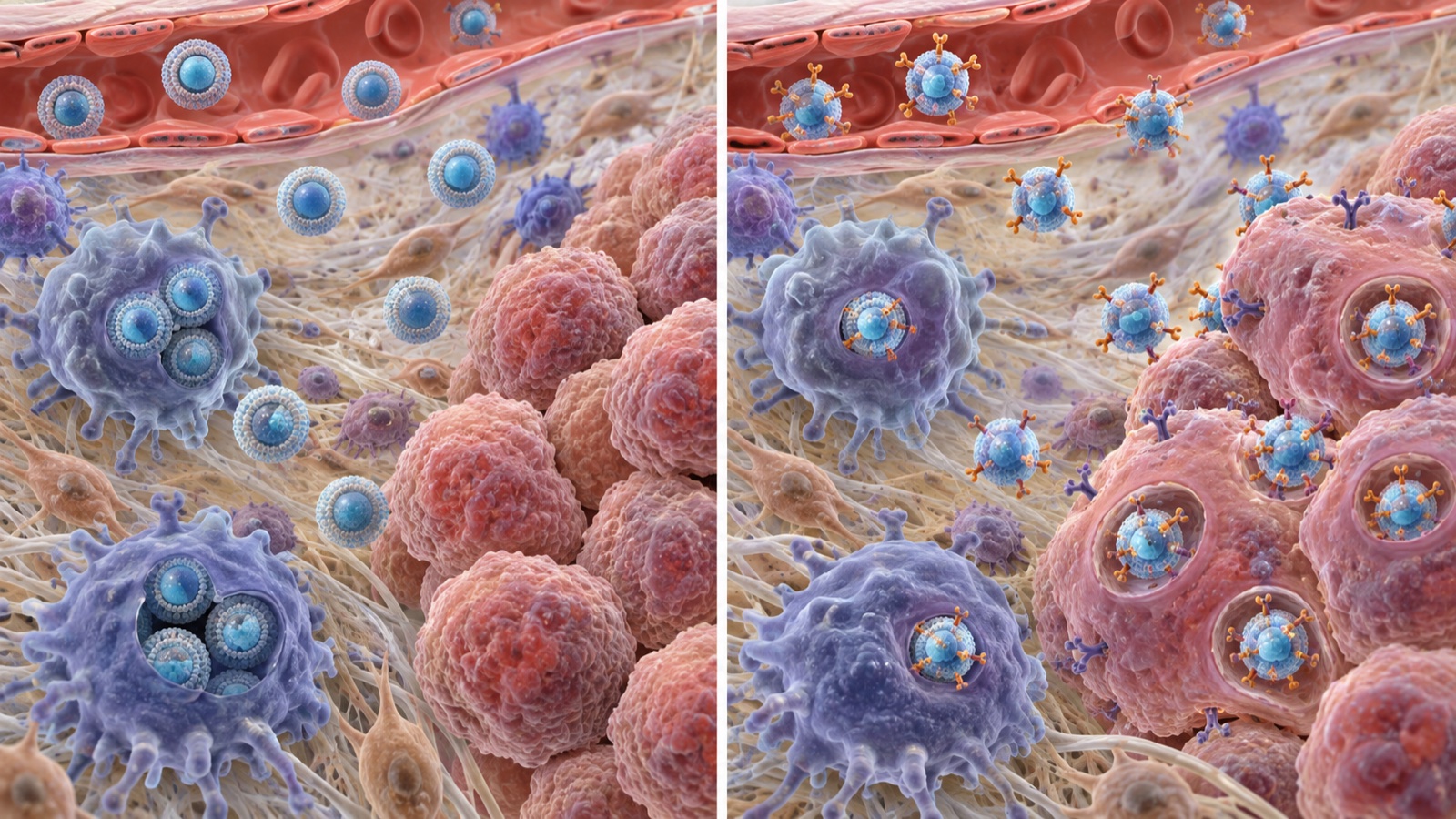
Show the original schematic with sources and numbers
Fig. 4 Conceptual diagram (vector drawing). Accumulation, distribution and uptake ratios are from the abstract of Kirpotin et al. (2006) [Ref. 6]. Particle numbers and positions are schematic, and the summary at the bottom is this article's. These are results in mice, and the same need not happen in humans.
Then there are the clinical results. Rosenblum and co-authors state that as of 2018, 15 passively targeted nanocarriers had been approved for clinical use, whereas no actively targeted one had gone beyond the clinical-trial stageSourced[Ref. 3]. For BIND-014, a docetaxel nanoparticle targeting PSMA, they note that two phase 2 trials did not meet their primary endpointsSourced[Ref. 3]. As background, the same paper lists barriers such as penetration into the tumor, tumor heterogeneity, hypoxia and escape from the endosome, together with the regulatory hurdles of actively targeted nanocarriers and their comparatively complex manufacturing scale-upSourced[Ref. 3].
Work on the manufacturing side has also been reported. Moreira and co-authors made micelles with a peptide ligand attached to the end of a PEG-lipid and tried "post-insertion", in which the ligand is transferred into already-formed liposomes. They obtained liposomes carrying up to about 0.3 µg of ligand per µmol of phospholipid, which showed cytotoxicity comparable to that with conventional coupling methodsSourced[Ref. 8]. The paper opens by saying that moving into clinical trials requires a simple way to manufacture ligand-targeted liposomes on a large scaleSourced[Ref. 8]. Whether the step that makes the particle can be separated from the step that loads the ligand is thus a point at issue in scale-up (this article's commentary).
8. For small molecules, active targeting has already become a product
Not every use of a ligand for targeting has failed. Where the ligand is bonded to the drug itself instead of being attached to a particle, there are approved products.
| Product (generic name) | Ligand | What is carried | First US approval |
|---|---|---|---|
| GIVLAARI (givosiran) | A ligand containing three N-acetylgalactosamine (GalNAc) residues, covalently attached to the siRNA for delivery to hepatocytes | siRNA (molecular weight about 16,000) | 2019 |
| PLUVICTO (lutetium Lu 177 vipivotide tetraxetan) | A PSMA-binding ligand, linked to radioactive lutetium through a DOTA chelator | The radionuclide 177Lu (molecular weight of the drug about 1,216) | 2022 |
| Antibody-drug conjugates (ADCs) | The antibody itself | A small-molecule drug | Covered in detail in our explainer on antibody drugs and ADCs |
The GIVLAARI and PLUVICTO entries are Sourced (FDA labels [Refs. 22, 23]; years from "Initial U.S. Approval" in each label). This article does not cover indications or clinical results. Oligonucleotide drugs that use GalNAc are covered in detail in our explainer on oligonucleotide therapeutics.
In the experiments of Chapter 7, what decided whether a particle reached the tumor was size and surface, and the ligand worked after arrival. Conversely, when what is carried is small and can itself spread through tissue, attaching a ligand is likely to bear more directly on where it goes. The molecular weight of GalNAc-siRNA is about 16,000Sourced, an order of magnitude different in size from a liposome of about 100 nm. However, no primary source that directly compares why it succeeded could be confirmed within the scope of this survey. The above is this article's reading of the published facts laid side by side.
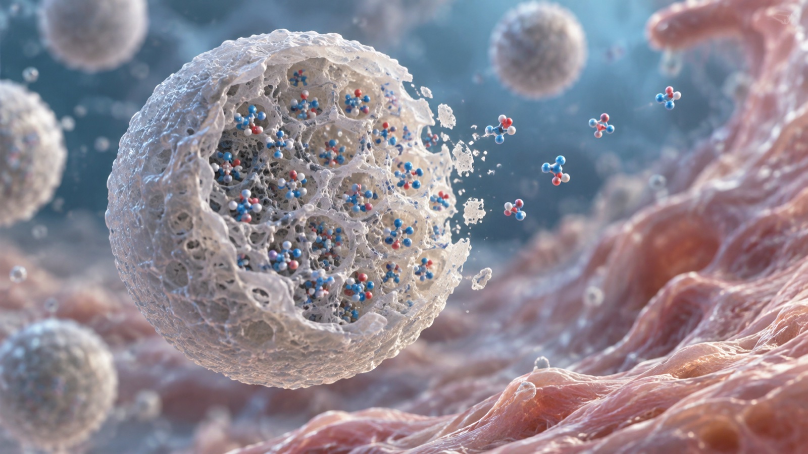
9. Controlled release — how a drug is held decides how it is released
The other pillar of DDS is controlled release. Reading the labels of approved products and the papers shows that the state in which the drug is held is completely different from product to product.
(1) Making the drug a "crystal" or a "salt" inside the liposome
Barenholz summarizes DOXIL as resting on three mutually independent principlesSourced[Ref. 11].
- PEG-modified nanoliposomes, which prolong circulation time in the blood and avoid the reticuloendothelial system
- "Remote loading" (active encapsulation) of doxorubicin, driven by an ammonium sulfate concentration gradient across the membrane, which gives a high and stable load and also allows release in the tumor
- A lipid bilayer in the "liquid-ordered phase", made from phosphatidylcholine with a high phase-transition temperature (53 °C) and cholesterol
On the encapsulated state, the FDA product-specific guidance says that inside the liposome, doxorubicin exists mainly in the form of doxorubicin sulfate crystalsSourced[Ref. 28]. In ONIVYDE, irinotecan is encapsulated gelled or precipitated as the sucrose octasulfate saltSourced[Ref. 17]. Keeping the drug in a nearly solid state inside the sac suppresses leakage: control of crystallization and salting-out is, in effect, release control (this article's commentary).
(2) Embedding the drug in microspheres of a biodegradable polymer
The label of LUPRON DEPOT 3.75 mg lists, as a lyophilized powder of microspheres, leuprolide acetate 3.75 mg, purified gelatin 0.65 mg, DL-lactic and glycolic acids copolymer (PLGA) 33.1 mg and D-mannitol 6.6 mgSourced[Ref. 21]. It is a formulation whose effect continues with one injection a monthSourced[Ref. 21]. Drugs@FDA lists the first approval of LUPRON DEPOT (NDA 019732) as 26 January 1989Sourced[Ref. 27].
On how release from PLGA works, the review by Fredenberg and co-authors says that PLGA is the biodegradable polymer most frequently used for controlled release of encapsulated drugs, but also that drug release from PLGA-based DDS is complex, and that this complexity makes it hard to generalize results or predict releaseSourced[Ref. 14].
(3) Attaching a polymer to change the size of the molecule itself
In ONCASPAR, 69 to 82 mPEG chains of about 5 kDa are attached to the enzymeSourced[Ref. 20]. This uses the property seen in Chapter 3, that "larger molecules leave the blood less readily", by making the molecule larger through attached polymer (this article's commentary). SMANCS, the starting point for Matsumura and Maeda, was also a protein with a polymer attachedSourced[Ref. 1].
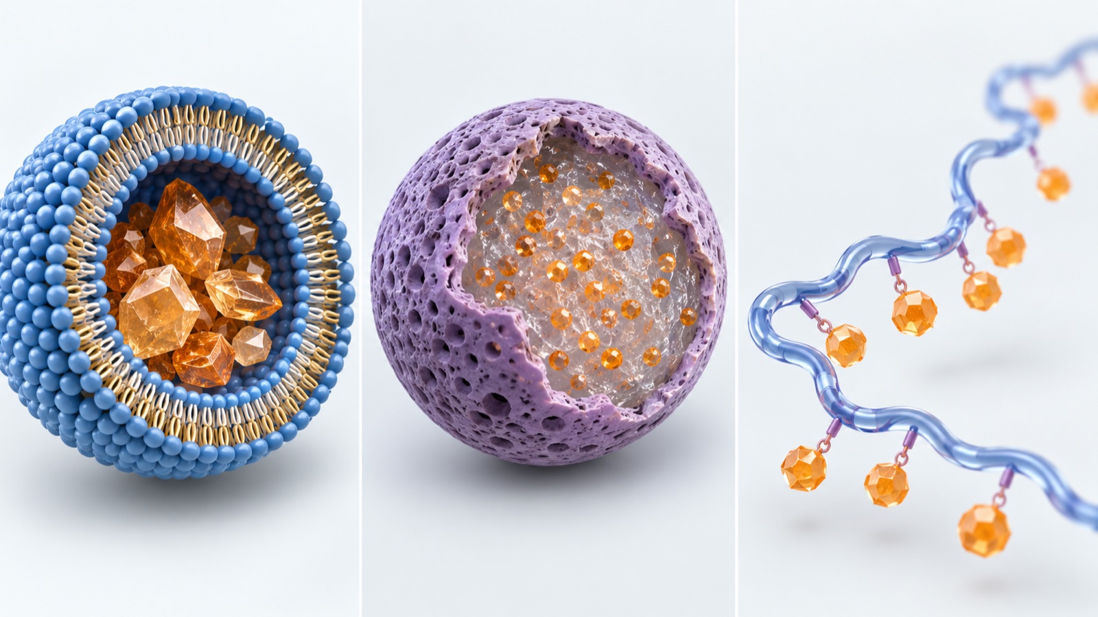
Show the original schematic with sources and numbers
Fig. 6 Conceptual diagram (vector drawing). Panel 1 is based on Barenholz (2012) [Ref. 11], the FDA product-specific guidance [Ref. 28] and the ONIVYDE label [Ref. 17]; panel 2 on the LUPRON DEPOT label [Ref. 21] and Fredenberg et al. (2011) [Ref. 14]; panel 3 on the ONCASPAR label [Ref. 20] and Matsumura and Maeda (1986) [Ref. 1]. The split into three types is this article's own, and the drawing is schematic.
10. Our calculation: how many times the drug's mass is the carrier?
- DOXIL (per mL): doxorubicin hydrochloride 2 mg / cholesterol 3.19 mg / HSPC 9.58 mg / MPEG-DSPE 3.19 mg / sucrose 94 mgSourced
- ONIVYDE (per mL): irinotecan (free base) 4.3 mg / DSPC 6.81 mg / cholesterol 2.22 mg / MPEG-2000-DSPE 0.12 mgSourced
- VYXEOS (per vial): daunorubicin 44 mg and cytarabine 100 mg / DSPC 454 mg, DSPG 132 mg, cholesterol 32 mg / sucrose 2,054 mgSourced
- ABRAXANE (per vial): paclitaxel 100 mg / human albumin about 900 mgSourced
- LUPRON DEPOT 3.75 mg: leuprolide acetate 3.75 mg / PLGA 33.1 mgSourced
- ONCASPAR: an enzyme of four 34.5 kDa subunits, with 69 to 82 mPEG chains of about 5 kDa attachedSourced
From the figures above, we convert how many times the drug's mass the carrier materials amount toOur calculation.
- DOXIL: total lipid 3.19 + 9.58 + 3.19 = 15.96 mg. 15.96 ÷ 2 = about 8.0 times. Of the lipid, PEG-lipid (MPEG-DSPE) is 3.19 ÷ 15.96 = about 20%
- ONIVYDE: total lipid 6.81 + 2.22 + 0.12 = 9.15 mg. 9.15 ÷ 4.3 = about 2.1 times. Of the lipid, PEG-lipid is 0.12 ÷ 9.15 = about 1.3%
- VYXEOS: total lipid 454 + 132 + 32 = 618 mg, total drug 44 + 100 = 144 mg. 618 ÷ 144 = about 4.3 times
- ABRAXANE: 900 ÷ 100 = about 9 times (albumin / paclitaxel)
- LUPRON DEPOT 3.75 mg: 33.1 ÷ 3.75 = about 8.8 times (PLGA / drug)
- ONCASPAR: enzyme 34.5 × 4 = 138 kDa, PEG 5 × 69 to 82 = 345 to 410 kDa. PEG is 345 ÷ 483 = 71% to 410 ÷ 548 = 75% of the whole conjugate, that is, roughly 70% or more of the mass is PEG
Premises and limits: DOXIL is compared by the mass of the hydrochloride and ONIVYDE by the mass of the free base, so the salt forms are not matched. The "about 900 mg" for ABRAXANE is the approximate figure in the label, and the albumin contains sodium caprylate and sodium acetyltryptophanate. The molecular weight of ONCASPAR is a simple sum of the subunit molecular weight in the label and the molecular weight of mPEG. All of these are for looking at the composition of the formulation from a materials viewpoint, not for comparing efficacy or safety.
11. A materials engineer's view 2: why generics took 17 years, read from FDA guidance
According to Drugs@FDA, DOXIL (NDA 050718) was approved on 17 November 1995 and the first generic (Sun Pharma, ANDA 203263) on 4 February 2013Sourced[Refs. 24, 25]. The gap is about 17 years 2 monthsOur calculation. Barenholz likewise wrote in his 2012 review that about two years after the relevant patents expired, there was still no FDA-approved generic version of DoxilSourced[Ref. 11].
Why does it take so long? The product-specific guidance for doxorubicin liposomes (a draft revised in May 2022) published by the FDA shows that what is required of a generic reads, in effect, as a materials and process specificationSourced[Ref. 28].
- Composition: the excipients must be qualitatively the same as the reference product (Q1), with concentrations within ±5% (Q2)
- Manufacturing method: it must be made by active loading with an ammonium sulfate gradient. The main steps are: forming liposomes containing ammonium sulfate, reducing particle size, forming the ammonium sulfate gradient, and actively loading the drug
- Lipid raw materials: the guidance says that lipid excipients are critical in liposomal formulations. The lipids must be of the same synthetic-route class (natural or synthetic) as in the reference product, and the comparison must go beyond meeting specifications to the distribution of molecular species
- Properties to compare: liposome composition, the state of the encapsulated drug (doxorubicin sulfate crystals), the internal environment (volume, pH, sulfate and ammonium ion concentrations), morphology and lamellarity, phase transition of the lipid bilayer, particle size distribution, thickness of the surface PEG layer, surface potential, and leakage under several conditions
- Particle size distribution: D10, D50 and D90 are measured, and equivalence is judged by D50 and SPAN [(D90 − D10)/D50], using at least 30 vials each of the reference and the generic
What is required here is not "the same ingredient list" but "the same internal structure and interfaces". The state of the crystal, the phase behavior of the membrane, the thickness of the polymer layer on the surface and the shape of the particle size distribution are all physical properties that move with process conditions, and the guidance itself recommends identifying critical process parameters and critical material attributes by evaluating the sensitivity of liposome propertiesSourced[Ref. 28].
For a lipid supplier, the point is that meeting the specification may not be enough: even the same distribution of molecular species as the reference product can be asked for. The origin of the raw material, whether natural or synthetic, thus becomes a condition of product equivalence (this article's commentary). Our explainer on biosimilars looks at how the same problem is handled for biologics.
12. Challenges, and what could not be confirmed
(1) The assessment of the EPR effect is unsettled
As seen in Chapter 4, papers differ widely in how they assess the significance of the EPR effect. Sindhwani and co-authors say that understanding the active route through endothelial cells could open up strategies to increase accumulation in tumorsSourced[Ref. 5], but as of this survey (September 2026) we could not confirm any product that has used that route to raise the amount delivered in the clinicNot yet confirmed.
(2) The clinical value of ligands attached to particles
Rosenblum and co-authors report no approved actively targeted nanocarrier as of 2018Sourced[Ref. 3], but whether any product of this kind has been approved by the FDA as of September 2026 was not checked exhaustively in this articleNot yet confirmed.
(3) Patient selection
Rosenblum and co-authors discuss a direction in which the degree of EPR is measured by imaging so that patients who are likely to respond to a nanocarrier can be selectedSourced[Ref. 3]. We could not find, within the scope of this survey, any primary source showing that this has been established as a standard clinical procedureNot yet confirmed.
- A DDS is a "how to carry it" technology. It includes liposomes, albumin particles, polymeric micelles and polymer conjugates, and each has approved productsSourced
- The EPR effect was reported in Japan in 1986. For large proteins, the tumor-to-blood concentration ratio reached 5 within 19 to 72 hoursSourced
- The amount that arrives is a median of 0.7% (pooled animal studies). The amount going elsewhere than the tumor is about 142 times that in the tumorOur calculation
- Attaching a ligand did not change the amount accumulating in the tumor but changed uptake into cells, according to a report in miceSourced
- In approved products, the carrier materials make up about 2 to 9 times the mass of the drugOur calculation
- For generics, equivalence of "internal structure and interfaces" is required, not just the ingredient list. For DOXIL, it took about 17 years 2 months until the first genericOur calculation
13. Glossary
- DDS
- Drug delivery system. A technology that controls where a drug goes and how fast it is released.
- Nanocarrier
- A nanometer-scale particle or polymer that carries a drug by enclosing or binding it.
- Liposome
- A small sac made of a lipid bilayer. The drug is placed in the inner water phase or within the membrane.
- Polymeric micelle
- A particle with a core and a shell, formed by the self-assembly of block copolymers.
- Polymer conjugate
- A drug or protein covalently bound to a polymer chain, such as a PEGylated protein.
- Passive targeting
- Using particle size and surface properties to make a drug gather more readily in a particular tissue.
- Active targeting
- Attaching a ligand so that particles bind to, and are taken up by, cells carrying a particular receptor.
- EPR effect
- Enhanced permeability and retention: the tendency of macromolecules and particles to leak into a tumor and stay there. Reported by Matsumura and Maeda in 1986.
- Ligand
- A component that binds selectively to a specific molecule such as a receptor. Antibodies, peptides and sugars are examples.
- Protein corona
- The layer of proteins that adsorbs onto a particle's surface in the blood and changes the particle's apparent properties.
- Mononuclear phagocyte system (MPS)
- The cellular system, in the liver, spleen and elsewhere, that takes up and removes foreign material.
- PEGylation
- Attaching polyethylene glycol. It suppresses aggregation and clearance and prolongs time in the blood.
- % ID/g
- A unit giving the percentage of the injected dose present per gram of tissue.
- Remote loading
- A loading method that uses an ion concentration difference across the membrane to draw a drug into already-formed liposomes.
- PLGA
- A copolymer of lactic and glycolic acids. A polymer that degrades in the body and is used in sustained-release formulations.
- Q1 / Q2
- Q1: the excipients of a generic are qualitatively the same as those of the reference product. Q2: their concentrations are within ±5%.
- SPAN
- An index of the breadth of a particle size distribution, calculated as (D90 − D10)/D50.
14. References (primary sources)
- Matsumura Y., Maeda H. "A new concept for macromolecular therapeutics in cancer chemotherapy: mechanism of tumoritropic accumulation of proteins and the antitumor agent smancs", Cancer Research 46(12 Pt 1), 6387–6392 (1986), PMID 2946403 — pubmed.ncbi.nlm.nih.gov
- Wilhelm S. et al. "Analysis of nanoparticle delivery to tumours", Nature Reviews Materials 1, 16014 (2016), DOI 10.1038/natrevmats.2016.14 — nature.com
- Rosenblum D., Joshi N., Tao W., Karp J. M., Peer D. "Progress and challenges towards targeted delivery of cancer therapeutics", Nature Communications 9, 1410 (2018), DOI 10.1038/s41467-018-03705-y — nature.com
- Danhier F. "To exploit the tumor microenvironment: Since the EPR effect fails in the clinic, what is the future of nanomedicine?", Journal of Controlled Release 244(Pt A), 108–121 (2016), DOI 10.1016/j.jconrel.2016.11.015 — pubmed.ncbi.nlm.nih.gov
- Sindhwani S. et al. "The entry of nanoparticles into solid tumours", Nature Materials 19(5), 566–575 (2020), DOI 10.1038/s41563-019-0566-2 — pubmed.ncbi.nlm.nih.gov
- Kirpotin D. B. et al. "Antibody targeting of long-circulating lipidic nanoparticles does not increase tumor localization but does increase internalization in animal models", Cancer Research 66(13), 6732–6740 (2006), DOI 10.1158/0008-5472.CAN-05-4199 — pubmed.ncbi.nlm.nih.gov
- Moreira J. N., Gaspar R., Allen T. M. "Targeting Stealth liposomes in a murine model of human small cell lung cancer", Biochimica et Biophysica Acta 1515(2), 167–176 (2001), DOI 10.1016/s0005-2736(01)00411-4 — pubmed.ncbi.nlm.nih.gov
- Moreira J. N., Ishida T., Gaspar R., Allen T. M. "Use of the post-insertion technique to insert peptide ligands into pre-formed stealth liposomes with retention of binding activity and cytotoxicity", Pharmaceutical Research 19(3), 265–269 (2002), DOI 10.1023/a:1014434732752 — pubmed.ncbi.nlm.nih.gov
- Choi H. S. et al. "Renal clearance of quantum dots", Nature Biotechnology 25(10), 1165–1170 (2007), DOI 10.1038/nbt1340 — pubmed.ncbi.nlm.nih.gov
- Charrois G. J., Allen T. M. "Rate of biodistribution of STEALTH liposomes to tumor and skin: influence of liposome diameter and implications for toxicity and therapeutic activity", Biochimica et Biophysica Acta 1609(1), 102–108 (2003), DOI 10.1016/s0005-2736(02)00661-2 — pubmed.ncbi.nlm.nih.gov
- Barenholz Y. "Doxil® — The first FDA-approved nano-drug: Lessons learned", Journal of Controlled Release 160(2), 117–134 (2012), DOI 10.1016/j.jconrel.2012.03.020 — pubmed.ncbi.nlm.nih.gov
- Duncan R. "The dawning era of polymer therapeutics", Nature Reviews Drug Discovery 2(5), 347–360 (2003), DOI 10.1038/nrd1088 — pubmed.ncbi.nlm.nih.gov
- Cabral H., Miyata K., Osada K., Kataoka K. "Block Copolymer Micelles in Nanomedicine Applications", Chemical Reviews 118(14), 6844–6892 (2018), DOI 10.1021/acs.chemrev.8b00199 — pubmed.ncbi.nlm.nih.gov
- Fredenberg S., Wahlgren M., Reslow M., Axelsson A. "The mechanisms of drug release in poly(lactic-co-glycolic acid)-based drug delivery systems — a review", International Journal of Pharmaceutics 415(1–2), 34–52 (2011), DOI 10.1016/j.ijpharm.2011.05.049 — pubmed.ncbi.nlm.nih.gov
- Anselmo A. C., Mitragotri S. "Nanoparticles in the clinic: An update", Bioengineering & Translational Medicine 4(3), e10143 (2019), DOI 10.1002/btm2.10143 — pmc.ncbi.nlm.nih.gov
- FDA / NLM DailyMed "DOXIL (doxorubicin hydrochloride liposome injection) prescribing information" — dailymed.nlm.nih.gov
- FDA / NLM DailyMed "ONIVYDE (irinotecan liposome injection) prescribing information" — dailymed.nlm.nih.gov
- FDA / NLM DailyMed "ABRAXANE (paclitaxel protein-bound particles) prescribing information" — dailymed.nlm.nih.gov
- FDA / NLM DailyMed "VYXEOS (daunorubicin and cytarabine liposome) prescribing information" — dailymed.nlm.nih.gov
- FDA / NLM DailyMed "ONCASPAR (pegaspargase) prescribing information" — dailymed.nlm.nih.gov
- FDA / NLM DailyMed "LUPRON DEPOT 3.75 mg (leuprolide acetate for depot suspension) prescribing information" — dailymed.nlm.nih.gov
- FDA / NLM DailyMed "GIVLAARI (givosiran) prescribing information" — dailymed.nlm.nih.gov
- FDA / NLM DailyMed "PLUVICTO (lutetium Lu 177 vipivotide tetraxetan) prescribing information" — dailymed.nlm.nih.gov
- FDA Drugs@FDA "NDA 050718 DOXIL (LIPOSOMAL)", approval history — accessdata.fda.gov
- FDA Drugs@FDA "ANDA 203263 Doxorubicin Hydrochloride (liposomal), Sun Pharma", approval history — accessdata.fda.gov
- FDA Drugs@FDA "NDA 207793 ONIVYDE", approval history — accessdata.fda.gov
- FDA Drugs@FDA "NDA 019732 LUPRON DEPOT", approval history — accessdata.fda.gov
- FDA "Draft Guidance on Doxorubicin Hydrochloride (Injectable, liposomal)", product-specific guidance, revised May 2022 (PDF) — accessdata.fda.gov
15. Claim-to-source audit
| Claim in the text | Basis | Class |
|---|---|---|
| The earlier finding that SMANCS (neocarzinostatin with a polymer attached) accumulates in tumor tissue more than the original neocarzinostatin. That, with proteins of molecular weight 12,000 to 160,000, many accumulated gradually in tumor tissue and the tumor-to-blood concentration ratio reached 5 within 19 to 72 hours. That neocarzinostatin (molecular weight 12,000) reached neither 1 nor 5. That the authors attributed this to abundant blood vessels, increased permeability to macromolecules and little recovery through blood vessels and lymphatics. The journal and year | Abstract of Matsumura and Maeda (1986) Reference 1 https://pubmed.ncbi.nlm.nih.gov/2946403/ | Sourced |
| That, in a pooled analysis of ten years of literature, a median of 0.7% of the administered nanoparticles reaches a solid tumor, and that this has adverse effects on manufacturing, cost, toxicity, and the performance of imaging and therapy | Abstract of Wilhelm et al. (2016) Reference 2 https://www.nature.com/articles/natrevmats201614 | Sourced |
| Examples of passively targeted nanocarriers in clinical use (Doxil, Abraxane, Marqibo, DaunoXome and Onivyde in the United States; Myocet and Mepact in Europe; Genexol-PM in South Korea; SMANCS in Japan). That the EPR effect was first described by Matsumura and Maeda in 1986. That 15 passively targeted types are approved and no actively targeted type has gone beyond clinical trials (as of 2018). That 0.7% is a value from a meta-analysis of preclinical data, that 0.7% is relatively higher than the delivery efficiency of conventional formulations, and the preclinical example of 0.6% ID versus 0.2% ID. Clearance by the MPS, trapping in liver sinusoids, uptake by hepatocytes and Kupffer cells. That the protein corona affects targeting efficiency. The definition of active targeting and ligand partners (HER2, folate receptor, CD44), and that efficient passive targeting is a prerequisite. The barriers (tumor penetration, heterogeneity, hypoxia, endosomal escape) and the regulatory hurdles and manufacturing scale-up complexity. That BIND-014 did not meet its primary endpoints in two phase 2 trials. That, in 19 patients with HER2-positive metastatic breast cancer, accumulation of 64Cu-labelled liposomes was quantified by PET/CT and peaked at 24 to 48 hours. Animal studies showing heterogeneity of accumulation. The direction of selecting patients by imaging the EPR effect | Rosenblum et al. (2018) Reference 3 https://www.nature.com/articles/s41467-018-03705-y | Sourced |
| That the verdict was that the EPR effect works in rodents but not in humans, and that the author says claims of improved efficacy through the EPR effect should be dropped | Abstract of Danhier (2016) Reference 4 https://pubmed.ncbi.nlm.nih.gov/27871992/ | Sourced |
| That gaps between endothelial cells of tumor vessels have been put at up to 2,000 nm, that those gaps are not responsible for transporting nanoparticles into the tumor, that up to 97% of nanoparticles enter tumors through an active process passing through endothelial cells, and that understanding that route opens strategies to increase accumulation | Abstract of Sindhwani et al. (2020) Reference 5 https://pubmed.ncbi.nlm.nih.gov/31932672/ | Sourced |
| Long-circulating liposomes with an anti-HER2 antibody fragment (Fab′ or scFv) attached to PEG chains: that biodistribution and circulation did not change, that tumor accumulation was similar at 7–8% ID/g with or without targeting, that the targeted ones were mainly inside cancer cells and the untargeted ones in stroma and macrophages, and that uptake into cancer cells was up to 6-fold | Abstract of Kirpotin et al. (2006) Reference 6 https://pubmed.ncbi.nlm.nih.gov/16818648/ | Sourced |
| That Stealth liposomes carrying antagonist G bound 30 to 44 times more strongly to cells from the tumor, that tumor accumulation depended on liposome size but not on the presence of the ligand, and that it was up to 2–4% ID/g | Abstract of Moreira et al. (2001) Reference 7 https://pubmed.ncbi.nlm.nih.gov/11718672/ | Sourced |
| That "post-insertion", which transfers a peptide ligand from micelles with the ligand at the end of PEG-lipid into pre-formed liposomes, gave liposomes carrying up to about 0.3 µg of ligand per µmol of phospholipid with cytotoxicity comparable to conventional coupling. That the paper says a simple way to manufacture ligand-targeted liposomes on a large scale is needed to move into clinical trials | Abstract of Moreira et al. (2002) Reference 8 https://pubmed.ncbi.nlm.nih.gov/11934232/ | Sourced |
| That rapid and efficient urinary excretion occurred at a final hydrodynamic diameter below 5.5 nm, that zwitterionic or neutral organic coatings prevented adsorption of serum proteins, that adsorption increased the hydrodynamic diameter by more than 15 nm and impeded renal excretion, and that these are results of giving quantum dots to rodents | Abstract of Choi et al. (2007) Reference 9 https://pubmed.ncbi.nlm.nih.gov/17891134/ | Sourced |
| That, comparing liposomes of 82, 101, 154 and 241 nm diameter in mice, the three smaller ones accumulated similarly and significantly more than the largest | Abstract of Charrois and Allen (2003) Reference 10 https://pubmed.ncbi.nlm.nih.gov/12507764/ | Sourced |
| That DOXIL was the first FDA-approved nanodrug (1995) and rests on three principles: extended circulation by PEG-modified nanoliposomes, remote loading by an ammonium sulfate gradient, and a liquid-ordered membrane of phosphatidylcholine with a high phase-transition temperature (53 °C) and cholesterol. That no generic existed about two years after patent expiry (as of 2012) | Abstract of Barenholz (2012) Reference 11 https://pubmed.ncbi.nlm.nih.gov/22484195/ | Sourced |
| That nano-sized (5–100 nm) polymeric drugs, "polymer therapeutics", include polymer–drug conjugates, polymer–protein conjugates, polymeric micelles with covalently bound drug and polyplexes for DNA delivery | Abstract of Duncan (2003) Reference 12 https://pubmed.ncbi.nlm.nih.gov/12750738/ | Sourced |
| That the review addresses micelles assembled through multimolecular interactions between block copolymers and their cargo, and that the type of interaction and the chemistry and engineering of the block copolymer are at the center of the design | Abstract of Cabral et al. (2018) Reference 13 https://pubmed.ncbi.nlm.nih.gov/29957926/ | Sourced |
| That PLGA is the biodegradable polymer most frequently used for controlled release of encapsulated drugs, and that release is complex and hard to generalize or predict | Abstract of Fredenberg et al. (2011) Reference 14 https://pubmed.ncbi.nlm.nih.gov/21640806/ | Sourced |
| That VYXEOS is a 100 nm bilamellar liposome | Anselmo and Mitragotri (2019) Reference 15 https://pmc.ncbi.nlm.nih.gov/articles/PMC6764803/ | Sourced |
| That DOXIL is doxorubicin hydrochloride encapsulated in a STEALTH liposome, containing per mL 2 mg doxorubicin hydrochloride, 3.19 mg cholesterol, 9.58 mg HSPC, 3.19 mg MPEG-DSPE and 94 mg sucrose, that more than 90% of the drug is encapsulated, and that Initial U.S. Approval is 1995 | DOXIL label Reference 16 https://dailymed.nlm.nih.gov/dailymed/drugInfo.cfm?setid=1c153e9e-4cf2-4ac7-9cf9-16f9b48d7dce | Sourced |
| That ONIVYDE is a unilamellar liposome of about 110 nm diameter with irinotecan encapsulated gelled or precipitated as the sucrose octasulfate salt, containing per mL 4.3 mg irinotecan (free base), 6.81 mg DSPC, 2.22 mg cholesterol and 0.12 mg MPEG-2000-DSPE | ONIVYDE label Reference 17 https://dailymed.nlm.nih.gov/dailymed/drugInfo.cfm?setid=c8b58efa-1820-48a4-b70d-62918fc4abfc | Sourced |
| That ABRAXANE is an albumin-bound particle with an average diameter of about 130 nm and amorphous paclitaxel, containing per vial 100 mg paclitaxel and about 900 mg human albumin and no solvent, and that Initial U.S. Approval is 2005 | ABRAXANE label Reference 18 https://dailymed.nlm.nih.gov/dailymed/drugInfo.cfm?setid=24d10449-2936-4cd3-b7db-a7683db721e4 | Sourced |
| That VYXEOS encapsulates daunorubicin and cytarabine at a 1:5 molar ratio, that the membrane is DSPC, DSPG and cholesterol at a 7:2:1 molar ratio, that one vial contains 44 mg daunorubicin, 100 mg cytarabine, 454 mg DSPC, 132 mg DSPG, 32 mg cholesterol, 100 mg copper gluconate and 2,054 mg sucrose, and that Initial U.S. Approval is 2017 | VYXEOS label Reference 19 https://dailymed.nlm.nih.gov/dailymed/drugInfo.cfm?setid=7ea701ce-e7d3-4349-a9c2-642a501d45c8 | Sourced |
| That ONCASPAR is L-asparaginase made of four 34.5 kDa subunits with 69 to 82 mPEG chains of about 5 kDa attached, and that Initial U.S. Approval is 1994 | ONCASPAR label Reference 20 https://dailymed.nlm.nih.gov/dailymed/drugInfo.cfm?setid=ac7b6a6f-6ddb-48c6-8088-d8a8e3fea6ce | Sourced |
| That LUPRON DEPOT 3.75 mg is a lyophilized microsphere powder containing 3.75 mg leuprolide acetate, 0.65 mg purified gelatin, 33.1 mg DL-lactic and glycolic acids copolymer and 6.6 mg D-mannitol, and that its effect continues with one injection a month | LUPRON DEPOT 3.75 mg label Reference 21 https://dailymed.nlm.nih.gov/dailymed/drugInfo.cfm?setid=00c486b0-bd7b-4898-834d-8c656e5e73cb | Sourced |
| That GIVLAARI is siRNA to which a ligand containing three GalNAc residues is covalently attached for delivery to hepatocytes, that the molecular weight of givosiran (free acid) is 16,300.34 Da, and that Initial U.S. Approval is 2019 | GIVLAARI label Reference 22 https://dailymed.nlm.nih.gov/dailymed/drugInfo.cfm?setid=167e663c-11e1-497b-a3fc-951d65d58eaa | Sourced |
| That PLUVICTO is a radioligand therapy in which a PSMA-binding ligand is linked to 177Lu through a DOTA chelator, that its molecular weight is 1216.06 g/mol, and that Initial U.S. Approval is 2022 | PLUVICTO label Reference 23 https://dailymed.nlm.nih.gov/dailymed/drugInfo.cfm?setid=14908037-2892-4d98-a053-253ce35afb1a | Sourced |
| That the first approval of DOXIL (NDA 050718) was on 17 November 1995 | Drugs@FDA Reference 24 https://www.accessdata.fda.gov/scripts/cder/daf/index.cfm?event=overview.process&ApplNo=050718 | Sourced |
| That Sun Pharma's doxorubicin hydrochloride liposome injection (ANDA 203263) was approved on 4 February 2013 | Drugs@FDA Reference 25 https://www.accessdata.fda.gov/scripts/cder/daf/index.cfm?event=overview.process&ApplNo=203263 | Sourced |
| That the first approval of ONIVYDE (NDA 207793) was on 22 October 2015 | Drugs@FDA Reference 26 https://www.accessdata.fda.gov/scripts/cder/daf/index.cfm?event=overview.process&ApplNo=207793 | Sourced |
| That the first approval of LUPRON DEPOT (NDA 019732) was on 26 January 1989 | Drugs@FDA Reference 27 https://www.accessdata.fda.gov/scripts/cder/daf/index.cfm?event=overview.process&ApplNo=019732 | Sourced |
| That a generic must have excipients qualitatively the same (Q1) and concentrations within ±5% (Q2), be made by active loading with an ammonium sulfate gradient (four main steps), use lipids of the same synthetic-route class and compare the distribution of molecular species because lipid excipients are critical, and compare the properties listed (composition, state of the doxorubicin sulfate crystals, internal environment, morphology and lamellarity, phase transition, particle size distribution, PEG layer thickness, surface potential, leakage under several conditions). That the MPEG coating protects against MPS clearance and prolongs circulation, and that the PEG layer thickness is thermodynamically limited and estimated at a few nm. The measurement of D10, D50 and D90 with judgement by D50 and SPAN, at least 30 vials each, and identifying critical process parameters and material attributes through the sensitivity of liposome properties. That the document is a draft revised in May 2022 | FDA product-specific guidance (doxorubicin hydrochloride liposome injection) Reference 28 https://www.accessdata.fda.gov/drugsatfda_docs/psg/PSG_050718.pdf | Sourced |
| That the amount going elsewhere than the tumor is 99.3%, about 142 times the tumor. 0.6% ÷ 0.2% = 3 times. DOXIL: total lipid 15.96 mg, about 8.0 times, PEG-lipid about 20%; ONIVYDE: total lipid 9.15 mg, about 2.1 times, PEG-lipid about 1.3%; VYXEOS about 4.3 times; ABRAXANE about 9 times; LUPRON DEPOT about 8.8 times; ONCASPAR PEG about 71–75% (about 2.5 to 3.0 times the enzyme). "Carrier materials are about 2 to 9 times the mass of the drug". About 17 years 2 months from the DOXIL reference approval to the first generic approval | Our calculation. 0.7% is a median from animal studies and not a human value. Salt forms are not matched. The molecular weight of ONCASPAR is a simple sum of the subunit and mPEG molecular weights. This does not compare efficacy or safety | Our calculation |
| Whether any product has used the active route through endothelial cells to raise the amount delivered in the clinic. Whether any actively targeted nanocarrier has been approved by the FDA since 2018. Whether patient selection by imaging has been established as a standard clinical procedure | As of this survey (September 2026), no primary source showing establishment or approval could be confirmed in this article (whether actively targeted types have been approved was not checked exhaustively) | Not yet confirmed |
| A comparison of why active targeting succeeded when a ligand is bonded to a small molecule | No primary source making a direct comparison could be confirmed within the scope of this survey. The text lays published facts side by side as this article's reading (commentary) | Commentary |
| The framing of DDS as a "how to carry it" technology. The split of nanocarriers into four types. Treating the EPR debate as a question of reproducibility and generality and likening it to a paint. The reading that what cells in the body see is a protein layer. Likening the PEG layer to dispersion stabilizers and anti-fouling coatings. The summary that a ligand acts after arrival in the tumor. The reading that holding the drug in a nearly solid state suppresses leakage. The split of controlled release into three types. The reading that what is required of generics is equivalence of "internal structure and interfaces". The reading that whether the particle-making step can be separated from the ligand-loading step is a point at issue in scale-up. The reading that for a lipid supplier the origin of the raw material becomes a condition of equivalence. The grouping of papers by position | This article's arrangement and commentary based on published content. Not views stated by the authors of the papers or by regulators | Commentary |
| The indications, clinical results, and the evaluation and comparison of safety of each product | This article is an explanation of materials and formulation technology and does not evaluate or compare therapeutic effect or safety. For approved products, only compositions and approval dates written in regulators' published materials are quoted | Commentary |
| That the figures combine AI-generated explanatory images (ImageGen) and vector drawings, kept distinct from real objects or measured images | Note by this article | Commentary |
Last updated 26 September 2026. Sources are limited to primary material (FDA-approved labels, Drugs@FDA, FDA product-specific guidance and peer-reviewed papers). Because the article includes readings of materials and process design, those are marked as Commentary and kept separate from sourced fact. Animal-study figures (0.7%, % ID/g and so on) are not human values. The clinical assessment of the EPR effect, the approval status of actively targeted nanocarriers and the establishment of patient selection by imaging could not be confirmed from primary sources and are therefore marked "Not yet confirmed". This article is an explanation of materials and formulation technology; it does not evaluate therapeutic effect or safety and is not medical advice. All figures are explanatory concept graphics. The figures combine AI-generated explanatory images (ImageGen) and vector drawings. The generated images are not real photographs, micrographs or measured data. The original schematics with numbers and sources are left in place, either shown directly or expandable beneath the new images.