TECHNOLOGY EXPLAINER
Bioprinting and Bioinks
— between "can be printed" and "stays alive and works" lie the walls of shear and blood vessels
Bioprinting is a technology for placing cells and materials at set positions in three dimensions. Making the shape itself is now possible with inkjet, extrusion, light-based fabrication and other methods. The hard parts are not damaging cells with the force of dispensing and building channels for blood inside thick tissue. Transplants into people so far amount to small clinical trials, and within the scope of this article there is no product approved by the FDA.
- What bioprinting is (the short version)
- Four methods — extrusion, inkjet, light-based and laser
- Bioink materials — collagen, alginate, GelMA and decellularized matrix
- A materials engineer's view (1): three time scales — during printing, after landing and in culture
- Cell viability and shear
- Our calculation: what order of magnitude is the shear stress in the nozzle?
- How to add a vascular network
- Clinical examples and their stage
- A materials engineer's view (2): a bioink is the raw material of a living product
- Open issues, and what this article could not confirm
- Glossary / References / Claim-to-source audit
Sourced = content stated in published material, papers or registry entries (link given)
Our calculation = a figure this article derived from stated assumptions
Not yet confirmed = research-stage, registered only, or an outlook, with no confirmed track record
Structural readings and interpretations from the materials and process side are marked separately as Commentary.
This article explains materials technology and manufacturing technology. Descriptions of clinical trials and cases introduce what registry entries and papers state; they do not assess the efficacy or safety of any treatment, recommend any treatment, or constitute medical advice. Nor is there any intention to compare the results of small trials with other treatments or other products.
1. What bioprinting is (the short version)
Bioprinting is a technology that places material containing cells at set positions in three dimensions, following design data. In a 2014 review [Source 22], Murphy et al. said that 3D bioprinting means printing biocompatible materials, cells and supporting components into complex, functional, living three-dimensional tissues, and that technical challenges around the choice of materials, cell types, growth and differentiation factors, and the sensitivities of living cells add a complexity that non-biological 3D printing does not haveSourced.
- What is printed: a formulation that contains cells and can be handled in an automated fabrication process is called a "bioink". One without cells is a "biomaterial ink" (the definition of Groll et al.) [Source 1]Sourced. This definition and the basics of the materials were covered in our explainer on hydrogels
- How it is printed: the review by Xu et al. divides the methods into four: inkjet, extrusion, laser-assisted and stereolithography [Source 3]Sourced
- What is hard: cells experience various stresses during printing, and the stronger and longer the stress, the lower the viability is said to be. And thick tissue dies from necrosis without blood vessels insideSourced
Before printing resolution, the range of force that cells can withstand sets the design conditions for the material. One review reports that the viability of mouse fibroblasts (L929) fell on average to 96%, 91% and 76% as the shear stress in the nozzle rose from under 5 kPa to 5 to 10 kPa and above 10 kPaSourced. From the materials side, a formulation that is both easy to flow (low shear stress) and holds its shape after landing (high yield stress) is the core of a bioink (commentary from this article).
2. Four methods — extrusion, inkjet, light-based and laser
The review by Xu et al. sorts, for each method, the main stress on cells, the suitable viscosity of the material and a guide to reported viability. Moroni et al. point out that terminology in the field of biofabrication is inconsistent, and give a guide that sets out how techniques and terms relate [Source 2]Sourced.
Extrusion — the most versatile, and the one that applies the most force
In 2016, Ozbolat et al. summarised extrusion-based bioprinting (EBB) [Source 4] as versatile enough to print a wide range of things, including cells, tissues, tissue constructs, organ modules and microfluidic devices, yet facing obstacles to organ fabrication, limits on the resolution of printed features, challenges to practical use, the development of new bioinks, and regulatory concerns on the way to commercialisationSourced. They list as forms of bioink tissue spheroids, tissue strands, cell pellets, decellularized matrix components, microcarriers and cell-laden hydrogelsSourced.
Inkjet — low viscosity and tiny droplets
In 2010, Cui et al. [Source 7] examined Chinese hamster ovary (CHO) cells printed with a thermal inkjet and reported viability of 89% after printing and 3.5% apoptosis, with transient pores forming in the cell membrane that were repaired within 2 hours. The optimum cell concentration was about 8 million cells/mLSourced. The review also says that cell damage grows when the nozzle temperature exceeds 37 °CSourced.
Stereolithography and laser — methods without a nozzle
In stereolithography (DLP and the like), almost no nozzle shear is applied, and the main damage is radiation stress from the light source and the cytotoxicity of the photoinitiatorSourced. The review's table lists viability of 85% to 91.5% for examples using GelMA and a photosensitizerSourced. Laser-assisted printing (LIFT) has no nozzle and so faces fewer viscosity limits, and there are examples with reported viability above 90%Sourced. Examples of making structures that include blood vessels by stereolithography are covered in chapter 7.
3. Bioink materials — collagen, alginate, GelMA and decellularized matrix
The basic properties of collagen, alginate and GelMA (origin, how they gel, elastic modulus, cell adhesion) were covered in our explainer on hydrogels. Here we sort out the points that bear on printing.
| Material | Role in printing (as the literature states it) | What becomes a problem in printing |
|---|---|---|
| Collagen | In 2019, Lee et al. reported controlling pH-driven gelation and, with the FRESH method (printing into a support bath of gelatin particles), achieving a filament resolution of 20 µm and making collagen structures at scales from capillaries to a whole heartSourced | A step that controls gelation by pH is needed (for the FRESH support bath, see the hydrogel explainer) |
| Alginate | Used in extrusion. Crosslinks with Ca²⁺ (the speed depends on the solubility of the Ca salt; see the hydrogel explainer). There are examples of mixing it with gelatin and controlling viscosity by temperature. Ouyang et al. reported that, with gelatin/alginate bioinks, a longer gelation time made printability worse, and a higher printing temperature and lower gelatin concentration gave higher viability of cells (embryonic stem cells)Sourced | Printability (holding shape) and viability (gentle force) trade off through temperature and gelatin concentration |
| GelMA (photocrosslinked) | Crosslinks by stereolithography or light exposure after the nozzle. The wavelength of the light source and the absorption of the photoinitiator must be matched in design (see the hydrogel explainer) | The load on cells from light and initiator is a main factor in viability, whether in photocrosslinked extrusion or in stereolithographySourced |
| Decellularized matrix (dECM) | In 2014, Pati et al. decellularized and solubilised fat, cartilage and heart tissue and printed the dECM as cell-laden bioinks, reporting high cell viability and functionSourced | Being tissue-derived, the composition is complex and lot-to-lot differences are likely. It is a solution at low temperature and gels at 37 °C, so handling under cooling is needed (Fig. 2) |
Collagen follows Lee et al. [Source 14], alginate Ouyang et al. [Source 8], dECM Pati et al. [Source 9], and the load of GelMA on cells the review by Xu et al. [Source 3]. The "what becomes a problem" column is this article's arrangement based on what the literature states.
Reading dECM bioink from the materials side
4. A materials engineer's view (1): three time scales — during printing, after landing and in culture
The requirements on a bioink are easier to sort out by time (commentary from this article).
- During printing (milliseconds to seconds): while flowing through the nozzle, low viscosity and small shear stress protect the cells. Shear thinning (viscosity falling when force is applied) helps. The dECM ink of Pati et al. [Source 9] was printed as a solution at or below 15 °C through a 250 µm nozzle at the low shear rate of about 0.6 to 2 s⁻¹Sourced
- Just after landing (seconds to minutes): to keep the shape it was placed in, the ink must set quickly (yield stress and storage modulus rise). dECM behaves like a crosslinked gel after 30 minutes at 37 °C, so it was printed together with a PCL frame (line width 100 to 200 µm) that supports the shapeSourced. Photocrosslinking sets within seconds, but puts a load of light and initiator on the cells
- In culture (days to weeks): so that cells can spread, multiply and function, the gel needs loosening, degradation and ease of adhesion (see the hydrogel explainer)
The three pull in opposite directions. It must flow easily (low viscosity), yet set once placed (high elasticity), and after setting let cells move (loosen moderately). The structure is the same as the design of industrial paints, adhesives and pastes that are "applied, then cured", but the difference is that living cells are inside, and the temperature, pH, light and solvents that can be used are limited.
In 2012, Aguado et al. [Source 6] examined viability when cells are injected through a needle, and reported that encapsulating cells in alginate gel (storage modulus G′ of 29.6 Pa) gave human umbilical vein endothelial cells a viability of 88.9±5.0%, higher than the 58.7±8.1% in buffer alone. The protective effect weakened whether G′ was raised or loweredSourced. The result shows that the material is both the object that is printed and a "cushion" that protects cells from force (commentary from this article).
5. Cell viability and shear
The review by Xu et al. sorts out that cell viability is determined mainly by the size and the duration of the stress received: the higher the stress and the longer the time, the lower the viability. In nozzle-based methods cells mainly receive shear stress on ejection, and in light-based methods they receive thermal and radiation stressSourced. The review also cites the view that if shear stretches the cell membrane by more than 5%, the membrane can no longer hold its shape, ruptures and the cell diesSourced.
- Effect of pressure (Nair et al., 2009) [Source 5]: varying pressure and nozzle diameter and examining cell damage, they reported that dispensing pressure affects viability more than nozzle diameter does, and that in structures printed at 40 psi the proportion of living cells was 38.75% lower than at 5 psiSourced
- Relation between shear stress and viability (Ouyang et al., 2016) [Source 8]: they reported an exponential relationship between embryonic stem cell viability and the shear stress induced, and that viability was higher at higher printing temperature and lower gelatin concentrationSourced
- Controlling shear (Blaeser et al., 2016) [Source 21]: using a microvalve-based device, they controlled the shear stress in the nozzle with a fluid-dynamics model and examined how the level of shear stress affects cell survival and proliferative capacitySourced
- Relation to injection (Aguado et al., 2012) [Source 6]: both experiment and theory suggest that the stretching flow at the needle inlet is the main cause of acute cell deathSourced
A caution is needed here. The viability figure changes with whether it is measured right after printing or several days later, with the stain used, and with the cells. Also, being "alive" is not the same as "functioning". The review by Xu et al. says that, besides viability, keeping the pluripotency and differentiation capacity of stem cells is a precondition for the function of engineered tissueSourced.
6. Our calculation: what order of magnitude is the shear stress in the nozzle?
Shear stress can be estimated roughly from the conditions of the material and the equipment. To get the order of magnitude, we use two relations.
Equation 1 (pressure balance): for steady, fully developed laminar flow in a circular tube, the wall shear stress is τw = ΔP × R ÷ (2L) (ΔP: pressure difference, R: nozzle radius, L: nozzle length). This holds regardless of the flow properties of the fluidOur calculation.
- ΔP = 5 psi (34.5 kPa), R = 0.2 mm, L = 12.7 mm → τw = 34.5 × 10³ × 0.2 × 10⁻³ ÷ (2 × 12.7 × 10⁻³) = about 270 Pa
- ΔP = 40 psi (276 kPa), R = 0.2 mm, L = 12.7 mm → about 2.2 kPa
- ΔP = 276 kPa, R = 0.1 mm, L = 3 mm (a short tip) → about 4.6 kPa
- ΔP = 500 kPa, R = 0.05 mm, L = 2 mm → about 6.3 kPa
Equation 2 (when the flow rate is fixed): for a Newtonian fluid, τw = 4μQ ÷ (πR³) = 4μv ÷ R (μ: viscosity, Q: flow rate, v: mean velocity). Doubling the viscosity doubles the stress, and halving the radius multiplies it by 8Our calculation.
- Assume a printing speed (mean velocity) v = 10 mm/s and R = 0.2 mm. The wall shear rate is 4v ÷ R = 200 s⁻¹
- μ = 1 Pa·s → τw = 4 × 1 × 0.01 ÷ 0.0002 = 200 Pa
- μ = 10 Pa·s → 2 kPa; μ = 50 Pa·s → 10 kPa
For the dECM ink of Pati et al.: τ = viscosity × shear rate = 2.8, 6.13 and 23.6 Pa·s × 2 s⁻¹ = 5.6, 12 and 47 Pa (values at 15 °C)Our calculation.
Premises and limits: Equation 2 assumes a Newtonian fluid; real bioinks show shear thinning, so the stress would be smaller than this. The pressures and nozzle dimensions in Equation 1 are this article's assumptions over typical ranges and are not values for a particular machine. The review's bands of 5 kPa and 10 kPa are reports of nozzle stress for L929 cells and do not apply unchanged to other cells or machines. This calculation is for grasping orders of magnitude.
What can be read from this is as follows (commentary from this article).
- The low-shear condition of Pati et al. (about 50 Pa) is two orders of magnitude below the review's "under 5 kPa" band. The design is to extrude cold at a low shear rate and support the shape with a frame until it sets
- Even a material of about 1 Pa·s can reach stresses of several kPa with a fine nozzle and fast printing. Narrowing the nozzle raises resolution, but the stress climbs steeply (it scales with the third power of the radius)
- The ways to lower stress come to four: lower the viscosity (temperature, concentration), strengthen shear thinning, widen the nozzle, or print more slowly. The first two are material design; the last two are a trade-off of equipment against resolution
7. How to add a vascular network
Thick tissue needs channels that carry oxygen and nutrients to its interior. In 2012, Miller et al. stated that without a perfusable vascular network, a three-dimensional tissue packed with cells soon develops a necrotic coreSourced. We calculated that the limit of oxygen diffusion is of the order of a few hundred µm in our explainer on organoids and spheroids. In bioprinting, making channels at the same time as the shape of the tissue becomes a central design problem.
Four ways to make perfusable channels
The left side of each image shows fabrication; the right side shows a cutaway of the result. Blue represents culture medium flowing through the open channel.
1. Embed and remove a sacrificial template (Miller et al.)
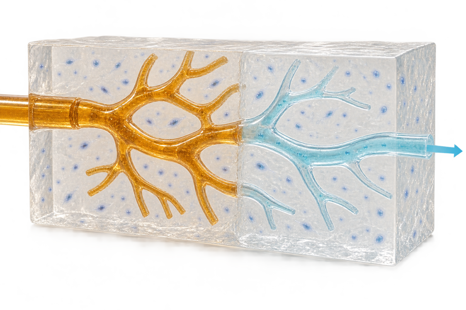
- Print sugar template
- Embed in cell-laden gel
- Remove template
Build the channel shape first, solidify material around it, then remove the template.
2. Co-print several materials (Kolesky et al.)
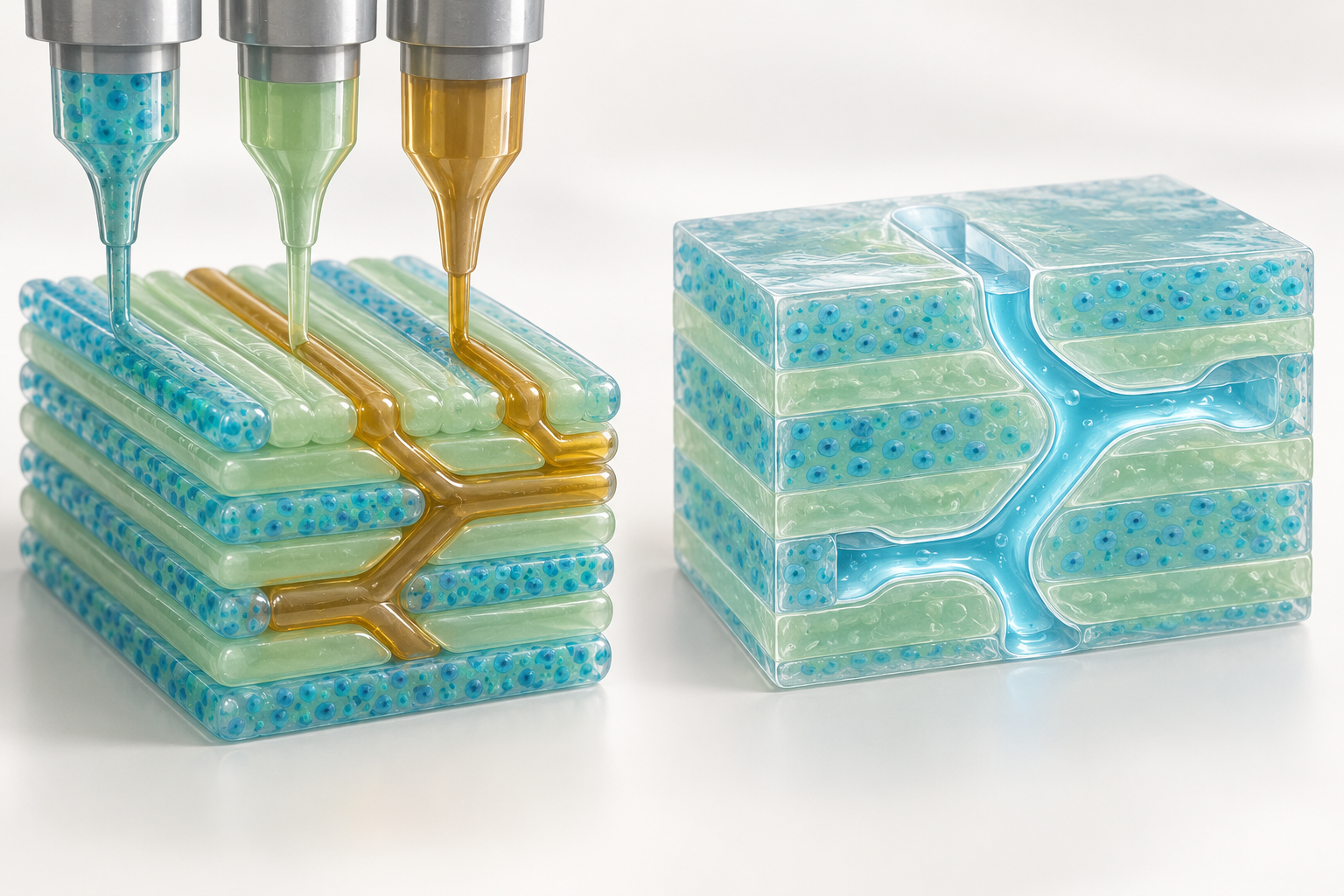
- Prepare bioinks
- Co-print cells and channel ink
- Remove channel ink
Place cell-containing material and a temporary space-holder for the channel together.
3. Shape the surrounding gel with light (Grigoryan et al.)
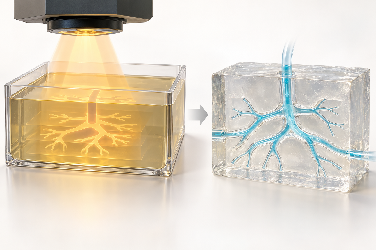
- Prepare photocurable gel
- Pattern it with light
- Wash out uncured gel
A light pattern defines the void without printing a solid sacrificial template.
4. Write inside densely packed cells (SWIFT)
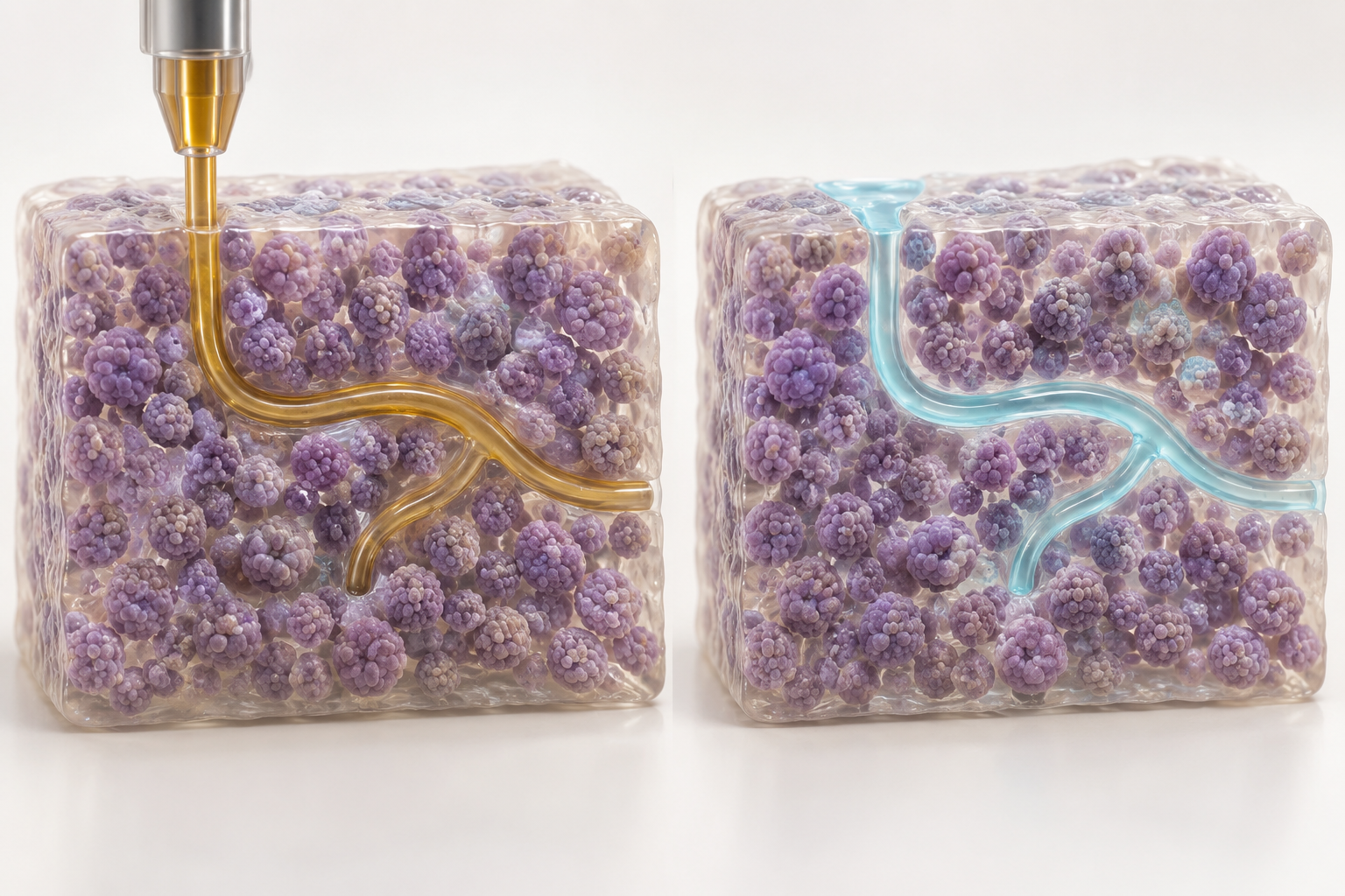
- Pack cell aggregates
- Write sacrificial ink inside
- Remove ink and perfuse
Write a route afterward through a material already rich in cells.
- Sacrificial mold (Miller et al., 2012): a rigid filament network of carbohydrate glass was printed and embedded in cell-laden tissue as a mold; it could be lined with endothelial cells, giving cylindrical networks that could be perfused with blood under high-pressure pulsatile flow. The perfused channels preserved the metabolic function of primary rat hepatocytes that was otherwise suppressed in the core of the tissue. The authors say the channel geometry, endothelialisation and tissue outside the channels can be controlled independently, and that it suits a range of cells, extracellular matrices and crosslinking methodsSourced
- Co-printing (Kolesky et al., 2014): they reported a method of printing several bioinks together in three dimensions to make heterogeneous tissue constructs with vessels, multiple cell types and extracellular matrixSourced
- Stereolithography (Grigoryan et al., 2019): adding food dyes to photopolymerizable hydrogels as biocompatible, powerful light absorbers, they used projection stereolithography to make, in minutes, transparent gels containing intravascular fluid mixers and bicuspid valves. They also show an example using a biodegradable hydrogel carrier in a rodent model of chronic liver injurySourced
- Embedded printing (Skylar-Scott et al., 2019): they reported the SWIFT method, which gathers hundreds of thousands of iPS cell-derived organoids into a dense cell mass of about 10⁸ cells/mL and writes perfusable channels into it. The heart tissue beat synchronously for 7 daysSourced
- Printing collagen (Lee et al., 2019): with the FRESH method, controlling gelation by pH, they printed collagen structures at scales from capillaries to a whole heart with a filament resolution of 20 µm. A ventricle seeded with human cardiomyocytes showed synchronised contraction and wall thickening (up to 14% at peak systole)Sourced
In almost every method, adding a vascular network comes down to designing either a material that is put in first and later disappears or a material that separates the parts to be set by light from the parts not to be (commentary from this article).
- A sacrificial material must be both strong enough to print and removable without harming cells. Miller et al. describe carbohydrate glass as a "cytocompatible sacrificial mold"Sourced
- The support of Skylar-Scott et al. is a self-healing viscoplastic body that flows as the writing needle passes and recovers afterwardsSourced. This is the same yield-stress and recovery rheology as the FRESH support bath in our hydrogel explainer
- The light absorber of Grigoryan et al. plays the same role as the absorbers and light blockers in UV-curable resins that decide "how deep to cure" (commentary from this article). Finding an absorber of low cytotoxicity, such as a food dye, is the task on the materials side
8. Clinical examples and their stage
| Trial / example | Product makeup (as stated) | Subjects and scale | What the registry entry and paper confirm |
|---|---|---|---|
| Kyoto University Hospital Bio 3D nerve conduit | The patient's own skin fibroblasts were expanded into spheroids about 500 µm in diameter and stacked on a needle array with Cyfuse's Bio 3D printer to make a tube without scaffold material. The spheroids fused in about a week and the needle array was removed. The tube was over 20 mm long and 2.5±1 mm in diameter | 3 patients aged 20 to 60 with a peripheral nerve defect of the hand (length 20 mm or less, nerve diameter 2 mm or less). Skin was harvested and the implant placed 6 weeks later. Followed for 48 weeks | jRCT2053200022 (registered 1 June 2020). A non-randomised safety and feasibility trial. No related adverse events were seen, and all 3 patients completed the trial. The authors state that recovery of the nerve, a secondary outcome, was also obtained [Source 16]Sourced |
| AuriNovo (3DBio Therapeutics) | A 3D bioprinted collagen hydrogel scaffold encapsulating the patient's own auricular cartilage cells, printed to match the size and shape of the opposite ear | Unilateral microtia (grade II to IV). Phase 1/2a, single-arm, open-label. Actual enrollment: 2 | NCT04399239. Started 9 August 2021. Status is terminated, and the reason for termination is recorded as a business decision unrelated to safety [Source 17]Sourced. We could not confirm any results |
| Seoul St. Mary's Hospital Trachea | A patient-specific trachea integrating a biopolymer, a hydrogel, nasal-derived stem cells and nasal septal chondrocytes by 3D bioprinting | Patients with a thyroid or airway disease needing partial resection. Phase 1/2, single-arm, actual enrollment: 1 | NCT06051747. Started 17 July 2023. Status is "unknown" (last verified status: active, not recruiting) [Source 18]. A commentary paper presents this trial as the first successful transplant of a bioprinted artificial trachea [Source 19]Sourced. We could not confirm details in a peer-reviewed original paperNot yet confirmed |
The Kyoto University Hospital example follows Ikeguchi et al. (2024) [Source 16]; the other two follow the ClinicalTrials.gov entries [Sources 17 and 18] and a commentary paper [Source 19]. Where the origin of the cells (autologous or allogeneic) is not clear from the registry entry, we have kept to what it says.
The Kyoto University Hospital trial is notable for stacking the spheroids themselves, without scaffold material (hydrogel). In 2015, Itoh et al., using the same needle-array method, made a tubular tissue of 1.5 mm inner diameter from 500 spheroids of endothelial cells, smooth muscle cells and fibroblasts, grafted it into the abdominal aorta of rats, and reported that every graft stayed open and a layer of endothelial cells was confirmed 5 days after graftingSourced.
As for approved products, none of the product names on the FDA list of approved cellular and gene therapy products (updated 17 September 2026) could be confirmed as made by bioprinting [Source 20]Sourced. We have not checked approved products in other countries and regions exhaustivelyNot yet confirmed.
9. A materials engineer's view (2): a bioink is the raw material of a living product
When a bioink becomes a real product, demands other than "can be printed" are added to the material (commentary from this article).
- Variation in biological raw materials: collagen comes from animals and dECM from tissue, materials whose composition is complex and which vary between lots. dECM is solubilised with acid and pepsin and used after adjusting the pH [Source 9]. The degree of solubilisation and the control of residues become central to the specification
- Printing conditions become the specification: temperature (dECM is a solution at or below 15 °C), pH, light wavelength and dose, pressure and nozzle diameter all affect cell viability (chapters 5 and 6). When the conditions change, it can become "a different product". The thinking in our explainer on QbD and CMC applies directly
- Wetted parts: as Equation 2 shows, the precision of the inner diameter of syringes, nozzles and cartridges, and the state of the inner surface, act on the wall shear stress through the inverse cube of the radius. The tolerance on inner diameter and the roughness and surface treatment of the inner surface are specifications bearing directly on cell viability
- Sterility: it is generally known that high-viscosity inks are hard to sterilise by filtration. Sterility control of raw materials and preparation in a closed system become important, but we could not confirm any public standard for bioinks. For sterility control of cell products, see our explainer on GMP
- Quality of scaffold-free methods: where spheroids are stacked, the uniformity of the spheroids (500±50 µm in diameter in the Kyoto University Hospital example) is central to quality instead of the ink [Source 16]. Quality control then comes closer to that of a cell product
10. Open issues, and what this article could not confirm
(1) Viability is no substitute for function
Many reports give viability after printing, but whether the cells keep their function and what happens in the long term need separate evaluation. The review by Xu et al. also says that, besides viability, the proliferation and differentiation capacity of stem cells must be kept [Source 3]Sourced.
(2) For vascular networks, there is a gap between making tubes and working as tissue
The reports in chapter 7 show that perfusable tubes can be made. As for the barrier function of the endothelial cells lining the tubes, long-term maintenance under blood flow, and coordination with surrounding tissue, we could not confirm enough within the scope of this surveyNot yet confirmed.
(3) Clinical evidence is at the small-study stage
The human trials we could confirm involve 3 patients, 2 (terminated) and 1. They are not on a scale that can evaluate efficacy, nor can they be compared with other treatments [Sources 16 to 18].
(4) Regulatory framework
The review by Ozbolat et al. lists regulatory concerns on the way to commercialisation as a challenge [Source 4]Sourced. We have not confirmed any FDA or PMDA guidance specific to bioprintingNot yet confirmed.
(5) What this article could not confirm
- Details of the Seoul St. Mary's Hospital tracheal transplant in a peer-reviewed original paper (only the registry entry and a commentary paper were confirmed)
- Results of the AuriNovo trial
- Approval status of bioprinted products in other countries and regions, including Japan and Europe
- Public specifications and standards for bioinks (such as standardised methods for evaluating printability)
- Market-size estimates (not covered, because we do not use market-research estimates)
- There are four methods: extrusion, inkjet, stereolithography and laser-assisted. The force on cells and the suitable viscosity differ by methodSourced
- Viability is set by the size and duration of stress. One report gives mean L929 viability of 96%, 91% and 76% for nozzle shear stress under 5 kPa, 5 to 10 kPa and above 10 kPaSourced
- Stress doubles when viscosity doubles, and rises 8-fold when the nozzle radius is halved. A low-shear dECM ink is about 50 Pa, and a material of 10 Pa·s can reach the order of several kPa depending on conditionsOur calculation
- Channels for blood are made with materials that are put in first and later disappear (sacrificial materials), or by designing light absorption. Perfusable tubes and heart tissue that beat for 7 days have been reportedSourced
- In humans, the scale is 3 patients for a nerve conduit, 2 (terminated) for ear reconstruction and 1 for a trachea. There is no approved product that this article could confirmSourced
11. Glossary
- Bioprinting
- Placing material containing cells in three dimensions following design data.
- Bioink
- A formulation that contains cells and can be handled in an automated fabrication process. One without cells is a biomaterial ink.
- Extrusion
- Pushing material out of a nozzle as a filament by pressure or a piston.
- Inkjet
- Ejecting droplets by heat or a piezoelectric element. Uses low-viscosity liquids.
- Stereolithography (DLP, etc.)
- Curing material one layer at a time with light. Almost no nozzle shear.
- Laser-assisted printing (LIFT)
- Pushing out droplets with the pressure of a laser pulse.
- Shear stress
- The force on a wall or a cell when layers of fluid slide past each other. Unit: Pa.
- Shear thinning
- The property of viscosity falling under force so that a material flows more easily.
- Yield stress
- The stress below which a material behaves like a solid. An indicator of shape retention.
- dECM
- Extracellular matrix from decellularized tissue. Solubilised and used as ink.
- Scaffold-free
- Making tissue from cell masses alone, without scaffold materials such as hydrogels.
- Sacrificial ink / sacrificial mold
- A material put in first and later dissolved away to leave a space such as a tube.
- Perfusion
- Flowing a liquid (culture medium or blood) through a tube.
- FRESH method
- Printing inside a support bath of gelatin particles, then warming to release the print.
- Spheroid
- A spherical mass formed by cells gathering together.
12. References
- Groll J et al. "A definition of bioinks and their distinction from biomaterial inks", Biofabrication 11:013001 (2018) — doi.org
- Moroni L et al. "Biofabrication: A Guide to Technology and Terminology", Trends Biotechnol 36:384 (2018) — doi.org
- Xu HQ et al. "A review on cell damage, viability, and functionality during 3D bioprinting", Mil Med Res 9:70 (2022) (PMC) — pmc.ncbi.nlm.nih.gov
- Ozbolat IT, Hospodiuk M "Current advances and future perspectives in extrusion-based bioprinting", Biomaterials 76:321 (2016) — doi.org
- Nair K et al. "Characterization of cell viability during bioprinting processes", Biotechnol J 4:1168 (2009) — doi.org
- Aguado BA et al. "Improving viability of stem cells during syringe needle flow through the design of hydrogel cell carriers", Tissue Eng Part A 18:806 (2012) — doi.org
- Cui X et al. "Cell damage evaluation of thermal inkjet printed Chinese hamster ovary cells", Biotechnol Bioeng 106:963 (2010) — doi.org
- Ouyang L et al. "Effect of bioink properties on printability and cell viability for 3D bioplotting of embryonic stem cells", Biofabrication 8:035020 (2016) — doi.org
- Pati F et al. "Printing three-dimensional tissue analogues with decellularized extracellular matrix bioink", Nat Commun 5:3935 (2014) (PMC) — doi.org
- Miller JS et al. "Rapid casting of patterned vascular networks for perfusable engineered three-dimensional tissues", Nat Mater 11:768 (2012) — doi.org
- Kolesky DB et al. "3D bioprinting of vascularized, heterogeneous cell-laden tissue constructs", Adv Mater 26:3124 (2014) — doi.org
- Grigoryan B et al. "Multivascular networks and functional intravascular topologies within biocompatible hydrogels", Science 364:458 (2019) — doi.org
- Skylar-Scott MA et al. "Biomanufacturing of organ-specific tissues with high cellular density and embedded vascular channels", Sci Adv 5:eaaw2459 (2019) — doi.org
- Lee A et al. "3D bioprinting of collagen to rebuild components of the human heart", Science 365:482 (2019) — doi.org
- Itoh M et al. "Scaffold-Free Tubular Tissues Created by a Bio-3D Printer Undergo Remodeling and Endothelialisation when Implanted in Rat Aortae", PLoS One 10:e0136681 (2015) — doi.org
- Ikeguchi R et al. "Peripheral nerve regeneration following scaffold-free conduit transplant of autologous dermal fibroblasts: a non-randomised safety and feasibility trial", Commun Med 4:12 (2024) (PMC) — doi.org
- ClinicalTrials.gov "NCT04399239: AuriNovo for Auricular Reconstruction" — clinicaltrials.gov
- ClinicalTrials.gov "NCT06051747: Patient-Customized Bioprinting Technology for Practical Regeneration of the Respiratory Tract (Trachea)" — clinicaltrials.gov
- Shah AM et al. "Artificial trachea transplant: Tale of two stories", Artif Organs 48:575 (2024) — doi.org
- US FDA "Approved Cellular and Gene Therapy Products" (updated 17 September 2026) — fda.gov
- Blaeser A et al. "Controlling Shear Stress in 3D Bioprinting is a Key Factor to Balance Printing Resolution and Stem Cell Integrity", Adv Healthc Mater 5:326 (2016) — doi.org
- Murphy SV, Atala A "3D bioprinting of tissues and organs", Nat Biotechnol 32:773 (2014) — doi.org
13. Claim-to-source audit
| Claim in the text | Basis | Label |
|---|---|---|
| The definition that a bioink is a formulation containing cells that can be handled in an automated fabrication process, and one without cells is a biomaterial ink | Reference 1 https://doi.org/10.1088/1758-5090/aaec52 | Sourced |
| That 3D bioprinting prints biocompatible materials, cells and supporting components into complex, functional, living three-dimensional tissues, and that challenges around materials, cell types, growth factors and the sensitivity of living cells add a complexity that non-biological 3D printing lacks | Reference 22 https://doi.org/10.1038/nbt.2958 | Sourced |
| That terminology in biofabrication is inconsistent and a guide setting out how techniques and terms relate is needed | Reference 2 https://doi.org/10.1016/j.tibtech.2017.10.015 | Sourced |
| The four-way classification of methods (inkjet, extrusion, laser-assisted, stereolithography), the suitable viscosity of each (inkjet 3 to 30 mPa·s, extrusion 30 to 6×10⁸ mPa·s, laser-assisted up to 300 mPa·s), and the guide to reported viability (inkjet above 85%, laser-assisted above 90%, GelMA stereolithography examples 85 to 91.5%). The kinds of stress on cells (shear for nozzle methods, heat and radiation for light-based methods). That viability is set by the size and duration of stress. That mean L929 viability was 96%, 91% and 76% at nozzle shear stress under 5 kPa, 5 to 10 kPa and above 10 kPa. The view that a cell membrane stretched by more than 5% ruptures. That damage grows when the nozzle exceeds 37 °C. That in stereolithography there is almost no nozzle shear and the main damage is radiation stress and photoinitiator cytotoxicity. That, besides viability, proliferation and differentiation capacity of stem cells must be kept | Reference 3 https://pmc.ncbi.nlm.nih.gov/articles/PMC9756521/ | Sourced |
| That extrusion bioprinting is versatile enough to print a wide range of things, yet faces challenges in organ fabrication, resolution, practical use, development of new bioinks and regulatory concerns; the forms of bioink (tissue spheroids, strands, cell pellets, dECM, microcarriers, cell-laden hydrogels) | Reference 4 https://doi.org/10.1016/j.biomaterials.2015.10.076 | Sourced |
| That dispensing pressure affects viability more than nozzle diameter, and that structures printed at 40 psi had a 38.75% lower proportion of living cells than at 5 psi; that an equation predicting viability as a function of shear stress was derived | Reference 5 https://doi.org/10.1002/biot.200900004 | Sourced |
| That endothelial cell viability on needle injection was 88.9±5.0% when encapsulated in alginate gel with a storage modulus of 29.6 Pa and 58.7±8.1% in buffer alone; that the protective effect weakened when the modulus was raised or lowered; that the stretching flow at the needle inlet is regarded as the main cause of acute cell death | Reference 6 https://doi.org/10.1089/ten.tea.2011.0391 | Sourced |
| That CHO cells printed with a thermal inkjet had 89% viability and 3.5% apoptosis, that transient membrane pores were repaired within 2 hours, and that the optimum cell concentration was about 8 million cells/mL | Reference 7 https://doi.org/10.1002/bit.22762 | Sourced |
| That with gelatin/alginate inks a longer gelation time worsened printability, and a higher printing temperature and lower gelatin concentration gave higher embryonic stem cell viability; that there is an exponential relationship between viability and the shear stress induced | Reference 8 https://doi.org/10.1088/1758-5090/8/3/035020 | Sourced |
| dECM bioink: dECM from fat, cartilage and heart solubilised at 3% (0.5 M acetic acid, pepsin, 48 hours, pH 2.8 to 3) and pH adjusted below 10 °C; a solution at or below 15 °C and gelling in 30 minutes at 37 °C; viscosity at 15 °C and 2 s⁻¹ of 2.8 (cartilage), 6.13 (fat) and 23.6 Pa·s (heart) (the unit is written Pa s⁻¹ in the paper but was treated as a viscosity value); shear rate of 0.64 to 2.048 s⁻¹ through a 250 µm nozzle; use with a PCL frame (cartilage 200 µm, fat 100 µm); high viability and function | Reference 9 https://doi.org/10.1038/ncomms4935 | Sourced |
| That tissue densely packed with cells develops a necrotic core without a perfusable vascular network; that a carbohydrate-glass sacrificial mold allowed endothelialisation and perfusion with blood under high-pressure pulsatile flow, and preserved the function of primary rat hepatocytes in the core; that it suits various cells, matrices and crosslinking methods | Reference 10 https://doi.org/10.1038/nmat3357 | Sourced |
| A method of co-printing several bioinks in three dimensions to make heterogeneous tissue constructs with vessels, multiple cell types and extracellular matrix | Reference 11 https://doi.org/10.1002/adma.201305506 | Sourced |
| That projection stereolithography with food dyes as light absorbers made transparent hydrogels containing intravascular fluid mixers and bicuspid valves in minutes; that a biodegradable hydrogel carrier was used in a rodent model of chronic liver injury | Reference 12 https://doi.org/10.1126/science.aav9750 | Sourced |
| That the SWIFT method wrote perfusable channels into a dense cell mass of about 10⁸ cells/mL gathered from hundreds of thousands of iPS cell-derived organoids, and heart tissue beat synchronously for 7 days; that the support behaves as a self-healing viscoplastic body | Reference 13 https://doi.org/10.1126/sciadv.aaw2459 | Sourced |
| That collagen was printed by the FRESH method, with pH-controlled gelation giving a filament resolution of 20 µm and structures from capillaries to a whole heart; that a ventricle seeded with human cardiomyocytes showed synchronised contraction and wall thickening of up to 14% at peak systole | Reference 14 https://doi.org/10.1126/science.aav9051 | Sourced |
| That a tubular tissue of 1.5 mm inner diameter made by the needle-array method from 500 spheroids of endothelial cells, smooth muscle cells and fibroblasts was grafted into rat abdominal aorta, all grafts stayed open, and an endothelial cell layer was confirmed after 5 days | Reference 15 https://doi.org/10.1371/journal.pone.0136681 | Sourced |
| The Kyoto University Hospital Bio 3D nerve conduit trial (jRCT2053200022, registered 1 June 2020): spheroids of 500±50 µm diameter made from the patient's own skin fibroblasts and made into a scaffold-free tube (length over 20 mm, diameter 2.5±1 mm) with Cyfuse's Bio 3D printer and a needle array; implanted in 3 patients with a peripheral nerve defect of the hand and followed for 48 weeks with no related adverse events; all 3 completed the trial; that the authors state recovery of the nerve, a secondary outcome, was also obtained; the eligibility criteria | Reference 16 https://doi.org/10.1038/s43856-024-00438-6 | Sourced |
| That the AuriNovo trial (NCT04399239) is a single-arm phase 1/2a trial using a 3D bioprinted collagen hydrogel scaffold encapsulating the patient's own auricular cartilage cells for microtia, started 9 August 2021, actual enrollment 2, status terminated, with the reason recorded as a business decision unrelated to safety | Reference 17 https://clinicaltrials.gov/study/NCT04399239 | Sourced |
| That the tracheal trial (NCT06051747) is a single-arm phase 1/2 trial transplanting a patient-specific trachea that integrates a biopolymer, a hydrogel, nasal-derived stem cells and nasal septal chondrocytes by 3D bioprinting, started 17 July 2023, actual enrollment 1, status "unknown" (last verified: active, not recruiting) | Reference 18 https://clinicaltrials.gov/study/NCT06051747 | Sourced |
| That a commentary paper presents the tracheal transplant as the first successful transplant of a bioprinted artificial trachea | Reference 19 https://doi.org/10.1111/aor.14758 | Sourced |
| That none of the product names on the FDA list of approved cellular and gene therapy products (updated 17 September 2026) could be confirmed as made by bioprinting | The result of this article checking the product-name list; a check by name only, which did not look into how each product is made. Reference 20 https://www.fda.gov/vaccines-blood-biologics/cellular-gene-therapy-products/approved-cellular-and-gene-therapy-products | Sourced |
| That a microvalve-based device controlled the shear stress in the nozzle with a fluid-dynamics model and the effect of shear stress on cell survival and proliferative capacity was examined | Reference 21 https://doi.org/10.1002/adhm.201500677 | Sourced |
| Estimates from wall shear stress τw = ΔP·R/(2L) (about 270 Pa at 5 psi, R = 0.2 mm, L = 12.7 mm; about 2.2 kPa at 40 psi; about 4.6 kPa at 276 kPa, R = 0.1 mm, L = 3 mm; about 6.3 kPa at 500 kPa, R = 0.05 mm, L = 2 mm). From τw = 4μv/R (v = 10 mm/s, R = 0.2 mm), 200 s⁻¹, and 200 Pa, 2 kPa and 10 kPa at μ = 1, 10 and 50 Pa·s. 5.6, 12 and 47 Pa for the dECM of Pati et al. Twice the stress at double the viscosity and 8 times at half the radius | Our calculation. Nozzle dimensions, pressures, printing speed and viscosity are this article's assumptions, and Equation 2 assumes a Newtonian fluid. The pressures of 5 psi and 40 psi use the conditions of Reference 5, viscosity and shear rate the values of Reference 9, and the 5 kPa and 10 kPa bands the values of Reference 3. It is for grasping orders of magnitude and not a measurement of any real machine or ink | Our calculation |
| Details in a peer-reviewed original paper of the Seoul St. Mary's Hospital tracheal transplant, results of the AuriNovo trial, the barrier function and long-term maintenance of endothelium in vessels, approval status in other regions including Japan and Europe, public specifications for bioinks, and regulatory guidance specific to bioprinting | No primary source could be confirmed within the scope of this article, so these are not covered or are treated as not yet confirmed | Not yet confirmed |
| The grouping into three time scales (during printing, after landing, in culture); the four ways to lower stress; the grouping of vascular-network methods into four; the arrangement of development stages into four; the readings on biological raw materials, wetted parts, sterility and quality of the spheroid method; the general view that high-viscosity inks are hard to sterilise by filtration | This article's own arrangement and commentary based on published content; not views expressed by any author or institution. The statement on sterilising filtration was not checked against a primary source for this article | Commentary |
| That Figs. 1, 2, 3, 5 and 6 are drawings made for explanation (Fig. 4 includes our calculation), and that the hero image is AI-generated | A note from this article | Commentary |
Last updated 1 October 2026. Sources are limited to primary material (peer-reviewed papers and reviews, registry entries on ClinicalTrials.gov and jRCT, and the FDA list of approved products). Market-size and share estimates are not used. Because the article includes structural readings and interpretations from the materials and process side, those are marked as Commentary and kept separate from sourced fact. Details of the Seoul St. Mary's Hospital tracheal transplant in an original paper, results of the AuriNovo trial, the barrier function and long-term maintenance of endothelium in vessels, approval status in other regions, public specifications for bioinks, and regulatory guidance specific to bioprinting could not be confirmed in published primary sources, so they are not covered or are treated as not yet confirmed. All figures are for explanation. Figs. 1, 2, 3, 5 and 6 are conceptual diagrams (vector drawings), Fig. 4 is a drawing that includes our calculation (vector drawing), and the hero image is AI-generated; none of them shows a real machine, tissue or cell. This article explains technology and is neither medical advice nor a recommendation of any particular treatment or product.