TECHNOLOGY EXPLAINER
Lipid Nanoparticles (LNPs)
— Performance is set by the lipid's pKa and by how it is mixed
An LNP is a particle made of four kinds of lipid. A paper that compared 56 lipids showed that, for the ionizable lipid at the center, a pKa shift of only 0.5 changes potency by orders of magnitude. And the step that makes the particle is simply "mixing two liquids". It is a material in which molecular design and mixing conditions are themselves product specification items.
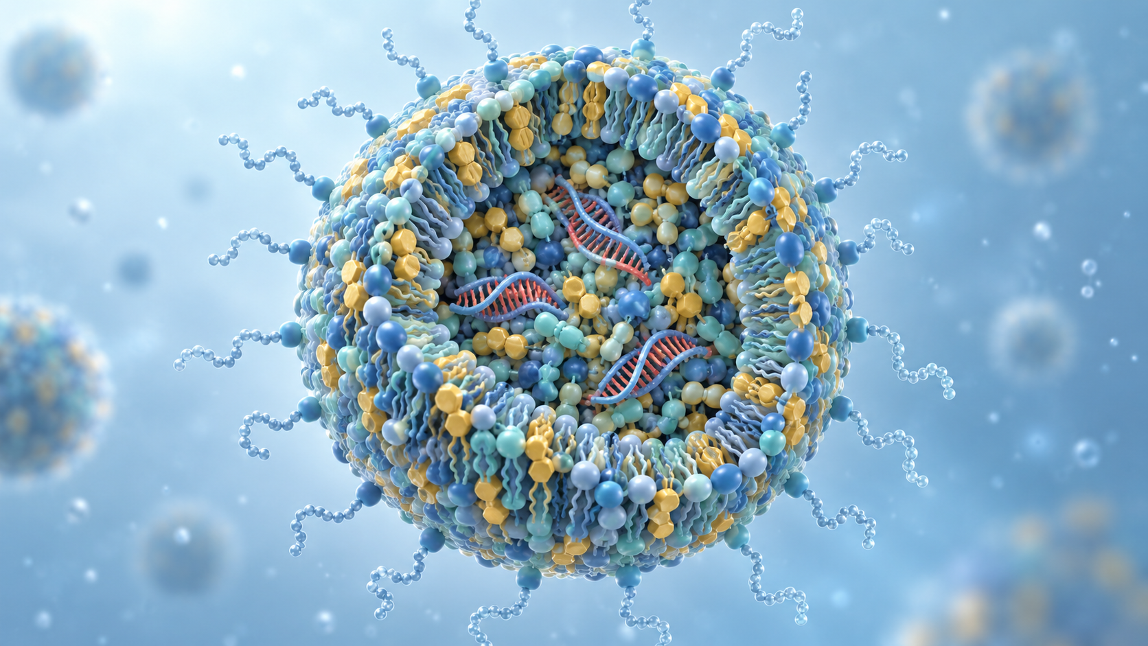
LNPs as a formulation for mRNA vaccines (the role of each of the four components, the composition of one dose, the cold chain) are covered in our explainer on mRNA medicines. To avoid overlapping with it, this article focuses on the molecular design of the ionizable lipid, the molar ratios of the lipids, the manufacturing process, the route taken in the body, and delivery beyond the liver. The chemistry of nucleic acids themselves is in our explainer on oligonucleotide therapeutics, and contract manufacturing of drug substances is in our explainer on CDMOs.
- What an LNP is (the short version)
- The ionizable lipid — pKa decides potency
- A materials engineer's view 1: pKa is not something you "adjust by formulation"
- Molar ratios of the four components — from 40/10/40/10 to 50/10/38.5/1.5
- Our calculation: working back to the molar ratio from the composition of an approved product
- PEG-lipid is a component designed to come off
- How it is made — from T-junction mixing to microfluidics
- Manufacturing process and specifications of an approved product — read from the EPAR
- A materials engineer's view 2: the specification items are the material's requirements
- The route in the body — apoE binds, and only 1–2% escapes the endosome
- Delivery beyond the liver — research-stage work
- Challenges, and what could not be confirmed
- Glossary / References / Claim-to-source audit
Sourced = stated in published material (link given)
Our calculation = a figure this article derived, with the assumptions spelled out
Not yet confirmed = a research-stage item or plan whose results have not been confirmed
Structural readings and interpretations of materials or process design are marked separately as Commentary. Inference is shown in bold.
This article is an explanation of materials and manufacturing technology. It does not evaluate the efficacy or safety of any treatment or vaccine, and it is not medical advice. Approval status and product compositions are quoted only where they appear in the published materials of regulators (FDA and EMA). Results in animals do not necessarily apply to humans.
1. What an LNP is (the short version)
- What it does: nucleic acids (siRNA and mRNA) are degraded in the body if given as they are, and cannot get into cells. An LNP is a material that wraps them in a lipid particle and carries them. For the lipid excipients of Onpattro, the EMA public assessment report says that they associate with the siRNA, protect it from immediate degradation in the circulation and help deliver it to the target site, the liverSourced[Ref. 7]
- What it is made of: four components: an ionizable lipid, a phospholipid (DSPC), cholesterol and a PEG-lipidSourced[Ref. 7]. In the approved product, the ionizable lipid and the PEG-lipid were assessed as "novel excipients"Sourced[Ref. 7]
- How it differs from a liposome: the EMA report states, from cryo-TEM images, that the particles of the patisiran formulation do not have the bilayer structure typical of ordinary liposomes but are spherical nanoparticles with an electron-dense coreSourced[Ref. 7]. It is a "filled particle", not a "hollow sac"
The performance of an LNP is set first by the pKa of the ionizable lipid, and only then by the lipid ratios. Jayaraman and co-authors compared 56 amino lipids and reported that pKa correlated strongly with the strength of gene silencing, with an optimum at pKa 6.2 to 6.5Sourced[Ref. 1]. This is a matter of molecular design, not formulation design, and moreover a value measured after the particle is made (this article's commentary).
2. The ionizable lipid — pKa decides potency
An ionizable lipid is a lipid that is positively charged in acid and carries no charge near neutral pH. The EMA report explains the significance of this property as followsSourced[Ref. 7].
The ionizable amino lipid DLin-MC3-DMA provides well-defined physicochemical properties that govern particle formation, siRNA encapsulation, cellular uptake, fusogenicity (the ability to fuse with the endosomal membrane) and release of the siRNA from the endosome. The lipid was chosen after extensive screening of related cationic lipidsSourced.
Being neutral at a biologically relevant pH (about 7.4) allows it to circulate without marked degradation. When it leaves that pH and enters the liver endosome (acidic, pH about 5), the LNP breaks down and the active substance is released as the endosomal membrane is disruptedSourced.
DLin-MC3-DMA is molecular formula C43H79NO2, molecular weight 642.09, a colorless to pale-yellow liquid with two linoleyl chains (C18:2) containing double bonds, freely soluble in organic solvents and practically insoluble in waterSourced.
So how high should the pKa be? The 2012 paper by Jayaraman and co-authors examined this systematically. They synthesized 56 amino lipids and studied the relation between pKa and the potency of silencing the mouse factor VII (FVII) gene (ED50)Sourced[Ref. 1].
| Item | What the paper says |
|---|---|
| Conclusion | A strong correlation was found between the lipid pKa and silencing of the mouse FVII gene (FVII ED50), and the optimal pKa range was 6.2 to 6.5 |
| How pKa was measured | By titrating the fluorescence of TNS (2-(p-toluidino)-6-naphthalenesulfonic acid) from pH 3 to 10 in steps of 0.5. TNS fluoresces when it associates with a positively charged membrane surface and does not when it is free in solution |
| Structure and pKa | Changing the distance between the ester and the amine by 1 to 5 methylene units moved the pKa across a range of 4.17 to 7.16 |
| Most active lipid | DLin-MC3-DMA (lipid 16): pKa 6.44, ED50 0.03 mg/kg. Activity fell sharply when the pKa shifted to either the acidic or the basic side |
| Conditions for comparison | Particles were extruded up to three times through an 80 nm membrane to give a diameter of 70–90 nm and a polydispersity of 0.11 ± 0.04 (n = 56). The siRNA-to-total-lipid weight ratio was about 0.05 |
All Sourced (Jayaraman et al. 2012 [Ref. 1]). These are results in mice, not values in humans.
3. A materials engineer's view 1: pKa is not something you "adjust by formulation"
With an ordinary formulated material, when the target property is not reached you can vary the mix ratio. The pKa of an ionizable lipid, however, is a property of that molecule. What Jayaraman and co-authors did was to rebuild the skeleton: lengthening the methylene chain between the ester and the amine one unit at a time, or changing the substituents on the nitrogenSourced[Ref. 1]. As a result the pKa moved from 4.17 to 7.16Sourced[Ref. 1].
That is, a requirement such as "raise the pKa by 0.3" becomes a change to the synthetic route (this article's commentary). The scale of synthesizing and comparing 56 compounds is exactly the typical way of materials development: filling in the structure-property relationship by experiment.
Another important point is how it is measured. The TNS assay makes the particle first and then probes the positive charge of its membrane surface by pH titrationSourced[Ref. 1]. It measures not the dissociation constant of the isolated molecule but an apparent pKa "as a particle surface", which is the same idea as measuring the zeta potential of a powder or the acid number of a resin surface (this article's commentary). The material is controlled not by its own value but by the value in the form in which it will be used.
This is why the comparison experiments were held to such tight conditions. Particles were brought to a diameter of 70–90 nm and a polydispersity of 0.11 ± 0.04 by extruding up to three times through an 80 nm membrane, and only then was the pKa variedSourced[Ref. 1]. It also means that if the particle size is left scattered, the effect of pKa cannot be seen (this article's commentary).
4. Molar ratios of the four components — from 40/10/40/10 to 50/10/38.5/1.5
Once the pKa is fixed, the next question is in what ratio to mix the four components. The paper by Jayaraman and co-authors records this progression clearly.
The standard composition for the comparison experiments was amino lipid / DSPC / cholesterol / PEG-lipid at a molar ratio of 40/10/40/10Sourced[Ref. 1].
The paper then says that the molar ratio of the four components (50/10/38.5/1.5) had been further optimized to raise in vivo activity, achieving an ED50 of 0.005 mg/kg in the mouse FVII model, which was a 6-fold improvement over the 40/10/40/10 compositionSourced[Ref. 1]. A study giving the same lipid composition to cynomolgus monkeys was also carried outSourced[Ref. 1].
What stands out is how unevenly the numbers moved. The ionizable lipid rose from 40 to 50 mol%, but the PEG-lipid fell from 10 mol% to 1.5 mol%, to less than one-seventh. Chapter 6 deals with the reason.
5. Our calculation: working back to the molar ratio from the composition of an approved product
Is the molar ratio the paper found optimal also used in an actual approved product? It can be checked from the figures in the label.
- patisiran (siRNA): 2 mg (equivalent to 2.1 mg as patisiran sodium)
- DLin-MC3-DMA: 13.0 mg
- Cholesterol: 6.2 mg
- DSPC: 3.3 mg
- PEG2000-C-DMG: 1.6 mg
- Potassium dihydrogen phosphate 0.2 mg / sodium chloride 8.8 mg / disodium hydrogen phosphate heptahydrate 2.3 mg / water for injection. The pH is about 7.0
- Molecular formula of patisiran sodium C412H480N148Na40O290P40, molecular weight 14,304 Da
All of these are as written in the FDA labelSourced[Ref. 6]. The molecular weights are 642.09 for DLin-MC3-DMA and 2,650 ± 300 Da for PEG2000-C-DMG (EMA public assessment report)Sourced[Ref. 7], and 386.7 for cholesterol and 790.1 for DSPC (PubChem)Sourced[Refs. 12, 13].
We convert the masses to moles and find the molar ratioOur calculation.
- DLin-MC3-DMA: 13.0 ÷ 642.09 = 20.2 µmol
- Cholesterol: 6.2 ÷ 386.7 = 16.0 µmol
- DSPC: 3.3 ÷ 790.1 = 4.18 µmol
- PEG2000-C-DMG: 1.6 ÷ 2,650 = 0.60 µmol
- Total 40.99 µmol. The mole percentages are ionizable lipid 49.3% / DSPC 10.2% / cholesterol 39.0% / PEG-lipid 1.5%
This nearly matches the 50/10/38.5/1.5 that the paper calls "optimized"Our calculation.
We also make a few other conversionsOur calculation.
- Mass ratio of lipid to siRNA: total lipid 6.2 + 13.0 + 3.3 + 1.6 = 24.1 mg. 24.1 ÷ 2 = about 12 times
- Weight ratio of siRNA to total lipid: 2 ÷ 24.1 = 0.083 (the paper's comparison experiments used about 0.05)
- Ionizable lipid per siRNA molecule: siRNA is 2 mg ÷ 14,304 = 0.140 µmol. 20.2 ÷ 0.140 = about 145 molecules
- Amine-to-phosphate ratio: the formula has 40 P, so one siRNA molecule carries about 40 negative charges. 145 ÷ 40 = about 3.6
Premises and limits: the molecular weight of PEG2000-C-DMG is a value with a range, 2,650 ± 300, so its mole percentage has a range too (1.7% at 2,350 and 1.4% at 2,950). The molecular weights of cholesterol and DSPC are the PubChem values. The amine-to-phosphate ratio is an upper limit, assuming that each ionizable lipid molecule has one amine and that all are charged; the actual degree of charging is set by pH and pKa. Nor does it indicate the ratio fed in during manufacturing.
6. PEG-lipid is a component designed to come off
As Chapter 4 showed, the PEG-lipid fell from 10 mol% to 1.5 mol%. Why did it have to be reduced? The EMA report states its role clearly.
The chain length of the PEG-lipid was chosen so that the PEG-lipid dissociates over time from the circulating particle, PEG-free particles appear, and these are taken up by hepatocytes, leading to release of the siRNASourced[Ref. 7].
PEG2000-C-DMG is molecular formula (C2H4O)n C36H73NO5 (n about 47), molecular weight 2,650 ± 300 Da, a white to off-white powder, and freely soluble in water, chloroform and acetonitrileSourced[Ref. 7].
Coming off is built into the design of the PEG-lipid. What sets the speed of shedding is the length of the hydrophobic chains, and Mui and co-authors studied this quantitativelySourced[Ref. 10].
For three PEG-lipids with dialkyl chains of 14, 16 and 18 carbons, they measured in vivo how fast the lipid transfers from the LNP to plasma lipoproteinsSourced[Ref. 10].
At 1.5 mol%, PEG-lipid is the threshold concentration at which chain length has the smallest effect on gene silencing in the liver, but it can still alter the pharmacokinetics and biodistribution of the LNPSourced[Ref. 10].
Raising the concentration to 2.5 mol% and 3.5 mol% greatly impairs hepatocyte gene silencing for PEG-lipid with distearyl (C18) chains, but has little effect with the shorter dimyristyl (C14) chainsSourced[Ref. 10].
The paper positions PEG-lipid as necessary to control the self-assembly process when the lipid particle forms, while it can negatively affect delivery to hepatocytes and gene silencing in the liverSourced[Ref. 10].
The PEG-lipid has an unusual standing. While the particle is being made, it is needed to stop self-assembly and set the particle size. But once in the body, it hinders uptake by cellsSourced[Ref. 10].
The solution adopted was two measures: "reduce the amount" and "make it easier to come off" (this article's framing). The PEG2000-C-DMG used in Onpattro has dimyristyl (C14) chainsSourced[Ref. 7]. In the results of Mui and co-authors, the C14 chain is on the fast-shedding side, since silencing is not easily impaired when its concentration is raisedSourced[Ref. 10].
For engineers who handle dispersants and emulsifiers, "an additive that is needed to form the film but that you do not want left in the final product" is not unusual. Normally one can evaporate it or burn it off, but that is impossible in the body. So the desorption rate is designed through the length of the hydrophobic chain, which builds the removal step into the molecular structure (this article's commentary).
7. How it is made — from T-junction mixing to microfluidics
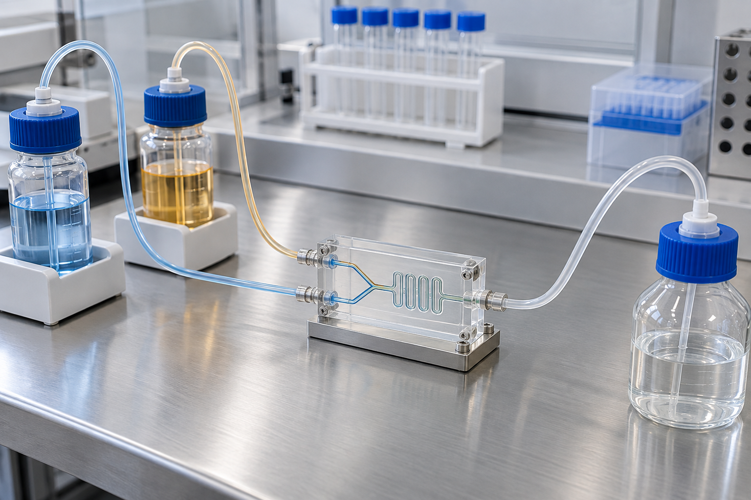
LNP manufacturing centers on a step in which an ethanol phase containing the lipids and an aqueous phase containing the nucleic acid are mixed. The way of mixing has changed over the past 20 years.
| Generation | Method | What was obtained (as described in the paper) |
|---|---|---|
| Jeffs et al. (2005) | A T-shaped mixing chamber merges a stream of DNA buffer with a stream of ethanol and lipid, lowering the ethanol concentration at once. A scalable method that does not use extrusion | Monodisperse vesicles smaller than 200 nm, with DNA encapsulation efficiency above 80%. cryo-TEM showed a mixture of unilamellar, bilamellar and multilamellar vesicles |
| Belliveau et al. (2012) | Microfluidic mixing, which allows mixing in milliseconds at the nanoliter scale | Reproducible production of "limit-size" LNPs of 20 nm and above with nearly complete siRNA encapsulation over a wide range of conditions. Polydispersity as low as 0.02. The optimized system gave 50% gene silencing in mouse hepatocytes at 10 µg/kg of siRNA |
All Sourced (Jeffs et al. 2005 [Ref. 2], Belliveau et al. 2012 [Ref. 3]). Belliveau and co-authors say that conventional macroscopic mixing gives systems 70 nm or larger in diameter, with variable siRNA encapsulation efficiency, uniformity and reproducibilitySourced[Ref. 3]. These are animal-study values.
8. Manufacturing process and specifications of an approved product — read from the EPAR
Onpattro (patisiran) was approved by the FDA on 10 August 2018Sourced[Ref. 14]. The EMA public assessment report describes the manufacture and quality control of the product in concrete terms.
The LNPs are formed by mixing an aqueous solution containing the siRNA (the patisiran active substance) with an ethanolic solution containing the four lipidsSourced[Ref. 7].
The manufacturing process of the drug product consists of five main stepsSourced[Ref. 7]: (1) preparing the solutions of the active substance and the lipids, (2) mixing the solutions to form LNPs, (3) ultrafiltration, buffer exchange and first concentration, (4) dilution to the final concentration and bioburden filtration, and (5) sterile filtration and filling into vials.
On how development proceeded, the report says that an iterative approach was used, examining at the same time the effect of using different ionizable lipids and the relative molar ratios of the four lipidsSourced[Ref. 7].
On particle size, it says that particle size of the product affects pharmacokinetic parameters, and is therefore controlled during manufacture throughout the LNP formation process, and that the particle size was chosen mainly to enable delivery to the liverSourced[Ref. 7]. The size of the particles is given as roughly 60–100 nmSourced[Ref. 7].
The specifications at release and over the shelf life include the following testsSourced[Ref. 7].
Appearance / identification by molecular weight (IPRP-HPLC MS) / identification by single-strand retention time (AX-HPLC UV) / purity and impurities / content (AX-HPLC UV) / lipid identification and lipid content (RP-HPLC ELSD) / encapsulation of the duplex (siRNA) (fluorescence assay) / pH / osmolality / particle size (DLS) / elemental impurities (ICP-MS) / residual ethanol (GC) / residual EDTA / subvisible particles / endotoxin / sterility / content uniformity / extractable volume / container closure integrity / in vitro release of siRNA (kinetic fluorescence assay)
The report also records a discussion of how the limits were set. The initially proposed statistically derived limits were not accepted by the CHMP, which asked for limits within ±3 SD or narrower to be proposed, and the report says that as a result the limits for some items were tightenedSourced[Ref. 7].
Another entry will interest materials engineers: the problem that a white film can form in the vial after transport. The report says this is an inherent property of LNPs as a response to mechanical stress, and that the label instructs users to draw the product up with a filter needle, not to shake or agitate the vial, and, after adding it to the infusion bag, to mix by gently invertingSourced[Ref. 7].
9. A materials engineer's view 2: the specification items are the material's requirements
If the underlined items among the specifications in Chapter 8 are lined up again, they turn out to be the quantities that can move in the process (this article's commentary).
- Lipid content (each of the four components): whether the formulation ratio is kept. As Chapter 5 showed, the molar ratio bears directly on potency
- Encapsulation: whether the nucleic acid is inside the particle. It moves with mixing conditionsSourced[Ref. 7]
- Particle size (DLS): the report itself says it is controlled during manufacture throughout the LNP formation processSourced[Ref. 7]
- Residual ethanol: the flip side of a process that uses a solvent
- In vitro release: it measures not only "is it in there" but "does it come out". According to the report, this item was added to the specification during the procedureSourced[Ref. 7]
This shows the character of LNPs as a material. Even with the same ingredient list, a different way of mixing changes particle size, encapsulation and release. That is why the specifications extend beyond "composition" to "structure" and "behavior" (this article's commentary). The pattern is exactly the same as the requirements for generic versions of liposomal products discussed in our explainer on drug delivery systems and targeting.
From the point of view of the lipid supplier there is one more important point. For DLin-MC3-DMA and PEG2000-C-DMG the EMA report says that they are chemically synthesized lipid compounds, manufactured from well-defined starting materials under acceptable specifications, and it confirms that critical quality attributes and critical process parameters are defined, and that information on process- and product-related organic impurities, residual solvents, inorganic impurities (including elemental impurities) and genotoxic impurities is sufficiently detailedSourced[Ref. 7]. For PEG2000-C-DMG it also states that no animal-derived or human-derived materials are used in the manufacturing processSourced[Ref. 7].
This means that lipids used as excipients are asked to be controlled in the manner of a drug substance (this article's commentary). In general industrial materials, it is like being required to control even auxiliaries and dispersants to the same impurity-profile standard as the main raw material.
10. The route in the body — apoE binds, and only 1–2% escapes the endosome
Why do LNPs go to hepatocytes? No liver-directed ligand is attached to the surface. Akinc and co-authors reported the mechanismSourced[Ref. 4].
They found that apolipoprotein E (apoE), which plays a large part in the physiological clearance of lipoproteins and their uptake into hepatocytes, acts as an endogenous targeting ligand for ionizable LNPs, but not for cationic LNPsSourced[Ref. 4].
This role was examined in in vitro experiments with recombinant apoE and in vivo experiments in wild-type and apoE-deficient mice, and dependence on the receptor was examined using low-density lipoprotein receptor (LDLR)-deficient miceSourced[Ref. 4].
The paper also developed a method that uses a multivalent GalNAc cluster as an exogenous ligand to bind the asialoglycoprotein receptor (ASGPR) of hepatocytes, and concludes that both the endogenous apoE-based and the exogenous GalNAc-based approaches appear to be effective strategies for delivering ionizable LNPs to the liverSourced[Ref. 4].
What happens after the particle enters the cell? Gilleron and co-authors followed it with fluorescence imaging and electron microscopySourced[Ref. 5].
LNPs enter cells by clathrin-dependent endocytosis and macropinocytosis, through both constitutive and inducible pathways depending on the cell typeSourced[Ref. 5].
By directly detecting colloidal-gold particles attached to the siRNA, the authors estimated that escape of siRNA from the endosome into the cytosol occurs only at low efficiency (1–2%), and only within a limited time while the LNP stays in a particular compartment that has features of both early and late endosomesSourced[Ref. 5].
The figure of 1–2% changes how goals are set in materials development (this article's commentary). Circulation in the blood and uptake are already happening quite efficiently. Even so, two orders of magnitude are lost at the last step, crossing the membrane. If this can be improved, the dose might be reduced: that is the line of reasoning.
In fact, Sabnis and co-authors, who developed a new amino lipid for mRNA, say in their abstract that mechanistic studies show that the gain in efficiency is attributable to increased endosomal escapeSourced[Ref. 11]. The paper reports new amino lipids that deliver mRNA efficiently in rodent and primate models, and says that one-month toxicity evaluations in rats and non-human primates found no adverse events with the new LNP systemSourced[Ref. 11].
The trade-off between "raising reactivity" and "keeping stability" is at the center here too. A lipid that breaks membranes easily also breaks them easily in the blood. Setting the boundary of response by pKa is a way of solving this contradiction by handing it over to the pH difference of the environment (this article's commentary). It is the same pattern as the design of cleavable linkers in our explainer on antibody drugs and ADCs.
11. Delivery beyond the liver — research-stage work
As Chapter 10 showed, LNPs go to the liver because apoE binds and carries them to a receptor. The flip side is that to reach anywhere other than the liver, a particle has to get away from this mechanism. All of the following are research-stage reports and not approved products.
| Study | Method | Reported result |
|---|---|---|
| Cheng et al. (2020) | SORT (selective organ targeting). Systematically designing several types of LNP so that tissues other than the liver can be edited, by adding a supplementary "SORT molecule" | Designed SORT lipid nanoparticles that target the lung, spleen and liver. Selectively edited cell types such as epithelial cells, endothelial cells, B cells, T cells and hepatocytes. Can be used with mRNA, Cas9 mRNA with guide RNA, and Cas9 ribonucleoprotein complexes |
| Rurik et al. (2022) | An LNP targeting CD5 delivers modified mRNA encoding a CAR to T lymphocytes. Injected into a mouse model of heart failure | Efficient delivery to T lymphocytes was observed, and transient, functional CAR-T cells were generated in the body. Fibrosis decreased and cardiac function recovered after injury |
All Sourced (abstracts of Cheng et al. 2020 [Ref. 8] and Rurik et al. 2022 [Ref. 9]). Both are animal-study reports and not approved treatments. CAR-T cells themselves are covered in our explainer on CAR-T cells.
12. Challenges, and what could not be confirmed
(1) Delivery beyond the liver is at the research stage
The reports in Chapter 11 are all animal studies. Whether any LNP product targeting an organ other than the liver has been approved by the FDA or EMA as of September 2026 was not checked exhaustively in this articleNot yet confirmed.
(2) The mixing method of the approved product could not be identified
The EMA report says that LNPs are formed by mixing an aqueous solution and an ethanolic solutionSourced[Ref. 7], but the public materials we consulted did not give the type of mixing device (T-junction, microfluidic and so on), the flow rates, the temperature or the lipid concentrations. The report itself says only that the critical process parameters and their acceptable ranges are "defined".
(3) How far the efficiency of endosomal escape can be raised
The 1–2% figure of Gilleron and co-authors is an estimate for a particular cell system and set of conditionsSourced[Ref. 5]. We could not find, within the scope of this survey, any primary source showing how far this has been improvedNot yet confirmed.
- An LNP is a four-component particle, not a hollow sac. cryo-TEM images are described as showing spherical particles with an electron-dense coreSourced
- The pKa of the ionizable lipid decides potency. Across 56 lipids the optimal range was 6.2 to 6.5, and DLin-MC3-DMA was 6.44Sourced
- The molar ratio moved from 40/10/40/10 to 50/10/38.5/1.5. Working back from the composition of an approved product also gives 49.3/10.2/39.0/1.5, a near matchOur calculation
- PEG-lipid is a component designed to come off. Chain length changes the shedding rate, and 1.5 mol% is described as the thresholdSourced
- Manufacturing centers on "mixing two liquids", and the specifications extend beyond composition to particle size, encapsulation and releaseSourced
- The hardest part is escape from the endosome, with an efficiency estimated at 1–2%Sourced
13. Glossary
- LNP (lipid nanoparticle)
- A nanoparticle made of four kinds of lipid that encloses a nucleic acid and carries it into cells.
- Ionizable lipid
- A lipid that is positively charged in acid and carries no charge near neutral pH. The main component of an LNP.
- pKa
- The pH at which dissociation is half complete. For an ionizable lipid, it means the apparent value measured on the particle by the TNS assay.
- TNS assay
- A method that probes the positive charge of a membrane surface by pH titration, using the fluorescence of 2-(p-toluidino)-6-naphthalenesulfonic acid.
- DLin-MC3-DMA
- The ionizable lipid used in Onpattro. Molecular weight 642.09; pKa reported as 6.44.
- DSPC
- Distearoylphosphatidylcholine. A phospholipid that supports the structure of the particle.
- PEG-lipid
- A lipid with polyethylene glycol attached. It controls particle formation and gradually dissociates in the body.
- Molar ratio
- The ratio of the amounts (in moles) of the components. For LNPs it is written in forms such as 50/10/38.5/1.5.
- Encapsulation efficiency
- The fraction of the added nucleic acid that is inside the particles. A release-specification item.
- Polydispersity index (PDI)
- An index of the spread of particle size. The smaller it is, the more uniform the size.
- Microfluidic mixing
- A method that merges two liquids in a fine channel and mixes them in milliseconds.
- Ultrafiltration
- An operation that uses a membrane to remove solvent and small molecules, exchanging the buffer or concentrating the product.
- apoE
- Apolipoprotein E. It binds ionizable LNPs in the blood and mediates their uptake into hepatocytes.
- Endosomal escape
- The release of the nucleic acid from the engulfed particle into the cytosol. Reported efficiency is 1–2%.
- cryo-TEM
- Cryogenic transmission electron microscopy: observing a frozen sample in a transmission electron microscope to study the internal structure of particles.
- Novel excipient
- An excipient with no record of use in an approved formulation. Regulators require more data for it.
- SORT
- Selective organ targeting. A research-stage method that changes the target organ by adding a supplementary lipid.
14. References (primary sources)
- Jayaraman M. et al. "Maximizing the potency of siRNA lipid nanoparticles for hepatic gene silencing in vivo", Angewandte Chemie International Edition 51(34), 8529–8533 (2012), DOI 10.1002/anie.201203263 — pmc.ncbi.nlm.nih.gov
- Jeffs L. B. et al. "A scalable, extrusion-free method for efficient liposomal encapsulation of plasmid DNA", Pharmaceutical Research 22(3), 362–372 (2005), DOI 10.1007/s11095-004-1873-z — pubmed.ncbi.nlm.nih.gov
- Belliveau N. M. et al. "Microfluidic Synthesis of Highly Potent Limit-size Lipid Nanoparticles for In Vivo Delivery of siRNA", Molecular Therapy — Nucleic Acids 1, e37 (2012), DOI 10.1038/mtna.2012.28 — pmc.ncbi.nlm.nih.gov
- Akinc A. et al. "Targeted delivery of RNAi therapeutics with endogenous and exogenous ligand-based mechanisms", Molecular Therapy 18(7), 1357–1364 (2010), DOI 10.1038/mt.2010.85 — pmc.ncbi.nlm.nih.gov
- Gilleron J. et al. "Image-based analysis of lipid nanoparticle-mediated siRNA delivery, intracellular trafficking and endosomal escape", Nature Biotechnology 31(7), 638–646 (2013), DOI 10.1038/nbt.2612 — pubmed.ncbi.nlm.nih.gov
- FDA / NLM DailyMed "ONPATTRO (patisiran) prescribing information" — dailymed.nlm.nih.gov
- European Medicines Agency (CHMP) "Onpattro — Assessment report", EMA/554262/2018 (PDF) — ema.europa.eu
- Cheng Q. et al. "Selective organ targeting (SORT) nanoparticles for tissue-specific mRNA delivery and CRISPR-Cas gene editing", Nature Nanotechnology 15(4), 313–320 (2020), DOI 10.1038/s41565-020-0669-6 — pmc.ncbi.nlm.nih.gov
- Rurik J. G. et al. "CAR T cells produced in vivo to treat cardiac injury", Science 375(6576), 91–96 (2022), DOI 10.1126/science.abm0594 — pubmed.ncbi.nlm.nih.gov
- Mui B. L. et al. "Influence of Polyethylene Glycol Lipid Desorption Rates on Pharmacokinetics and Pharmacodynamics of siRNA Lipid Nanoparticles", Molecular Therapy — Nucleic Acids 2, e139 (2013), DOI 10.1038/mtna.2013.66 — pmc.ncbi.nlm.nih.gov
- Sabnis S. et al. "A Novel Amino Lipid Series for mRNA Delivery: Improved Endosomal Escape and Sustained Pharmacology and Safety in Non-human Primates", Molecular Therapy 26(6), 1509–1519 (2018), DOI 10.1016/j.ymthe.2018.03.010 — pmc.ncbi.nlm.nih.gov
- NIH / NCBI PubChem "Cholesterol (CID 5997)", molecular formula and weight — pubchem.ncbi.nlm.nih.gov
- NIH / NCBI PubChem "1,2-Distearoyl-sn-glycero-3-phosphocholine (CID 94190)", molecular formula and weight — pubchem.ncbi.nlm.nih.gov
- FDA Drugs@FDA "NDA 210922 ONPATTRO", approval history — accessdata.fda.gov
15. Claim-to-source audit
| Claim in the text | Basis | Class |
|---|---|---|
| That, comparing 56 amino lipids, there was a strong correlation between lipid pKa and mouse FVII gene silencing (ED50), with an optimal pKa range of 6.2 to 6.5. That DLin-MC3-DMA (lipid 16) has a pKa of 6.44 and an ED50 of 0.03 mg/kg. That activity falls sharply when the pKa shifts to the acidic or basic side. The method of titrating TNS (2-(p-toluidino)-6-naphthalenesulfonic acid) fluorescence from pH 3 to 10 in 0.5 steps, and that TNS fluoresces when associated with a positively charged membrane surface but not when free. That changing the distance between ester and amine by 1 to 5 methylene units moves the pKa from 4.17 to 7.16. The comparison conditions (extrusion up to three times through an 80 nm membrane, diameter 70–90 nm, polydispersity 0.11 ± 0.04, n = 56, siRNA-to-total-lipid weight ratio about 0.05). That the standard composition was 40/10/40/10 and the optimized molar ratio 50/10/38.5/1.5, that an ED50 of 0.005 mg/kg and a 6-fold improvement were obtained in the mouse FVII model, and that the same lipid composition was given to cynomolgus monkeys | Jayaraman et al. (2012) Reference 1 https://pmc.ncbi.nlm.nih.gov/articles/PMC3470698/ | Sourced |
| That a T-shaped mixing chamber merges a DNA buffer stream with an ethanol-lipid stream in a scalable method without extrusion. That monodisperse vesicles under 200 nm were obtained with DNA encapsulation efficiency above 80%. That cryo-TEM showed a mixture of unilamellar, bilamellar and multilamellar vesicles | Abstract of Jeffs et al. (2005) Reference 2 https://pubmed.ncbi.nlm.nih.gov/15835741/ | Sourced |
| That conventional macroscopic mixing gives systems 70 nm or larger with variable encapsulation efficiency, uniformity and reproducibility. That microfluidic mixing allows mixing in milliseconds at the nanoliter scale. That limit-size LNPs of 20 nm and above were produced reproducibly with nearly complete encapsulation over a wide range of conditions, with polydispersity as low as 0.02. That the optimized system achieved 50% gene silencing in mouse hepatocytes at 10 µg/kg of siRNA | Abstract of Belliveau et al. (2012) Reference 3 https://pmc.ncbi.nlm.nih.gov/articles/PMC3442367/ | Sourced |
| That apoE acts as an endogenous targeting ligand for ionizable LNPs but not for cationic LNPs. In vitro experiments with recombinant apoE, in vivo experiments in wild-type and apoE-deficient mice, and examination of receptor dependence with LDLR-deficient mice. That a method using multivalent GalNAc clusters as an exogenous ligand binding ASGPR was developed, and that both the endogenous apoE-based and the exogenous GalNAc-based approaches appear effective for delivery to the liver | Abstract of Akinc et al. (2010) Reference 4 https://pmc.ncbi.nlm.nih.gov/articles/PMC2911264/ | Sourced |
| That LNPs enter cells by clathrin-dependent endocytosis and macropinocytosis, through both constitutive and inducible pathways depending on the cell type. That direct detection of gold-labelled siRNA led to an estimate that escape from the endosome into the cytosol occurs only at low efficiency (1–2%) and only within a limited time in a compartment with features of both early and late endosomes | Abstract of Gilleron et al. (2013) Reference 5 https://pubmed.ncbi.nlm.nih.gov/23792630/ | Sourced |
| The composition of ONPATTRO per mL (patisiran 2 mg, equivalent to 2.1 mg of patisiran sodium; cholesterol 6.2 mg; DLin-MC3-DMA 13.0 mg; DSPC 3.3 mg; PEG2000-C-DMG 1.6 mg; potassium dihydrogen phosphate 0.2 mg; sodium chloride 8.8 mg; disodium hydrogen phosphate heptahydrate 2.3 mg; water for injection) and a pH of about 7.0. The molecular formula of patisiran sodium, C412H480N148Na40O290P40, and its molecular weight of 14,304 Da. That it is formulated as a lipid complex for delivery to hepatocytes | ONPATTRO label Reference 6 https://dailymed.nlm.nih.gov/dailymed/drugInfo.cfm?setid=e87ec36f-b4b4-49d4-aea4-d4ffb09b0970 | Sourced |
| That the lipid excipients associate with the siRNA, protect it from degradation in the circulation and help delivery to the liver. That DLin-MC3-DMA and PEG2000-C-DMG are novel excipients. That DLin-MC3-DMA provides properties governing particle formation, encapsulation, cellular uptake, fusogenicity and endosomal release, and was chosen after extensive study of related cationic lipids. That being neutral at about pH 7.4 allows distribution through the circulation and that the particle breaks down in the endosome at about pH 5, releasing the active substance. DLin-MC3-DMA: formula C43H79NO2, molecular weight 642.09, two linoleyl chains, a colorless to pale-yellow liquid, practically insoluble in water. PEG2000-C-DMG: formula, n of about 47, molecular weight 2,650 ± 300 Da, a white to off-white powder, soluble in water and other solvents, no animal- or human-derived materials. That the PEG-lipid chain length was chosen so that it dissociates in circulation and PEG-free particles are taken up by hepatocytes. That DSPC is a structural lipid that optimizes stability and encapsulation and cholesterol is a structural lipid that affects membrane permeability. That cryo-TEM images show no bilayer structure but spherical particles with an electron-dense core. That the particle size is roughly 60–100 nm. That particle size affects pharmacokinetics, is controlled during manufacture and was chosen mainly to enable delivery to the liver. That LNPs are formed by mixing an aqueous and an ethanolic solution, and that an iterative approach examined different ionizable lipids and the relative molar ratios of the four lipids together. That the manufacturing process has five main steps. The list of specification items, the addition of the in vitro siRNA release test to the specification, and that limits within ±3 SD or narrower were requested and some were tightened. That both lipids are chemically synthesized from well-defined starting materials under acceptable specifications, that critical quality attributes and critical process parameters are defined and that impurity information is detailed. That the white film after transport is an inherent property of LNPs as a response to mechanical stress, and that use of a filter needle and gentle inversion is instructed | EMA public assessment report (Onpattro, EMA/554262/2018) Reference 7 https://www.ema.europa.eu/en/documents/assessment-report/onpattro-epar-public-assessment-report_en.pdf | Sourced |
| That SORT (selective organ targeting) is a method that systematically designs several types of LNP to edit tissues other than the liver by adding supplementary SORT molecules. That SORT lipid nanoparticles targeting the lung, spleen and liver were designed and selectively edited epithelial cells, endothelial cells, B cells, T cells and hepatocytes. That it can be used with mRNA, Cas9 mRNA with guide RNA, and Cas9 ribonucleoprotein complexes | Abstract of Cheng et al. (2020) Reference 8 https://pmc.ncbi.nlm.nih.gov/articles/PMC7735425/ | Sourced |
| That a CD5-targeted LNP delivered CAR-encoding modified mRNA to T lymphocytes, that transient, functional CAR-T cells were generated in the body in a mouse heart-failure model, and that fibrosis decreased and cardiac function recovered after injury | Abstract of Rurik et al. (2022) Reference 9 https://pubmed.ncbi.nlm.nih.gov/34990237/ | Sourced |
| That, for PEG-lipids with dialkyl chains of 14, 16 and 18 carbons, the rate of transfer from the LNP to plasma lipoproteins was measured in vivo. That 1.5 mol% is the threshold concentration at which chain length has the smallest effect, but can still alter pharmacokinetics and biodistribution. That raising the concentration to 2.5 and 3.5 mol% greatly impairs hepatocyte gene silencing with C18 chains but has little effect with C14 chains. That PEG-lipid is needed to control self-assembly at particle formation but can negatively affect delivery to hepatocytes and gene silencing | Abstract of Mui et al. (2013) Reference 10 https://pmc.ncbi.nlm.nih.gov/articles/PMC3894582/ | Sourced |
| That a new amino lipid system for mRNA delivery gave efficient delivery and optimal pharmacokinetics in rodent and primate models. That one-month toxicity evaluations in rats and non-human primates found no adverse events. That the gain in efficiency is attributable to increased endosomal escape | Abstract of Sabnis et al. (2018) Reference 11 https://pmc.ncbi.nlm.nih.gov/articles/PMC5986714/ | Sourced |
| The molecular formula of cholesterol, C27H46O, and its molecular weight of 386.7 | PubChem CID 5997 Reference 12 https://pubchem.ncbi.nlm.nih.gov/compound/5997 | Sourced |
| The molecular formula of DSPC (1,2-distearoyl-sn-glycero-3-phosphocholine), C44H88NO8P, and its molecular weight of 790.1 | PubChem CID 94190 Reference 13 https://pubchem.ncbi.nlm.nih.gov/compound/94190 | Sourced |
| That ONPATTRO (NDA 210922) was approved by the FDA on 10 August 2018 | Drugs@FDA Reference 14 https://www.accessdata.fda.gov/scripts/cder/daf/index.cfm?event=overview.process&ApplNo=210922 | Sourced |
| The moles of each lipid (MC3 20.2 µmol, cholesterol 16.0 µmol, DSPC 4.18 µmol, PEG-lipid 0.60 µmol) and the mole percentages (49.3 / 10.2 / 39.0 / 1.5), and that they nearly match the paper's 50/10/38.5/1.5. Total lipid 24.1 mg, lipid-to-siRNA mass ratio about 12, siRNA-to-total-lipid weight ratio 0.083, siRNA 0.140 µmol, about 145 ionizable lipid molecules per siRNA, amine-to-phosphate ratio about 3.6. The mole percentages of PEG-lipid at molecular weights of 2,350 and 2,950 (1.7% and 1.4%) | Our calculation. The masses in the label divided by the molecular weight of each component. The molecular weight of the PEG-lipid has a range of ±300 Da. The amine-to-phosphate ratio is an upper limit assuming one amine per ionizable lipid molecule, all of it charged, while the actual degree of charging is set by pH and pKa. It does not indicate the ratio fed in during manufacturing | Our calculation |
| The approval status of LNP products targeting organs other than the liver. How far endosomal escape efficiency has improved from 1–2% | As of this survey (September 2026), no primary source showing approval or achievement could be confirmed in this article (approval status was not checked exhaustively) | Not yet confirmed |
| The mixing-device type, flow rates, temperature, lipid concentrations and other specific conditions used to make ONPATTRO | The EMA public assessment report says only that LNPs are formed by mixing an aqueous and an ethanolic solution, and the public materials we consulted gave no specific conditions, so none are stated (commentary) | Commentary |
| The reading that pKa tuning is a matter of molecular design and not formulation. Likening the TNS assay to measuring the zeta potential of a powder or the acid number of a resin. Describing the PEG-lipid as an additive that is essential in the process but a hindrance in use, with the removal step built into the molecular structure. Reading "quantities that move in the process" from the specification items. The reading that lipids used as excipients are asked to be controlled in the manner of a drug substance. The framing of the rate-limiting step as lying in how the membrane is broken, and likening it to cleavable linkers in ADCs. The split of delivery beyond the liver into two ideas | This article's arrangement and commentary based on published content. Not views stated by the authors of the papers or by regulators | Commentary |
| The efficacy, clinical results and safety evaluation of any product | This article is an explanation of materials and manufacturing technology and does not evaluate the efficacy or safety of treatments or vaccines. For approved products, only compositions, manufacturing and quality descriptions found in regulators' published materials are quoted | Commentary |
| That the figures combine AI-generated explanatory images (ImageGen) and vector drawings, kept distinct from real objects or measured images | Note by this article | Commentary |
Last updated 26 September 2026. Sources are limited to primary material (FDA-approved labels, EMA public assessment reports, Drugs@FDA, PubChem and peer-reviewed papers). Because the article includes readings of materials and process design, those are marked as Commentary and kept separate from sourced fact. The relation between pKa and potency, the endosomal escape efficiency and the results for SORT and CD5-targeted LNPs are all values from animal or cell experiments, not values in humans. The manufacturing conditions of the approved product (mixing method, flow rate, temperature and so on) and the approval status of LNP products targeting organs other than the liver could not be confirmed from published primary sources and are therefore not stated. This article is an explanation of materials and manufacturing technology; it does not evaluate the efficacy or safety of treatments or vaccines and is not medical advice. All figures are explanatory concept graphics. The figures combine AI-generated explanatory images (ImageGen) and vector drawings. The generated images are not real photographs, micrographs or measured data. The original schematics with numbers and sources are left in place, either shown directly or expandable beneath the new images.