TECHNOLOGY EXPLAINER
Wearable Biosensors
— Materials and signal quality for measuring on the skin, while moving
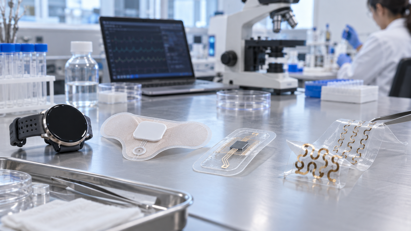
Optical sensing on a watch (PPG), electrical sensing with patches on the chest or forearm (ECG), electrochemical sensing of sweat: in wearable biosensors, a different principle means different disturbances to the signal and different materials challenges. What they share is the difficulty of getting a stable signal for hours from soft, moving skin. From a materials engineer's viewpoint, this article sets out the principle and error sources of each approach, the materials for flexible substrates, stretchable interconnects and encapsulation, wear and signal quality, and what can be confirmed as in practical use from regulatory documents versus what is still research.
- What wearable biosensors are (the short version)
- A map of three sensing approaches: light, electricity, chemistry
- Optical sensing (PPG): principle and what disturbs the signal
- Electrical sensing (ECG): between electrode and skin
- Our calculation: how much electrode contact impedance reduces an ECG signal
- A materials engineer's view (1): flexible substrates, stretchable interconnects, encapsulation
- Where sweat sensors fit
- A materials engineer's view (2): wear and signal quality are matters of materials and geometry
- What is in practical use (as far as official documents confirm) and what is research
- What is still hard, and what could not be confirmed
- Glossary / References / Claim-to-source audit
Sourced = stated in a published source (link given)
Our calculation = a value this article calculated from stated assumptions
Not yet confirmed = a plan, a target, or something for which no result could be confirmed
Interpretation of material design is marked "Commentary" and kept separate.
This article explains materials, principles, signal quality and regulation. It is not advice on diagnosis, treatment or medication, or on how to take a measured value or notification, and it does not rank any product or company.
1. What wearable biosensors are (the short version)
- What they measure: signals that can be taken from outside the body: changes in blood volume (light), the heart's electrical activity (electricity), constituents of sweat (chemistry) and so on.
- What is hard: a review says that to obtain more than heart rate from a wearable, one must account for noise sources from individual variation (skin tone, obesity, age, gender), physiology (respiration, venous pulsation, body site, body temperature) and external perturbations of the device (motion artifact, ambient light, applied pressure on the skin) Sourced [Reference 2].
- Practical use: the FDA classified an ECG app and a PPG-based irregular-rhythm notification feature by De Novo in 2018 Sourced (Section 9). On the other hand, the FDA in 2025 warned about unauthorized software features on smartwatches and similar devices that claim to measure blood pressure Sourced.
A sensor being "small and thin" does not mean "the signal is stable on the skin". Signal quality is set by principle, materials, geometry and contact with the skin together (Commentary).
2. A map of three sensing approaches: light, electricity, chemistry
3. Optical sensing (PPG): principle and what disturbs the signal
Photoplethysmography (PPG) is a simple, low-cost optical technique that detects blood volume changes in the microvascular bed of tissue. The PPG waveform comprises a pulsatile ('AC') component synchronous with the heartbeat, superimposed on a slowly varying baseline ('DC') with lower-frequency components attributed to respiration, sympathetic nervous system activity and thermoregulation Sourced [Reference 1].
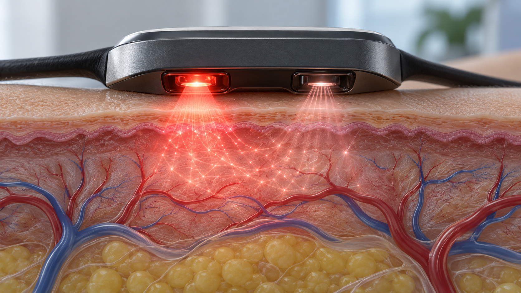
View the original SVG schematic, source notes and numerical details
Wavelength and skin tone: an optical-materials issue
Fine and colleagues' review relates skin tone to melanin content and notes that melanin absorption falls gradually from the ultraviolet through the visible range, which affects how light travels through skin [Reference 2]. Two reported examples are cited Sourced.
- A dorsal-wrist heart-rate monitor with a single 535 nm (green) LED, validated in 256 subjects (54 "fair", 181 "moderate", 21 "dark" skin), showed a tendency for heart-rate error to increase with darker skin tone
- A multi-wavelength PPG device with a 590 nm (yellow-orange) LED (20 subjects, Fitzpatrick II to IV) showed that for Fitzpatrick IV the perfusion index (AC/DC) rose by a factor of 1.2 to 7.1
Both are individual studies cited by the review, with limited devices, subjects and conditions. What follows is that source wavelength, window transmission and detector layout are design variables that shape differences by skin tone (Commentary).
Sjoding and colleagues analyzed pairs of arterial blood gas and pulse oximetry measurements in hospital patients. Among measurements in which pulse oximetry read 92 to 96%, arterial oxygen saturation was below 88% (occult hypoxemia) in 11.7% (88/749) of Black patients and 3.6% (99/2778) of White patients in the University of Michigan cohort, and in 17.0% (160/939) and 6.2% (546/8795) in the multicenter cohort Sourced [Reference 3]. The authors state that Black patients had nearly three times the frequency of occult hypoxemia not detected by pulse oximetry. This is a study of hospital pulse oximeters and does not directly show the performance of wearable devices. It is, however, an important report that differences by skin tone can be a real issue in optical measurement.
4. Electrical sensing (ECG): between electrode and skin
An electrocardiogram (ECG) picks up, with two or more electrodes, the tiny potentials on the skin surface produced by the heart's electrical activity. The challenge for wearables is contact between electrode and skin.
- Gel-type Ag/AgCl electrodes: the review by Meziane and colleagues says that conventional disposable Ag/AgCl electrodes give excellent signal quality but are irritating for long-term use, and that skin preparation such as shaving and cleansing with alcohol is usually required Sourced [Reference 4].
- Dry electrodes: according to the same review, dry electrodes that work without gel, adhesive or skin preparation have been studied for decades but, while used in research, have yet to achieve acceptance for medical use (as of 2013) [Reference 4].
- Thin electronics laminated on skin: Kim and colleagues reported electronic systems with thickness, effective elastic modulus, bending stiffness and areal mass density matched to the epidermis. Laminated on skin, they achieve conformal contact and adhesion based on van der Waals interactions alone, and were used to measure electrical activity of the heart, brain and skeletal muscles Sourced [Reference 5].
Among standards, the basic safety and essential performance requirements for ambulatory electrocardiographic systems are set out in IEC 60601-2-47:2012 (Medical electrical equipment – Part 2-47: Particular requirements for the basic safety and essential performance of ambulatory electrocardiographic systems) Sourced [Reference 19] (the text is paid, so only the title and scope are confirmed here).
5. Our calculation: how much electrode contact impedance reduces an ECG signal
Treat the electrode–skin contact impedance Ze and the amplifier input impedance Zin as forming a voltage divider Our calculation. The fraction of the signal reaching the amplifier is Zin ÷ (Zin + Ze).
- Assumptions: Zin = 10 MΩ (a value assumed here); Ze = 10 kΩ, 100 kΩ, 1 MΩ and 5 MΩ (illustrative values)
- Results: 10 kΩ → 99.9%, 100 kΩ → 99.0%, 1 MΩ → 90.9%, 5 MΩ → 66.7%
- Difference between the two electrodes: if the two electrodes' Ze differ by ΔZ, part of a common-mode disturbance (such as mains pickup) reaching both is converted to a differential signal, roughly ΔZ ÷ Zin. For ΔZ = 100 kΩ that is 1%; for a common-mode disturbance of 1 V (assumed), a spurious differential signal of about 10 mV
- Comparison: if an ECG signal is assumed to be of the order of 1 mV, this spurious signal is larger
Assumptions and limits: Zin, Ze, the size of the common-mode disturbance and the ECG amplitude are all illustrative values assumed here, not values of any real device or person. Real amplifiers include circuits that suppress common-mode disturbance (CMRR, right-leg drive and so on). All the calculation shows is the structure: both the size of the contact impedance and the difference between the two sides bear on signal quality (Commentary).
6. A materials engineer's view (1): flexible substrates, stretchable interconnects, encapsulation
The wearable challenge is how to connect hard electronic parts to stretching skin. The materials strategies that can be confirmed in papers are organized layer by layer.
| Layer / element | Materials and design confirmed in papers | Points for materials engineers (Commentary) |
|---|---|---|
| Substrate | 100-µm moisture-resistant PET (sweat sensor) [Reference 10]. A flexible SEBS elastomer (sweat cortisol sensor) [Reference 11]. Circuits built on 1-µm polymer foil with an areal mass of 3 g/m² [Reference 9]. Ultrathin systems matched to the modulus and bending stiffness of the epidermis [Reference 5] | The substrate's thickness and modulus decide how well it adheres to skin and follows movement. Fundamentals of polyesters, elastomers and polyimides are in the polymers explainers of this series |
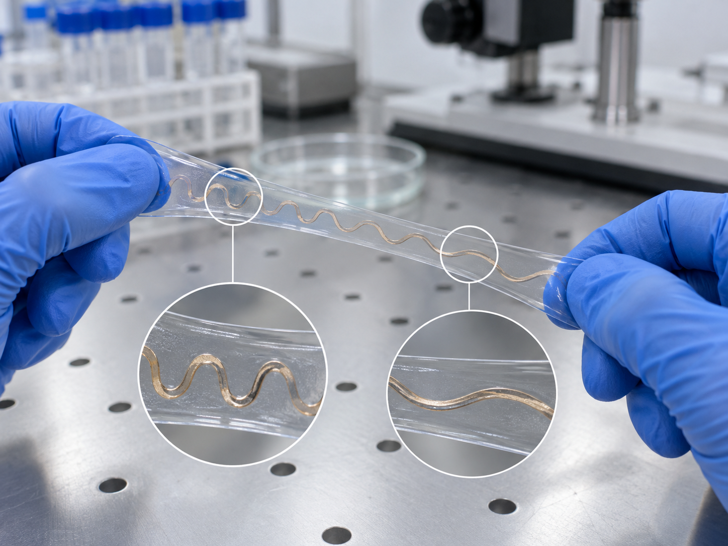
| Interconnect | Rigid functional elements embedded in an ultralow-modulus soft medium, surviving strain through controlled mechanical buckling and similar means [Reference 7]. A strategy of combining microstructured inorganic and organic materials with elastomeric substrates [Reference 6]. A printable elastic conductor in which silver nanoparticles form in situ from silver flakes, fluorine rubber and surfactant: above 4,000 S/cm at 0% strain and 935 S/cm when stretched up to 400% (highest value 6,168 S/cm) [Reference 8] | The challenge is keeping conductivity and stretchability together. Metal lines let strain escape through their shape (wavy, serpentine); conductive fillers keep stretchability through how particles are arranged (this article's summary). Filler–resin relations are in the explainer on polymer blends and fillers |
| Electrode and skin | Gel-type Ag/AgCl gives excellent signals but irritates in long-term use; dry electrodes were not accepted for medical use as of 2013 [Reference 4]. Thin devices matched to the epidermis adhere through van der Waals interactions [Reference 5] | Quality of contact is a matter of adhesive, gel and shape. Skin adhesion is in the explainer on patch sensors and skin adhesion; adhesives in the explainers on adhesives and tapes |
| Insulation and protection | 500-nm parylene C insulation (sweat sensor) [Reference 10]. A polyethylene-based hydrophobic protection layer (sweat cortisol sensor) [Reference 11] | Both encapsulation against sweat, moisture and washing and an opening that lets sweat in are needed. Barrier properties are in the explainer on barrier properties and coatings; silicones in the explainer on silicones |
| Safety of skin-contact materials | The framework for biological evaluation of medical devices is ISO 10993-1:2025; skin sensitization tests are ISO 10993-10:2021 and irritation tests ISO 10993-23:2021 [References 20 to 22] | For a material worn on skin for long periods, the adhesive, substrate and electrode materials can all be subject to evaluation (Commentary). How it applies depends on the product and use |
All Sourced (papers and the official pages of standards bodies). The right column is this article's Commentary. For the standards, only titles and editions were confirmed; the texts were not.
7. Where sweat sensors fit
For electrochemical wearables that measure sweat lactate, electrolytes and the like, Gao and colleagues reported an example integrating a sensor array on a PET substrate with skin-temperature compensation Sourced [Reference 10]. Parlak and colleagues reported a cortisol sensor with a built-in MIP together with microcapillary uptake of sweat [Reference 11]. How each fluid relates to blood, the conditions of collection, dilution, pH and temperature, the stack of an electrochemical sweat sensor and the status of practical use are covered in detail in the explainer on biomarkers in body fluids in this series, so they are not repeated here. This article also could not confirm regulatory authorization of a wearable sensor measuring glucose or lactate in sweat.
8. A materials engineer's view (2): wear and signal quality are matters of materials and geometry
For each approach, the factors that move signal quality tie back to materials and geometry (the factors come from papers; the ties are this article's Commentary).
- PPG (light): applied pressure and motion [Reference 2] are set by band material, tightness and case shape. Ambient light is a matter of light-shielding structure, and skin tone of wavelength and the optical properties of the window
- ECG (electrical): electrode–skin contact [Reference 4] is a matter of electrode material, gel or adhesive, skin preparation and how well the substrate conforms. As the calculation in Section 5 shows, the size of the contact impedance and the difference between the two sides bear directly on signal quality
- Sweat (chemical): skin-surface contamination and temperature [Reference 10] are matters of channel design, temperature compensation, and cleaning and adhesion
- Long wear: gel-type electrodes irritate in long-term use [Reference 4]. Biological evaluation of the materials on skin (adhesive, substrate, electrode) under the ISO 10993 series constrains material selection [References 20 to 22]
It is the same pattern as "the interface decides performance" in adhesion and encapsulation. The knowledge in the explainers on adhesives, tapes, silicones and barrier properties and coatings applies directly here.
9. What is in practical use (as far as official documents confirm) and what is research
| Approach / function | What regulatory documents and standards confirm | Class and source |
|---|---|---|
| ECG (OTC software) | ECG App: De Novo DEN180044, classification order dated 11 September 2018. A software-only mobile medical application for use with the Apple Watch that "create[s], record[s], store[s], transfer[s], and display[s] a single channel electrocardiogram (ECG) similar to a Lead I ECG" and determines the presence of atrial fibrillation (AFib) or sinus rhythm. Over the counter (OTC). The data are for informational use only, and "the user is not intended to interpret or take clinical action based on the device output without consultation of a qualified healthcare professional". Not for people under 22; not recommended for users with other known arrhythmias. Classification: 21 CFR 870.2345 (electrocardiograph software for over-the-counter use, Class II) | FDA classification order [Reference 12], U.S. Code of Federal Regulations [Reference 15]. De Novo classifies a new type of device |
| Irregular-rhythm notification from PPG (OTC) | Irregular Rhythm Notification Feature: De Novo DEN180042, dated 11 September 2018. Analyzes pulse rate data to identify episodes of irregular heart rhythms suggestive of AFib and provides a notification. It is "not intended to provide a notification on every episode" and "the absence of a notification is not intended to indicate no disease process is present". Data are captured only when the user is still. Not for people under 22 or previously diagnosed with AFib. Classification: 21 CFR 870.2790 (photoplethysmograph analysis software for over-the-counter use, Class II). A Fitbit Loss of Pulse Detection (510(k) K242967, 25 February 2025) is recorded in the same category | FDA classification order [Reference 13], U.S. Code of Federal Regulations [Reference 16], FDA 510(k) [Reference 23] |
| Pulse oximeters | 21 CFR 870.2700: an oximeter transmits radiation of known wavelength(s) through blood and measures oxygen saturation from the reflected or scattered radiation; Class II. The standard is ISO 80601-2-61:2026 (basic safety and essential performance of pulse oximeter equipment, third edition). A product code for over-the-counter pulse oximeters (OLK) exists | U.S. Code of Federal Regulations [Reference 17], ISO [Reference 18]. The product code is this article's own check (openFDA) |
| Standard for ambulatory ECG systems | IEC 60601-2-47:2012 (basic safety and essential performance of ambulatory electrocardiographic systems, second edition) | IEC [Reference 19] |
| Blood pressure (software features on wearables) | In a safety communication of 16 September 2025 the FDA said that many over-the-counter blood pressure devices lack FDA marketing authorization and, for unauthorized software features on smartwatches and smart rings that claim to measure or estimate blood pressure, safety and effectiveness have not been evaluated. Authorized devices will generally have the product code DXN. The FDA says it is taking steps against unauthorized blood pressure devices, including software features on wearables | FDA safety communication [Reference 14] |
| Wearable sweat sensors | Authorization could not be confirmed (see the result in the explainer on biomarkers in body fluids in this series). Research reports: Gao et al., Parlak et al. | [References 10 and 11] |
Clearance or approval is a judgment for a specific intended use and does not mean "safe and accurate however it is used". FDA 510(k) clearance, De Novo authorization and PMA approval are different systems. Product names are given as they appear in FDA documents, as fact, not as a ranking.
10. What is still hard, and what could not be confirmed
(1) Differences by skin tone
Differences by skin tone have been reported for hospital pulse oximetry [Reference 3], and the PPG review also lists skin tone as a noise source [Reference 2]. This article could not confirm a primary source that systematically compared the performance of wearable devices across skin tones. Not yet confirmed
(2) Long wear and effects on skin
Gel-type electrodes are said to irritate in long-term use [Reference 4], and dry electrodes had not been accepted for medical use as of 2013. This article has not confirmed the latest primary sources on the situation since then.
(3) Long-term reliability of stretchable interconnects
Values for the conductivity and stretchability of elastic conductors have been reported [Reference 8], but this article could not confirm a primary source that quantified degradation under repeated stretching, washing and long exposure to sweat in real use. Not yet confirmed
(4) How to take values and notifications
This article does not cover how to take measured values or notifications, or reference ranges. The FDA indications also state that the output is informational and that medical judgment presupposes consulting a healthcare professional [References 12 and 13].
- A different approach means different noise sources and materials challenges. Light: motion, pressure, skin tone; electricity: electrode–skin contact; chemistry: dilution, contamination, temperature Sourced
- Contact impedance bears on both the signal fraction and the difference between sides. In the illustration, at 5 MΩ the signal falls to 66.7% Our calculation
- The materials strategy is a thin soft substrate, stretchable interconnects and encapsulation together. An elastic conductor is reported at 935 S/cm at 400% stretch Sourced
- What regulatory documents confirm includes OTC ECG software and PPG irregular-rhythm notification (De Novo, 2018) and the pulse-oximeter category Sourced
- In 2025 the FDA warned about unauthorized blood pressure features and the like Sourced
- Authorization of wearable sweat sensors could not be confirmed Sourced
11. Glossary
- PPG (photoplethysmography)
- Detecting changes in tissue blood volume with light. Used to detect heartbeat and pulse.
- AC / DC component
- The heartbeat-synchronous pulsatile part (AC) and the slowly varying baseline (DC) of a PPG waveform.
- Perfusion index
- The ratio of the AC to the DC component of PPG; a guide to signal strength.
- Fitzpatrick scale
- A scale of 1 to 6 for skin colour, originally devised to express sensitivity to ultraviolet light.
- ECG (electrocardiogram)
- A recording of the potentials on the skin surface produced by the heart's electrical activity.
- Ag/AgCl electrode
- A silver/silver-chloride electrode, standard for biopotential measurement, used with a gel.
- Dry electrode
- An electrode that touches the skin directly, without gel or adhesive.
- Contact impedance
- A measure of the electrical resistance at the electrode–skin contact; bears directly on signal quality.
- Common-mode disturbance
- Noise that enters both electrodes in the same way, such as pickup from mains power.
- Elastomer
- A rubber-like polymer that stretches and recovers; used for substrates and encapsulation.
- Elastic conductor
- A material that stays conductive when stretched; some are made by dispersing conductive filler in an elastomer.
- Parylene C
- A thin polymer insulating film deposited from the gas phase.
- De Novo
- The FDA pathway that newly classifies a new type of lower-risk device into Class I or II.
- OTC
- Over the counter: available and usable without a prescription.
- ISO 10993
- A family of standards for biological evaluation of medical devices; relevant to materials that touch the skin.
12. References
- Allen J. "Photoplethysmography and its application in clinical physiological measurement", Physiol Meas 2007;28:R1-R39 — doi.org
- Fine J et al. "Sources of inaccuracy in photoplethysmography for continuous cardiovascular monitoring", Biosensors 2021;11:126 — doi.org
- Sjoding MW et al. "Racial bias in pulse oximetry measurement", N Engl J Med 2020;383:2477-2478 — doi.org
- Meziane N et al. "Dry electrodes for electrocardiography", Physiol Meas 2013;34:R47-R69 — doi.org
- Kim DH et al. "Epidermal electronics", Science 2011;333:838-843 — doi.org
- Rogers JA et al. "Materials and mechanics for stretchable electronics", Science 2010;327:1603-1607 — doi.org
- Xu S et al. "Soft microfluidic assemblies of sensors, circuits, and radios for the skin", Science 2014;344:70-74 — doi.org
- Matsuhisa N et al. "Printable elastic conductors by in situ formation of silver nanoparticles from silver flakes", Nat Mater 2017;16:834-840 — doi.org
- Kaltenbrunner M et al. "An ultra-lightweight design for imperceptible plastic electronics", Nature 2013;499:458-463 — doi.org
- Gao W et al. "Fully integrated wearable sensor arrays for multiplexed in situ perspiration analysis", Nature 2016;529:509-514 — doi.org
- Parlak O et al. "Molecularly selective nanoporous membrane-based wearable organic electrochemical device for noninvasive cortisol sensing", Sci Adv 2018;4:eaar2904 — doi.org
- U.S. Food and Drug Administration (FDA) De Novo DEN180044 (ECG App): classification order, 11 September 2018 (PDF) — www.accessdata.fda.gov
- FDA De Novo DEN180042 (Irregular Rhythm Notification Feature): classification order, 11 September 2018 (PDF) — www.accessdata.fda.gov
- FDA "Do Not Use Unauthorized Devices for Measuring Blood Pressure: FDA Safety Communication", 16 September 2025 — www.fda.gov
- U.S. Code of Federal Regulations 21 CFR 870.2345, Electrocardiograph software for over-the-counter use — www.ecfr.gov
- U.S. Code of Federal Regulations 21 CFR 870.2790, Photoplethysmograph analysis software for over-the-counter use — www.ecfr.gov
- U.S. Code of Federal Regulations 21 CFR 870.2700, Oximeter — www.ecfr.gov
- ISO ISO 80601-2-61:2026, Medical electrical equipment - Part 2-61: Particular requirements for basic safety and essential performance of pulse oximeter equipment (3rd edition) — www.iso.org
- IEC IEC 60601-2-47:2012, Medical electrical equipment - Part 2-47: Particular requirements for the basic safety and essential performance of ambulatory electrocardiographic systems — webstore.iec.ch
- ISO ISO 10993-1:2025, Biological evaluation of medical devices - Part 1 (6th edition) — www.iso.org
- ISO ISO 10993-10:2021, Biological evaluation of medical devices - Part 10: Tests for skin sensitization — www.iso.org
- ISO ISO 10993-23:2021, Biological evaluation of medical devices - Part 23: Tests for irritation — www.iso.org
- FDA 510(k) database: K242967 (Loss of Pulse Detection, Fitbit), 25 February 2025 — www.accessdata.fda.gov
13. Claim-to-source audit
| Claim in the text | Basis | Label |
|---|---|---|
| That PPG is a technique for detecting blood volume changes in the microvascular bed of tissue and that its waveform comprises a heartbeat-synchronous pulsatile (AC) component and a slowly varying baseline (DC) | Abstract of the Physiol Meas 2007 review[Reference 1] https://doi.org/10.1088/0967-3334/28/3/R01 | Sourced |
| The noise sources of wearable PPG (individual: skin tone, obesity, age, gender; physiology: respiration, venous pulsation, body site, temperature; external: motion, ambient light, applied pressure; the device's hardware and software). A 535 nm single-LED wrist heart-rate monitor (256 subjects: 54 fair, 181 moderate, 21 dark) with greater error at darker skin tones; a 590 nm multi-wavelength PPG device (20 subjects, Fitzpatrick II to IV) with perfusion index up 1.2 to 7.1 times at Fitzpatrick IV | Abstract and text of the Biosensors 2021 review (open-access version)[Reference 2] https://doi.org/10.3390/bios11040126 | Sourced |
| Occult hypoxemia (arterial saturation below 88% with pulse oximetry at 92 to 96%): 11.7% (88/749) of Black and 3.6% (99/2778) of White patients in the University of Michigan cohort, 17.0% (160/939) and 6.2% (546/8795) in the multicenter cohort; the authors' statement of nearly three times the frequency | Text of the N Engl J Med 2020 letter (open-access version)[Reference 3] https://doi.org/10.1056/NEJMc2029240 | Sourced |
| That Ag/AgCl electrodes give excellent signal quality but irritate in long-term use and need skin preparation; that dry electrodes have been studied for decades, are used in research, and had not achieved acceptance for medical use (as of 2013) | Abstract of the Physiol Meas 2013 review[Reference 4] https://doi.org/10.1088/0967-3334/34/9/R47 | Sourced |
| That electronic systems matched in thickness, modulus, bending stiffness and areal mass density to the epidermis adhere by van der Waals interactions alone and were used to measure electrical activity of the heart, brain and skeletal muscles | Abstract of the Science 2011 paper[Reference 5] https://doi.org/10.1126/science.1206157 | Sourced |
| A strategy combining microstructured inorganic and organic materials with elastomeric substrates for stretchable electronics | Abstract of the Science 2010 review[Reference 6] https://doi.org/10.1126/science.1182383 | Sourced |
| Embedding rigid functional elements in an ultralow-modulus soft system using soft microfluidics, structured adhesive surfaces and controlled mechanical buckling | Abstract of the Science 2014 paper[Reference 7] https://doi.org/10.1126/science.1250169 | Sourced |
| A printable elastic conductor in which silver nanoparticles form in situ from silver flakes, fluorine rubber and surfactant: above 4,000 S/cm at 0% strain (highest 6,168 S/cm) and 935 S/cm at 400% stretch | Abstract of the Nat Mater 2017 paper[Reference 8] https://doi.org/10.1038/nmat4904 | Sourced |
| Circuits on 1-µm polymer foils with an areal mass of 3 g/m² | Abstract of the Nature 2013 paper[Reference 9] https://doi.org/10.1038/nature12314 | Sourced |
| The substrate (moisture-resistant PET, 100 µm), insulation (parylene C, 500 nm) and skin-temperature compensation of the sweat sensor array | Text of the Nature 2016 paper (open-access version)[Reference 10] https://doi.org/10.1038/nature16521 | Sourced |
| The substrate (SEBS elastomer), polyethylene-based hydrophobic protection layer and microcapillary channels of the sweat cortisol sensor | Text of the Sci Adv 2018 paper (open-access version)[Reference 11] https://doi.org/10.1126/sciadv.aar2904 | Sourced |
| ECG App: De Novo DEN180044, 11 September 2018; software for use with the Apple Watch; single-channel ECG similar to Lead I; determination of AFib or sinus rhythm; OTC; informational use; not for people under 22; 21 CFR 870.2345, Class II | FDA classification order and U.S. Code of Federal Regulations[Reference 12] https://www.accessdata.fda.gov/cdrh_docs/pdf18/DEN180044.pdf[Reference 15] https://www.ecfr.gov/current/title-21/section-870.2345 | Sourced |
| Irregular Rhythm Notification Feature: De Novo DEN180042, 11 September 2018; identifies episodes of irregular rhythm suggestive of AFib from pulse rate data and notifies; not intended to notify on every episode; data captured only when still; not for people under 22 or previously diagnosed with AFib; 21 CFR 870.2790, Class II | FDA classification order and U.S. Code of Federal Regulations[Reference 13] https://www.accessdata.fda.gov/cdrh_docs/pdf18/DEN180042.pdf[Reference 16] https://www.ecfr.gov/current/title-21/section-870.2790 | Sourced |
| That the FDA, in a safety communication of 16 September 2025, warned about unauthorized blood pressure devices, including software features on smartwatches and smart rings; that authorized devices generally have product code DXN | FDA safety communication[Reference 14] https://www.fda.gov/medical-devices/safety-communications/do-not-use-unauthorized-devices-measuring-blood-pressure-fda-safety-communication | Sourced |
| The identification of an oximeter in 21 CFR 870.2700 and its Class II; that ISO 80601-2-61:2026 (third edition) covers pulse oximeter equipment | U.S. Code of Federal Regulations and the ISO official page[Reference 17] https://www.ecfr.gov/current/title-21/section-870.2700[Reference 18] https://www.iso.org/standard/84595.html | Sourced |
| That IEC 60601-2-47:2012 (second edition) sets particular requirements for the basic safety and essential performance of ambulatory electrocardiographic systems | The IEC Webstore official page[Reference 19] https://webstore.iec.ch/en/publication/2666 | Sourced |
| The titles and editions of ISO 10993-1:2025 (6th edition), ISO 10993-10:2021 (skin sensitization) and ISO 10993-23:2021 (irritation) | The ISO official pages[Reference 20] https://www.iso.org/standard/10993-1[Reference 21] https://www.iso.org/standard/75279.html[Reference 22] https://www.iso.org/standard/74151.html | Sourced |
| Fitbit Loss of Pulse Detection: 510(k) K242967, 25 February 2025, product code SDY (the 21 CFR 870.2790 category) | FDA 510(k) database record[Reference 23] https://www.accessdata.fda.gov/scripts/cdrh/cfdocs/cfpmn/pmn.cfm?ID=K242967 | Sourced |
| The voltage-divider fraction (with Zin = 10 MΩ assumed, 99.9%, 99.0%, 90.9% and 66.7% for Ze = 10 kΩ, 100 kΩ, 1 MΩ and 5 MΩ), the common-mode conversion ΔZ/Zin (100 kΩ/10 MΩ = 1%, about 10 mV for 1 V), and the comparison assuming an ECG amplitude of about 1 mV | Our calculation. Zin, Ze, the size of the common-mode disturbance and the ECG amplitude are illustrative values assumed here, not values of real devices or people. Real amplifiers include circuits that suppress common-mode disturbance | Our calculation |
| Regulatory authorization of wearable sweat sensors | Not confirmed within the scope of this article (see the result in the explainer on biomarkers in body fluids in this series) | Not yet confirmed |
| Systematic comparison of wearable-device performance across skin tones, quantification of long-term degradation of elastic conductors under repeated stretching, washing and sweat exposure, and the acceptance of dry electrodes since 2013 | Not confirmed in the primary sources consulted for this article | Not yet confirmed |
| The readings that the signal comes from the sensor-and-skin system, that wavelength, window material and detector layout are design variables behind skin-tone differences, that metal lines keep stretchability through shape and conductive fillers through particle arrangement, the right-hand column of the materials table, and the ties between each approach's factors and materials or geometry | This article's own organization and interpretation of published content; not views expressed by the authors of the papers or by the regulator | Commentary |
| That Figs. 1, 2, 4 and 6 are explanatory drawings, that Fig. 3 includes our calculation, and that the hero image and Fig. 5 are AI-generated images | Our note | Commentary |
Last updated 3 October 2026. Sources are limited to primary material (peer-reviewed papers, official FDA documents, the U.S. Code of Federal Regulations and the official pages of standards bodies). Interpretation of material design is marked Commentary and kept separate from sourced fact. Regulatory authorization of wearable sweat sensors, a systematic comparison of wearable-device performance across skin tones, quantification of long-term degradation of elastic conductors, and the acceptance of dry electrodes since 2013 are not stated, because no primary source could be confirmed. For standards (ISO and IEC) only titles and editions were confirmed, not the texts. The calculation in Section 5 uses illustrative values to explain structure. This article does not cover how to take measured values or notifications. All figures are explanatory concept graphics. Figs. 1, 2, 3, 4 and 6 are vector drawings; the hero image and Fig. 5 are AI-generated images, and none of them shows a real product, skin structure or measurement result.