TECHNOLOGY EXPLAINER
Nanoparticles and Nanozymes
— nanomaterials as labels, separators, emitters and stand-ins for enzymes
In biosensors, nanoparticles take on many roles. Gold nanoparticles are the colour label in lateral flow strips (the format of a pregnancy test). Magnetic nanoparticles are tools for pulling a target out with a magnet and detecting it with a magnetic sensor. Quantum dots serve as bright fluorescent labels, nanozymes (nanomaterials with enzyme-like catalytic activity) as colour-producing catalysts in place of enzymes, and MOFs (metal-organic frameworks) as protective shells for enzymes and as supports for catalysts. For a materials engineer the point is that, even though all are called “nanoparticles”, the properties required differ by role (stability, surface, toxicity, reproducibility). This article sets out the role and the problems of each material, based on reviews, original papers, standards and regulatory documents.

- What nanoparticles and nanozymes are, in three lines
- Gold nanoparticles: the label in lateral flow
- Our calculation: what is one 20 nm gold nanoparticle?
- Magnetic nanoparticles: separation, and detection by magnetism
- Quantum dots: bright fluorescent labels, and the toxicity problem
- Choosing a label: four materials side by side
- Nanozymes: nanomaterials that stand in for enzymes
- MOFs: wrapping enzymes, and serving as catalyst supports
- A materials engineer's view: “stable” means different things for each role
- Toxicity, reproducibility and regulatory position
- What we could not confirm / Glossary / References / Claim-to-source audit
Sourced = stated in published material or a paper (link given)
Our calculation = a figure this article derived, with the assumptions spelled out
Not yet confirmed = at the research stage or an outlook, with no confirmed practical record
Structural readings and materials-design interpretations are marked separately as Commentary.
This article explains materials, principles, evaluation and regulation. It is not medical advice on diagnosis, treatment or how to read test results, and it does not recommend any product.
How whole test strips are used is covered in our explainer on point-of-care testing, enzyme–electrode interfaces in our explainer on enzymes and electrode interface materials, and regulatory classes in our explainer on medical device classification and home use.
1. What nanoparticles and nanozymes are, in three lines
- Nanoparticles as labels: a review of lateral flow assays says “nowadays colloidal gold is the most widely used label in commercial LFIA”, with an intense colour, no development step, and high stability in both liquid and dried forms Sourced
- Nanozymes: in 2007 Gao and co-workers reported that magnetite (Fe3O4) nanoparticles possess an intrinsic enzyme-mimetic activity like that of natural peroxidases. Until then such particles were generally considered biologically and chemically inert and were coated with metal catalysts, antibodies or enzymes Sourced. A 2018 paper says that since then more than 40 types of nanozymes with peroxidase-, oxidase-like and other activities have been reported Sourced
- Where it is hard: toxicity (cadmium in quantum dots, for example), batch-to-batch reproducibility, quantitative comparison of nanozyme activity, and the stability of MOFs in water are the shared problems. On the regulatory side there are frameworks for evaluating devices containing nanoscale materials (Chapter 10)
“Nanoparticle” describes only size, not role. Gold is a label for colour, magnetic particles for separation and magnetism, quantum dots for light, nanozymes are catalysts, and MOFs provide protection and support; what makes a good material is different for each. When comparing, first fix which role is being discussed (commentary).
2. Gold nanoparticles: the label in lateral flow
A lateral flow assay (LFA) is a format in which the sample flows by capillary force through a paper-like porous membrane. The review states that results are displayed within 5–30 min after the sample is placed, and that LFAs are widely used in hospitals, physicians' offices and clinical laboratories for qualitative and quantitative detection of antigens and antibodies Sourced.
A summary of the review Sourced.
- Sample pad: impregnated with buffer salts and surfactants that make the sample suitable for interaction with the detection system
- Conjugate release pad: holds antibodies specific to the target, conjugated to coloured or fluorescent particles—most commonly colloidal gold and latex microspheres
- Membrane (usually nitrocellulose): the detection zone with antibodies or antigens immobilized in lines. The test line responds to the target and the control line shows that the liquid flowed properly
- Absorbent pad: maintains flow and prevents backflow. The read-out is assessed by eye or with a dedicated reader
(1) What a label must provide
The review lists the most important requirements of the nanoparticle label Sourced.
- Colloidal stability in solution under various conditions and temperatures
- Detectability over a large and useful dynamic range
- Efficiency and reproducibility of conjugation, without loss of the antibody's chemical and biological integrity and activity
- Very low non-specific binding, ensuring a high signal-to-noise ratio
- Commercial availability at low cost, and an easy and scalable conjugation procedure
It adds that colloidal gold has an intense colour needing no development process, is highly stable in both liquid and dried forms, and “can be prepared in the laboratory at low cost, [and] there are many commercial sources” Sourced. For gold colloid synthesis there is the 1951 study of nucleation and growth by Turkevich and co-workers, and Frens reported in 1973 a way to regulate particle size by controlling nucleation in monodisperse gold suspensions Sourced (both by title). The SERS recommendations paper takes particles of 20–50 nm made by the Turkevich method (gold, citrate) as standard colloids Sourced (Reference 5).
(2) Limits of sensitivity
Because LFA is an antibody-based technique, the review writes, specificity and sensitivity may be affected by other chemicals with similar structures, leading to false positives. It says sensitivity is limited by the dissociation constant (Kd) of the antibody–antigen pair and by the colorimetric read-out, and that readers and new biochemical techniques have been developed to overcome these limits Sourced. It also notes that carbon nanotube labels have been shown to give a limit of detection 10-fold lower than gold, and that the high background noise that quantum dots may give has been overcome by polymer encapsulation and surface blocking Sourced.
The review says the most common difficulties in manufacturing LFA devices come from the hidden complexity of the device. Because a strip has many elements, material incompatibility, flaws in the connection of overlapping elements and imperfect material characteristics cause problems. Development attention tends to go to the detection method or the choice of antibody, but unless attention is also paid to basic components such as the backing card, adhesive strip and cover tape, a consistent, high-quality product is not obtained Sourced.
This connects directly to the adhesives and encapsulants part of this series (tapes, adhesive strength testing and so on). Conditions such as not letting the adhesive leach the label's antibody and not disturbing capillary flow in the membrane are also imposed on the adhesive materials (commentary).

3. Our calculation: what is one 20 nm gold nanoparticle?
We assume a spherical gold nanoparticle 20 nm in diameter (using the known gold density of 19.3 g/cm³) Our calculation.
- Volume: radius 10 nm = 1×10⁻⁶ cm. 4/3 × π × (1×10⁻⁶)3 = 4.19×10⁻¹⁸ cm³
- Mass: 4.19×10−18 × 19.3 = 8.1×10⁻¹⁷ g (about 0.081 fg)
- Particles per µg: 10−6 / 8.1×10−17 = about 1.2×10¹⁰
- Surface area: 4π × (10 nm)² = about 1,260 nm²
- Antibodies that can bind: assuming one IgG molecule occupies 100 nm² of surface (about 10 nm × 10 nm), 1,260 / 100 = about 12 as an upper guide
Assumptions and limits: the particle is a perfect sphere, the density is the bulk value and the antibody footprint is an assumption. The real number depends greatly on antibody orientation, density and the particle size distribution. The difficulty of conjugating antibodies “efficiently and reproducibly” (the requirement above) relates to how few antibodies one particle carries (commentary).
4. Magnetic nanoparticles: separation, and detection by magnetism
Magnetic nanoparticles (mostly iron oxide) have three roles (our grouping): separating the target with a magnet, detecting it with a magnetic sensor, and acting as a catalyst (the nanozymes of Chapter 7).
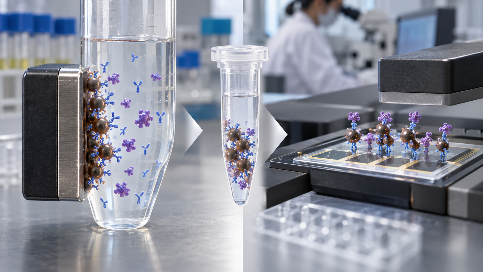
View the original SVG schematic, source notes and numerical details
(1) Magnetic nanotags and magnetic sensors
Osterfeld and co-workers demonstrated in 2008 multiplex protein assays for potential cancer markers using magnetic nanotags, a promising alternative to fluorescent labels whose minute quantities can be detected with inexpensive giant magnetoresistive (GMR) sensors. They report detection at subpicomolar concentrations with a dynamic range of more than four decades, extending to the low fM range with nanotag amplification, and call the technology a strong contender for versatile, portable molecular diagnostics in both research and clinical settings Sourced (a research-stage report).
The LFA review cautions that fluorescent dyes or paramagnetic particles cannot be detected directly by the naked eye and require dedicated readers for quantitative analysis Sourced. A magnetic label is a material to be thought of together with the design of the instrument (commentary).
(2) Reproducibility of magnetic particles: an example with micrometre beads
For separation in immunoassays, micrometre-sized magnetic beads are often used rather than nanoparticles. A 2025 PLOS ONE paper reports monodisperse streptavidin-functionalized magnetic beads, made by forming iron oxide nanoparticles inside porous polystyrene spheres and attaching streptavidin, used in an electrochemiluminescent (ECL) immunoassay for CEA (a tumour marker). Across three batches made under identical conditions, the coefficient of variation (CV) of size was below 5% and the batch-to-batch difference in ECL response was below 6.2%. The linear range was 0.05 to 1000 ng/mL with a limit of detection of 0.02383 ng/mL, and the authors also compared with a commercial assay kit Sourced. These are values for laboratory-scale, in-house beads, not the quality of commercial products or performance in actual diagnosis (commentary).
5. Quantum dots: bright fluorescent labels, and the toxicity problem
Quantum dots (QDs) are semiconductor nanocrystals, bright fluorophores whose emission colour changes with particle size. Their use as biological labels was reported by two papers in 1998 (Bruchez et al., “Semiconductor Nanocrystals as Fluorescent Biological Labels”, and Chan and Nie, “Quantum Dot Bioconjugates for Ultrasensitive Nonisotopic Detection”; both by title) Sourced. A review by Resch-Genger and co-workers describes QDs as inorganic nanocrystals “with unique optical and chemical properties but complicated surface chemistry” and compares their physicochemical properties with those of traditional organic dyes, setting out the advantages and limitations of both Sourced.
Derfus and co-workers reported in 2004, using primary hepatocytes, that CdSe-core QDs were acutely toxic under certain conditions. The toxicity was modulated by processing parameters during synthesis, exposure to ultraviolet light and surface coatings, and correlated with the liberation of free Cd2+ ions due to deterioration of the CdSe lattice. When appropriately coated, the QDs could be rendered non-toxic and used to track cell migration in vitro Sourced. The authors say this informs design criteria for QDs in vitro and especially in vivo, where deterioration over time may occur. So the safety of a QD is a question not of the material's name but of the reliability of the coating (commentary). Within the scope of this article we could not confirm any official review document for a quantum-dot label in a diagnostic use.
6. Choosing a label: four materials side by side
7. Nanozymes: nanomaterials that stand in for enzymes
Nanozymes are nanomaterials with enzyme-like catalytic activity. In 2007 Gao and co-workers reported that magnetic Fe3O4 nanoparticles in fact possess an intrinsic enzyme-mimetic activity similar to that of natural peroxidases. On this basis they built a new immunoassay in which antibody-modified magnetite nanoparticles provide three functions: capture, separation and detection Sourced.
(1) Breadth of research
A 2019 review by Wu and co-workers describes nanozymes as “nanomaterials with enzyme-like characteristics developed to address the limitations of natural enzymes and conventional artificial enzymes”, and sets out the types and representative materials, catalytic mechanisms, strategies for modulating activity and selectivity, applications from biomedical analysis and imaging to theranostics and environmental protection, and current challenges Sourced. Other reviews in the field are the 2013 review by Wei and Wang and the 2019 review by Huang and co-workers (classification, catalytic mechanisms, activity regulation, applications) (both by title) Sourced.
(2) An example nanozyme strip, and our calculation
Duan and co-workers reported in 2015 an immunochromatographic strip (Nanozyme-strip) using Fe3O4 magnetic nanoparticles as the nanozyme probe. It detects the glycoprotein of Ebola virus as low as 1 ng/mL, which is 100-fold more sensitive than the standard strip method; its sensitivity and diagnostic accuracy for New Bunyavirus clinical samples are comparable with ELISA, but it is within 30 min and needs no specialist facilities Sourced (a research-stage report). Back-calculating the detection level of the standard strip gives 1 ng/mL × 100 = about 100 ng/mL Our calculation (a conversion based on the paper's “100-fold”, not a value the paper states directly).
A 2018 paper by Jiang and co-workers says that, because of the complex interdependence of physicochemical properties and catalytic characteristics, a standard is needed by which the catalytic activities and kinetics of various nanozymes can be quantitatively compared, and presents a protocol for measuring and defining the catalytic activity units and kinetics of peroxidase nanozymes, the most widely used type. The catalytic-activity and kinetics assays can be performed within 4 hours, and they say nanozyme-based detection is repeatable and reliable when guided by this standard Sourced.
In the world of enzymes the “unit of activity” is a common language. With nanomaterials, activity changes with particle size, surface and coexisting surfactants, so particles with the same name do not have the same activity (that activity changes with size and so on is our commentary). The appearance of a standardization protocol can itself be read as the reverse side of the reproducibility problem.
8. MOFs: wrapping enzymes, and serving as catalyst supports
A metal-organic framework (MOF) is a porous crystal in which metal ions are linked by organic molecules. The 2013 review by Furukawa and co-workers says MOFs are crystalline, formed by strong bonds between inorganic and organic units, and can have ultrahigh porosity and high thermal and chemical stability; their interior can be chemically altered for use in gas separation, gas storage and catalysis, among other applications Sourced. On 8 October 2025 the Royal Swedish Academy of Sciences announced that the Nobel Prize in Chemistry 2025 would be awarded to Susumu Kitagawa, Richard Robson and Omar M. Yaghi “for the development of metal–organic frameworks” Sourced.
(1) A shell that protects enzymes: biomimetic mineralization
Liang and co-workers reported in 2015 a method, inspired by biomineralization, of encapsulating proteins, enzymes and DNA in a protective MOF coating. The biomolecules themselves induce the formation of the MOF coating under physiological conditions, and the resulting biocomposite is stable under conditions that would normally decompose biological macromolecules. For example, urease protected within a MOF shell retained bioactivity after treatment at 80 °C, and horseradish peroxidase after boiling in dimethylformamide (153 °C) Sourced.
(2) Weakness in water, and large-scale synthesis
A review of the chemical, thermal and mechanical stabilities of MOFs exists (Howarth et al., 2016, by title) Sourced. On the practical problems in biosensors, a 2025 review (MOF-based nanozymes for microbial biosensing) lists the following Sourced.
- Long-term stability and reusability: many MOFs are prone to structural degradation and loss of catalytic activity after repeated use or when exposed to moisture, extreme pH levels and microbial metabolites. Ligand engineering, post-synthetic modification and composites with graphene or polymers are named as countermeasures
- Specificity: most MOF-nanozyme biosensors rely largely on generalized catalytic activity, which makes it hard to identify specific microbial strains in complex matrices. Combining with aptamers, antibodies or molecularly imprinted polymers is proposed
- Large-scale synthesis: although MOFs can be synthesized relatively easily in the laboratory, scale-up needs high-purity metal precursors, energy-intensive synthesis and complex purification, which leads to batch-to-batch inconsistencies
- Turning the weakness to use: an assay in which phosphate-induced MOF decomposition amplifies the signal (reported to detect bacteria down to 10 CFU/mL within 2.5 h) is described (a research group's report, as introduced by the review)
So a MOF is a material that can use both “stability” and “decomposition” in its design. But long-term stability in aqueous blood or buffers is not a general assumption; it is an item to be confirmed case by case (commentary).
9. A materials engineer's view: “stable” means different things for each role
| Material (role) | What “stable” or “reproducible” means | Statements or figures in the literature |
|---|---|---|
| Gold nanoparticles (colour label) | Colloidally stable in solution and dried; antibody conjugation reproducible without loss of activity; little non-specific binding | Requirements listed by the review; stable in liquid and dried forms Sourced (Reference 1) |
| Magnetic beads (separation) | Narrow size distribution; consistent response between batches | For in-house laboratory-scale beads, size CV under 5% and batch-to-batch response difference under 6.2% Sourced (Reference 12) |
| Quantum dots (light label) | The coating stays intact and the core metal does not dissolve | Toxicity varies with synthesis, UV and coating and correlates with free Cd2+ Sourced (Reference 15) |
| Nanozymes (catalyst) | The “unit” of catalytic activity is aligned and can be compared | A standard assay for activity and kinetics was proposed (within 4 hours) Sourced (Reference 3) |
| MOFs (shell, support) | The structure survives water, phosphate and pH; uniform even when made in bulk | Degradation with moisture and extreme pH, and batch inconsistency, are problems Sourced (Reference 24) |
Each row comes from a separate paper or review and is not a comparison under the same conditions.
Reading the table down the columns shows that the “stability” required differs by material. For gold, it is “can be stored dry”; for magnetic beads, “uniform”; for quantum dots, “does not break down and release harmful components”; for nanozymes, “activity can be compared numerically”; for MOFs, “keeps its shape in water”. That requirement links directly to at which step (synthesis, purification, storage, use) quality is verified, that is, to the quality design of a materials supplier (commentary).
10. Toxicity, reproducibility and regulatory position
Nanomaterials are also seen by regulators within separate frameworks. What follows is a summary of what each document says; it does not judge the class of any specific product.
- EU Medical Device Regulation (Regulation (EU) 2017/745): Article 2 defines “nanomaterial” as a natural, incidental or manufactured material containing particles in an unbound state or as an aggregate or agglomerate where, for 50% or more of the particles in the number size distribution, one or more external dimensions is in the size range 1–100 nm, and states that fullerenes, graphene flakes and single-wall carbon nanotubes are also deemed nanomaterials. Rule 19 in Annex VIII classifies devices incorporating or consisting of nanomaterial as class III if they present a high or medium potential for internal exposure, class IIb if low, and class IIa if negligible Sourced
- EU In Vitro Diagnostic Regulation (Regulation (EU) 2017/746): searching the text as published in the Official Journal, we did not find the word “nanomaterial” Sourced (our check). We therefore make no judgement on whether Rule 19 above applies to gold nanoparticles or magnetic beads used as labels in test strips
- European Commission Recommendation on the definition of nanomaterial (10 June 2022, 2022/C 229/01): defines a nanomaterial as consisting of solid particles for which 50% or more in the number-based size distribution meet conditions such as 1–100 nm, and excludes materials with a volume-specific surface area below 6 m²/cm³. The Recommendation says the definition should neither affect nor reflect the scope of application of any instrument of Union legislation Sourced
- ISO/TR 10993-22:2017: a technical report setting out how to approach the biological evaluation of devices containing nanomaterials or generating nano-objects, which gives no detailed test procedures Sourced
- FDA guidance (June 2014, nonbinding recommendations): FDA asks whether a material or product is engineered to have an external dimension or structure of about 1–100 nm, and whether it is engineered to show dimension-dependent properties (up to about 1 µm). The products covered include medical devices and in vitro diagnostic devices Sourced
- U.S. 21 CFR 862.1155 (human chorionic gonadotropin [HCG] test system): an HCG test system for the early detection of pregnancy is class II; one for any other use (such as an aid in the diagnosis of certain tumours) is class III, requiring premarket approval Sourced. The regulation classifies by intended use and does not mention the label material (such as gold nanoparticles) (our reading of the text)
The frameworks above show how to think about evaluation when a product contains nanoscale materials; they do not mean that “using gold nanoparticles gets you approved” or “prevents approval”. The class of an individual product depends on its intended use, whether it is inside or outside the body, duration of contact and whether material is released. Nanoparticles introduced into the body (contrast agents, drug delivery and so on) fall in other regulatory areas and are not covered here. See our explainer on medical device classification and home use.
11. What we could not confirm
- Specific examples of in vitro diagnostic devices that use quantum dots as labels, or of diagnostic devices using nanozymes or MOFs, reviewed by regulators such as FDA or PMDA (not confirmed from primary sources within the scope of this article). Nanozyme strips are Not yet confirmed (at the research-report stage)
- Published figures for gold nanoparticle size, number of antibodies per particle and drying conditions for commercial lateral flow strips
- Primary sources on the in-body behaviour and toxicity of magnetic nanoparticles (this article is limited to ex vivo use as labels and for separation)
- Quantitative data on shelf life and signal change of gold colloid labels
- The individual conditions and particle-size figures of the Turkevich and Frens methods (we confirmed only the titles)
- How a gold nanoparticle label is treated under the In Vitro Diagnostic Regulation (we confirmed only that the word “nanomaterial” does not appear in the text)
- Choose nanoparticles by role. Gold = colour, magnetic = separation and magnetism, quantum dots = light, nanozymes = catalysis, MOFs = protection and support Sourced
- Colloidal gold is the most widely used label in commercial lateral flow. A strip is a combination of many parts, and the basic parts need quality Sourced
- A 20 nm gold particle carries only about a dozen antibodies. Conjugation reproducibility is directly the stability of the signal Our calculation
- Magnetic nanotags detected subpicomolar levels in multiplex with GMR (research). In-house beads varied less than 6.2% between batches Sourced
- Quantum-dot toxicity is set by the coating and by free Cd2+. Sourced
- Nanozymes need a standard to align the “unit of activity”. Fe3O4 made a strip 100 times more sensitive than the standard (research) Sourced
- MOFs can be shells that protect enzymes, but stability in water and batch-to-batch uniformity are problems. Sourced
- Regulation depends on intended use. Frameworks exist for evaluating devices containing nanomaterials, but we found no provision that classifies a gold-nanoparticle label itself Sourced
12. Glossary
- Lateral flow assay (LFA)
- A test format in which the sample flows by capillary force through a porous membrane.
- Colloidal gold
- A liquid with gold nanoparticles dispersed in water. Used as the colour label in strips.
- Conjugate
- An antibody or similar molecule attached to a particle.
- Test line / control line
- The line that responds to the target / the line that confirms the liquid flowed properly.
- Magnetic nanotag
- A magnetic nanoparticle used as a label and detected with a magnetoresistive sensor.
- GMR sensor
- A giant magnetoresistive sensor. Its resistance changes with the field of nearby magnetic particles.
- Quantum dot (QD)
- A semiconductor nanocrystal; a bright fluorophore whose colour changes with size.
- Nanozyme
- A nanomaterial with enzyme-like catalytic activity.
- Peroxidase-like activity
- Catalytic activity that uses hydrogen peroxide to oxidize a substrate and produce colour.
- MOF
- Metal-organic framework. A porous crystal of metal ions linked by organic molecules.
- Biomimetic mineralization
- Biomolecules inducing formation of an inorganic shell. Used to wrap enzymes in MOFs.
- Nanomaterial (EU definition)
- For example, a material in which 50% or more of particles by number have an external dimension of 1–100 nm.
- Kd
- Dissociation constant. A measure of the strength of antibody–antigen binding; smaller is stronger.
13. References
- Koczula KM, Gallotta A "Lateral flow assays", Essays Biochem 2016;60:111–120 — doi.org/10.1042/EBC20150012
- Gao L et al. "Intrinsic peroxidase-like activity of ferromagnetic nanoparticles", Nat Nanotechnol 2007;2:577–583 — doi.org/10.1038/nnano.2007.260
- Jiang B et al. "Standardized assays for determining the catalytic activity and kinetics of peroxidase-like nanozymes", Nat Protoc 2018;13:1506–1520 — doi.org/10.1038/s41596-018-0001-1
- Duan D et al. "Nanozyme-strip for rapid local diagnosis of Ebola", Biosens Bioelectron 2015;74:134–141 — doi.org/10.1016/j.bios.2015.05.025
- Bell SEJ et al. "Towards Reliable and Quantitative Surface-Enhanced Raman Scattering (SERS): From Key Parameters to Good Analytical Practice", Angew Chem Int Ed 2020;59:5454–5462 — doi.org/10.1002/anie.201908154
- Frens G "Controlled Nucleation for the Regulation of the Particle Size in Monodisperse Gold Suspensions", Nature Physical Science 1973;241:20–22 (cited by title only) — doi.org/10.1038/physci241020a0
- Turkevich J, Stevenson PC, Hillier J "A study of the nucleation and growth processes in the synthesis of colloidal gold", Discuss Faraday Soc 1951;11:55–75 (cited by title only) — doi.org/10.1039/DF9511100055
- Parolo C et al. "Tutorial: design and fabrication of nanoparticle-based lateral-flow immunoassays", Nat Protoc 2020;15:3788–3816 — doi.org/10.1038/s41596-020-0357-x
- Quesada-González D, Merkoçi A "Nanoparticle-based lateral flow biosensors", Biosens Bioelectron 2015;73:47–63 — doi.org/10.1016/j.bios.2015.05.050
- Osterfeld SJ et al. "Multiplex protein assays based on real-time magnetic nanotag sensing", Proc Natl Acad Sci USA 2008;105:20637–20640 — doi.org/10.1073/pnas.0810822105
- Gijs MAM, Lacharme F, Lehmann U "Microfluidic applications of magnetic particles for biological analysis and catalysis", Chem Rev 2010;110:1518–1563 (further reading; no figures are quoted from it here) — doi.org/10.1021/cr9001929
- Lei H, Tang Y "A novel preparation method for monodisperse streptavidin magnetic beads and its application in electrochemiluminescent immunoassay for CEA", PLOS ONE 2025;20:e0338214 — doi.org/10.1371/journal.pone.0338214
- Chan WCW, Nie S "Quantum Dot Bioconjugates for Ultrasensitive Nonisotopic Detection", Science 1998;281:2016–2018 (cited by title only) — doi.org/10.1126/science.281.5385.2016
- Bruchez M Jr, Moronne M, Gin P, Weiss S, Alivisatos AP "Semiconductor Nanocrystals as Fluorescent Biological Labels", Science 1998;281:2013–2016 (cited by title only) — doi.org/10.1126/science.281.5385.2013
- Derfus AM, Chan WCW, Bhatia SN "Probing the Cytotoxicity of Semiconductor Quantum Dots", Nano Lett 2004;4:11–18 — doi.org/10.1021/nl0347334
- Resch-Genger U et al. "Quantum dots versus organic dyes as fluorescent labels", Nat Methods 2008;5:763–775 — doi.org/10.1038/nmeth.1248
- Wu J et al. "Nanomaterials with enzyme-like characteristics (nanozymes): next-generation artificial enzymes (II)", Chem Soc Rev 2019;48:1004–1076 — doi.org/10.1039/C8CS00457A
- Wei H, Wang E "Nanomaterials with enzyme-like characteristics (nanozymes): next-generation artificial enzymes", Chem Soc Rev 2013;42:6060–6093 (cited by title only) — doi.org/10.1039/c3cs35486e
- Huang Y, Ren J, Qu X "Nanozymes: Classification, Catalytic Mechanisms, Activity Regulation, and Applications", Chem Rev 2019;119:4357–4412 (cited by title only) — doi.org/10.1021/acs.chemrev.8b00672
- Furukawa H, Cordova KE, O'Keeffe M, Yaghi OM "The Chemistry and Applications of Metal-Organic Frameworks", Science 2013;341:1230444 — doi.org/10.1126/science.1230444
- Nobel Foundation (Royal Swedish Academy of Sciences) "Press release: The Nobel Prize in Chemistry 2025", 8 October 2025 — nobelprize.org
- Liang K et al. "Biomimetic mineralization of metal-organic frameworks as protective coatings for biomacromolecules", Nat Commun 2015;6:7240 — doi.org/10.1038/ncomms8240
- Howarth AJ et al. "Chemical, thermal and mechanical stabilities of metal–organic frameworks", Nat Rev Mater 2016;1:15018 (cited by title only) — doi.org/10.1038/natrevmats.2015.18
- Kidanemariam D, Cho S "Recent Advances in Metal-Organic Framework-Based Nanozymes for Intelligent Microbial Biosensing: A Comprehensive Review of Biomedical and Environmental Applications", Biosensors (Basel) 2025;15(7):437 — doi.org/10.3390/bios15070437
- U.S. Code of Federal Regulations 21 CFR 862.1155 "Human chorionic gonadotropin (HCG) test system" — ecfr.gov
- European Parliament and Council Regulation (EU) 2017/745 (Medical Device Regulation, MDR) — eur-lex.europa.eu
- European Parliament and Council Regulation (EU) 2017/746 (In Vitro Diagnostic Regulation, IVDR) — eur-lex.europa.eu
- European Commission Commission Recommendation of 10 June 2022 on the definition of nanomaterial (2022/C 229/01) — eur-lex.europa.eu
- ISO ISO/TR 10993-22:2017, "Biological evaluation of medical devices — Part 22: Guidance on nanomaterials" — iso.org
- U.S. Food and Drug Administration "Considering Whether an FDA-Regulated Product Involves the Application of Nanotechnology", Guidance for Industry, June 2014 — fda.gov
14. Claim-to-source audit
| Claim in the text | Basis | Label |
|---|---|---|
| That LFA is a format in which the sample flows by capillary force through a paper-like porous membrane, with results in 5–30 min; the structure and roles of the sample pad, conjugate pad, membrane (mostly nitrocellulose) and absorbent pad; that colloidal gold and latex are the most common particles in the conjugate; the direct (sandwich) format (hCG and HIV p24 as examples) and the competitive format; the list of requirements for the nanoparticle label; that colloidal gold is the most widely used label with an intense colour, no development step, stability in liquid and dried forms, and availability from the laboratory or commercial sources; false positives from structurally similar chemicals and sensitivity limited by Kd and the colorimetric read-out; that carbon nanotube labels gave a 10-fold lower limit of detection than gold; that quantum-dot background was overcome by polymer encapsulation and blocking; that fluorescent dyes and paramagnetic particles cannot be seen by eye and need readers; that manufacturing difficulty arises from the hidden complexity of the device, including the backing card, adhesive strip and cover tape | Koczula and Gallotta (Essays Biochem 2016)[Reference 1] https://doi.org/10.1042/EBC20150012 | Sourced |
| That magnetic Fe3O4 nanoparticles have an intrinsic peroxidase-like catalytic activity; that such particles had been considered inert and coated with metal catalysts, antibodies or enzymes; and an immunoassay in which antibody-modified magnetite nanoparticles provide capture, separation and detection | Gao et al. (Nat Nanotechnol 2007)[Reference 2] https://doi.org/10.1038/nnano.2007.260 | Sourced |
| That more than 40 types of nanozymes (peroxidase-, oxidase-, haloperoxidase- and SOD-like) have been reported since 2007; that a standard is needed to compare activity and kinetics; that a protocol defines the activity units and kinetics of peroxidase-like nanozymes; that the assays can be run within 4 hours; and that detection with a nanozyme strip is repeatable and reliable when guided by the standard | Jiang et al. (Nat Protoc 2018)[Reference 3] https://doi.org/10.1038/s41596-018-0001-1 | Sourced |
| That a nanozyme strip using Fe3O4 magnetic nanoparticles detects Ebola glycoprotein down to 1 ng/mL, 100-fold more sensitive than the standard strip method, with sensitivity and diagnostic accuracy for New Bunyavirus clinical samples comparable with ELISA, within 30 min and with no specialist facilities | Duan et al. (Biosens Bioelectron 2015)[Reference 4] https://doi.org/10.1016/j.bios.2015.05.025 | Sourced |
| That the SERS recommendations paper takes 20–50 nm particles made by the Turkevich method (citrate, gold) as standard colloids | Bell et al. (Angew Chem 2020)[Reference 5] https://doi.org/10.1002/anie.201908154 | Sourced |
| That Frens reported in 1973 regulating particle size by controlling nucleation in monodisperse gold suspensions (by title) | Frens (1973)[Reference 6] https://doi.org/10.1038/physci241020a0 | Sourced |
| That Turkevich et al. studied nucleation and growth in the synthesis of colloidal gold in 1951 (by title) | Turkevich et al. (1951)[Reference 7] https://doi.org/10.1039/DF9511100055 | Sourced |
| That a tutorial on the design and fabrication of nanoparticle-based lateral-flow immunoassays exists (abstract) | Parolo et al. (Nat Protoc 2020)[Reference 8] https://doi.org/10.1038/s41596-020-0357-x | Sourced |
| That nanoparticle-based lateral flow biosensors use gold nanoparticles, carbon nanotubes, quantum dots, up-converting phosphors and latex beads as labels, with colorimetric, fluorescent, electrochemical and magnetic detection and signal-enhancement strategies (abstract) | Quesada-González and Merkoçi (Biosens Bioelectron 2015)[Reference 9] https://doi.org/10.1016/j.bios.2015.05.050 | Sourced |
| That magnetic nanotags detected with GMR sensors gave multiplex protein assays for potential cancer markers at subpicomolar levels over more than four decades, extending to the low fM range with amplification | Osterfeld et al. (PNAS 2008)[Reference 10] https://doi.org/10.1073/pnas.0810822105 | Sourced |
| That a review of microfluidic applications of magnetic particles exists (by title) | Gijs et al. (Chem Rev 2010)[Reference 11] https://doi.org/10.1021/cr9001929 | Sourced |
| That streptavidin magnetic beads made by forming iron oxide nanoparticles inside porous polystyrene spheres had a size CV under 5% over three batches, a batch-to-batch ECL response difference under 6.2%, a linear range of 0.05–1000 ng/mL and an LOD of 0.02383 ng/mL | Lei and Tang (PLOS ONE 2025)[Reference 12] https://doi.org/10.1371/journal.pone.0338214 | Sourced |
| That quantum-dot use as biological labels was reported in 1998 (by title) | Chan and Nie (1998)[Reference 13] https://doi.org/10.1126/science.281.5385.2016 | Sourced |
| That semiconductor nanocrystals as fluorescent biological labels were reported in 1998 (by title) | Bruchez et al. (1998)[Reference 14] https://doi.org/10.1126/science.281.5385.2013 | Sourced |
| That CdSe-core quantum dots were acutely toxic to primary hepatocytes under certain conditions; that toxicity varied with synthesis, UV exposure and coating and correlated with free Cd2+; and that appropriate coating could render them non-toxic for tracking cells in vitro | Derfus et al. (Nano Lett 2004)[Reference 15] https://doi.org/10.1021/nl0347334 | Sourced |
| That quantum dots are inorganic nanocrystals with unique optical and chemical properties but complicated surface chemistry, compared in their properties with organic dyes | Resch-Genger et al. (Nat Methods 2008)[Reference 16] https://doi.org/10.1038/nmeth.1248 | Sourced |
| That nanozymes are enzyme-like nanomaterials developed to address the limits of natural and conventional artificial enzymes, with types, mechanisms, modulation of activity and selectivity, applications and challenges reviewed | Wu et al. (Chem Soc Rev 2019)[Reference 17] https://doi.org/10.1039/C8CS00457A | Sourced |
| That the 2013 review by Wei and Wang and the 2019 review by Huang et al. exist (by title) | Wei and Wang (2013)[Reference 18] https://doi.org/10.1039/c3cs35486e; Huang et al. (2019)[Reference 19] https://doi.org/10.1021/acs.chemrev.8b00672 | Sourced |
| That MOFs can have ultrahigh porosity and high thermal and chemical stability, can be chemically altered inside, and are used in gas separation, storage and catalysis | Furukawa et al. (Science 2013)[Reference 20] https://doi.org/10.1126/science.1230444 | Sourced |
| That on 8 October 2025 the Nobel Prize in Chemistry 2025 was announced for Kitagawa, Robson and Yaghi "for the development of metal-organic frameworks" | Nobel Foundation press release[Reference 21] https://www.nobelprize.org/prizes/chemistry/2025/press-release/ | Sourced |
| That proteins, enzymes and DNA induce formation of a protective MOF coating under physiological conditions, and that urease kept activity after 80 °C and horseradish peroxidase after boiling DMF (153 °C) | Liang et al. (Nat Commun 2015)[Reference 22] https://doi.org/10.1038/ncomms8240 | Sourced |
| That a review of the thermal, chemical and mechanical stabilities of MOFs exists (by title) | Howarth et al. (Nat Rev Mater 2016)[Reference 23] https://doi.org/10.1038/natrevmats.2015.18 | Sourced |
| The problems of natural-enzyme biosensors (environmental instability, high cost, short shelf life through denaturation); the problems of MOF nanozymes (structural degradation and loss of activity from moisture, extreme pH and metabolites; reliance on generalized catalytic activity and combination with recognition molecules; batch inconsistency in large-scale synthesis); and an example in which phosphate-induced MOF decomposition amplifies a signal (10 CFU/mL, 2.5 h) | Kidanemariam and Cho (Biosensors 2025)[Reference 24] https://doi.org/10.3390/bios15070437 | Sourced |
| That an HCG test system for early detection of pregnancy is class II and one for other uses is class III requiring PMA | 21 CFR 862.1155[Reference 25] https://www.ecfr.gov/current/title-21/chapter-I/subchapter-H/part-862/subpart-B/section-862.1155 | Sourced |
| The definition of nanomaterial in Article 2 of the EU Medical Device Regulation and Rule 19 of Annex VIII | Regulation (EU) 2017/745[Reference 26] https://eur-lex.europa.eu/legal-content/EN/TXT/?uri=CELEX:32017R0745 | Sourced |
| That the word "nanomaterial" was not found in the Official Journal text of the In Vitro Diagnostic Regulation | Search of the text of Regulation (EU) 2017/746 (our check)[Reference 27] https://eur-lex.europa.eu/legal-content/EN/TXT/?uri=CELEX:32017R0746 | Sourced |
| That the Commission Recommendation of 10 June 2022 defines nanomaterial for solid particles at 50% or more by number and so on, excludes materials with a volume-specific surface area below 6 m2/cm3, and says the definition neither affects nor reflects the scope of any Union legislation | Commission Recommendation 2022/C 229/01[Reference 28] https://eur-lex.europa.eu/legal-content/EN/TXT/?uri=CELEX:32022H0614(01) | Sourced |
| That ISO/TR 10993-22:2017 is a technical report setting out how to approach biological evaluation of devices containing nanomaterials and gives no detailed test procedures | ISO standard page[Reference 29] https://www.iso.org/standard/65918.html | Sourced |
| That the June 2014 FDA guidance (nonbinding) asks about external dimensions or structure of about 1–100 nm and dimension-dependent properties (up to 1 micrometre), and covers medical devices and in vitro diagnostic devices | FDA guidance[Reference 30] https://www.fda.gov/media/88423/download | Sourced |
| The volume, mass (about 0.081 fg), number per µg (about 1.2×10¹⁰), surface area (about 1,260 nm²) and number of bindable antibodies (about 12) of a 20 nm gold nanoparticle; and the back-calculated detection level of the standard strip (1 ng/mL × 100 = about 100 ng/mL) | Our calculation. A sphere, the bulk gold density of 19.3 g/cm³ and an antibody footprint of 100 nm² are assumptions; the standard-strip value is a conversion from the paper's "100-fold" and is not a value the paper states directly | Our calculation |
| That gold is a colour label, magnetic particles serve separation and magnetism, quantum dots light, nanozymes catalysis and MOFs protection and support; that "stable" means different things by role; that strip parts impose conditions on adhesive materials; that stability of MOFs in water is to be confirmed case by case; the last sentence of Fig. 5; and the reading that the regulation does not mention the label material | Our commentary based on the statements in the references; not values checked against an individual primary source | Commentary |
| Examples of review of diagnostic devices using quantum-dot labels, nanozymes or MOFs; published particle size, antibody count and drying conditions for commercial strips; in-body toxicity of magnetic nanoparticles; shelf life of gold colloid labels; the individual conditions of the Turkevich and Frens methods; how the IVDR treats gold nanoparticle labels | Could not be confirmed from primary sources within the scope of this article, so none is stated | Not yet confirmed |
| That Figs. 1, 3, 4, 5, 6 and 7 are explanatory drawings rather than measured data, and that the hero image and Fig. 2 are AI-generated images | Our note | Commentary |
Last updated October 2026. Sources are limited to primary material (peer-reviewed papers, EU regulations and a Commission Recommendation, U.S. federal regulations and FDA guidance, an ISO standard page, and a Nobel Foundation announcement). Because the article includes structural readings and materials-design interpretations, those are marked as Commentary and kept separate from sourced fact. Examples of regulator-reviewed diagnostic devices using quantum-dot, nanozyme or MOF labels, the specifications of commercial strip labels, in-body toxicity of magnetic nanoparticles, changes in gold colloid labels in storage, and the treatment of labels under the IVDR are not stated here because no primary source could be confirmed. Research reports (nanozyme strips, magnetic nanotags, MOF protection of enzymes) do not show performance usable for diagnosis. This article is not medical advice. Figs. 1, 3, 4, 5, 6 and 7 are vector drawings; the hero image and Fig. 2 are AI-generated images, and none of them shows a real product, material or measurement result.