TECHNOLOGY EXPLAINER
Patch Sensors and Skin Adhesion
— making a patch stick and peel off cleanly on living skin
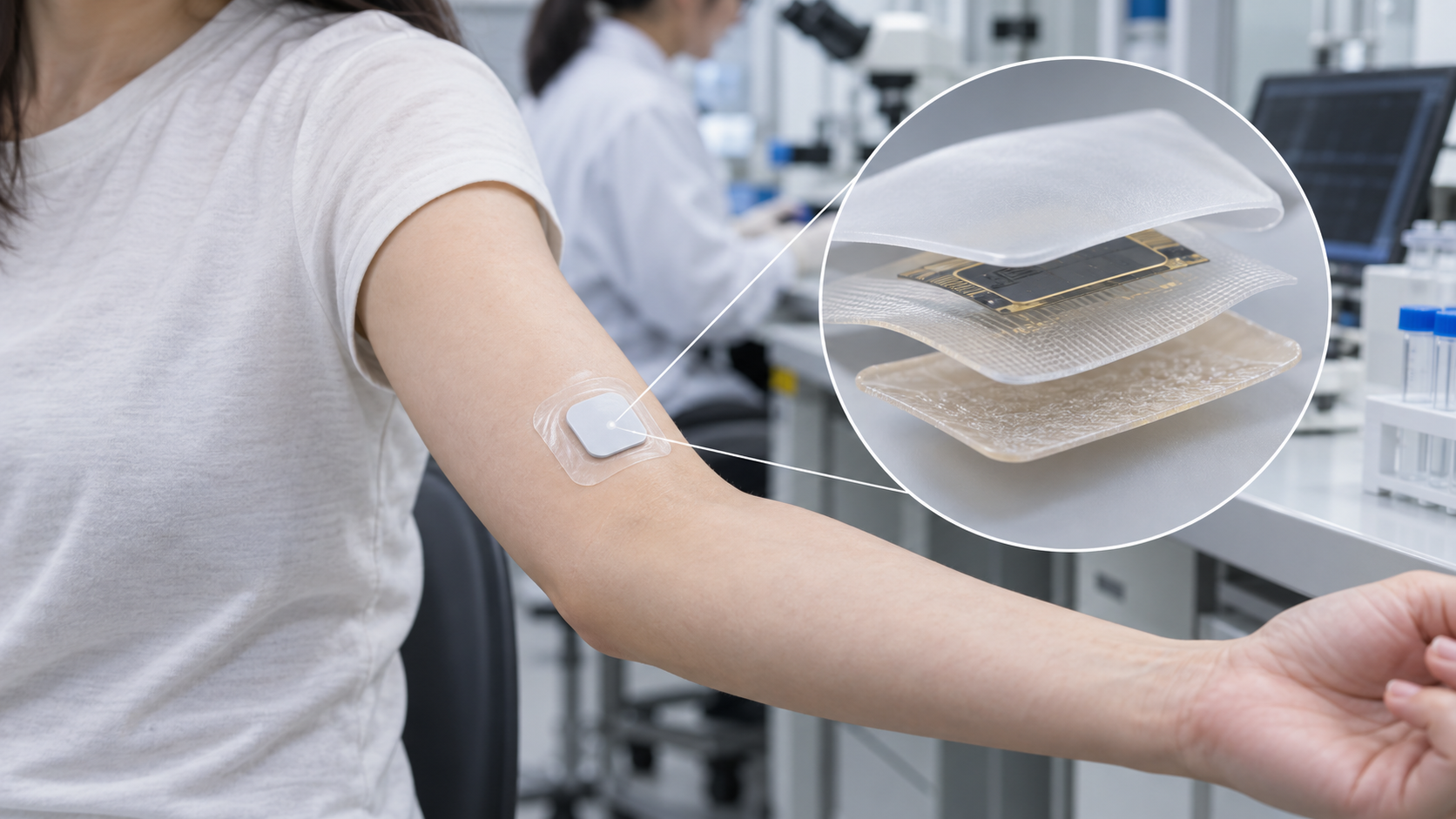
A sensor worn on the skin needs more than the sensing element. It needs an adhesive layer that stays put for days and then comes off without hurting the skin. Skin, unlike a steel plate, sweats, carries oil and hair, and constantly sheds its outer layer. This article walks through skin adhesive families, wear testing, the classification of adhesive-related skin injury, and biocompatibility evaluation under ISO 10993, drawing on manufacturers' technical papers, peer-reviewed studies and FDA guidance.
- What patch-sensor adhesion is (the short version)
- The layers of a patch: body, tie layer, backing, adhesive
- Skin adhesive families: acrylic, silicone, hydrocolloid and others
- A materials engineer's view 1: skin is not a steel plate
- Wear time and removal: a 14-day wear study as an example
- What happens at removal: skin injury (MARSI) and the force numbers
- Our calculation: unit conversions and the order of magnitude of moisture transmission
- Sweat and moisture: between breathability and adhesion
- A materials engineer's view 2: four demands that work against each other
- Evaluating irritation and sensitization: ISO 10993 and FDA guidance
- A materials engineer's view 3: FDA treats adhesives as a separate case
- Adhesion test methods: steel, skin and wear studies
- Open issues and what could not be confirmed / Glossary / References / Claim-to-source audit
Sourced = a statement in published material (link given)
Our calculation = a figure this article derived, with the assumptions spelled out
Not yet confirmed = a plan or target with no confirmed record
Structural readings and materials-design interpretations are marked separately as Commentary.
It explains materials, evaluation and regulation. It does not recommend using any particular product, and it does not say how to respond to any skin symptom. Values such as adhesion force or wear time are given together with what was tested, on what, and under what conditions (test method, subjects, conditions, source), and figures from different studies are never lined up to rank products. Values from manufacturers' documents are labelled as the manufacturer's stated values.
1. What patch-sensor adhesion is (the short version)
In this article a patch sensor means a small wearable sensor stuck to the skin to measure things such as glucose in interstitial fluid, sweat, the electrocardiogram or temperature (our own working definition). How the sensing works is covered in other articles in this series. Here we focus on the layer that holds the sensor to the skin.
- Role: 3M's technical paper says that the success of stick-to-skin wearable monitoring devices depends heavily on accuracy and design, and on secure attachment to the body for the whole required wear time Sourced
- Why it is hard: A review by Heikenfeld and colleagues (2018) says that many of the difficulties of wearable sensors become understandable once the skin is seen as "more of an information barrier than an information source" Sourced. For adhesion too, skin moves, sweats, produces oil, carries hair and renews its surface
- The trade-off: The adhesive must hold while the patch is worn and be gentle when it is removed. "Sticks" and "peels cleanly" are two sides of the same material properties (Commentary)
Stronger adhesion is not automatically better. 3M's paper says the adhesive strength should match the product's required wear time, and that a strong adhesive chosen for short wear can cause unnecessary pain or discomfort at removal Sourced. As later chapters show, the force needed to peel and the amount of stratum corneum pulled off the skin do not line up neatly Sourced.
2. The layers of a patch: body, tie layer, backing, adhesive
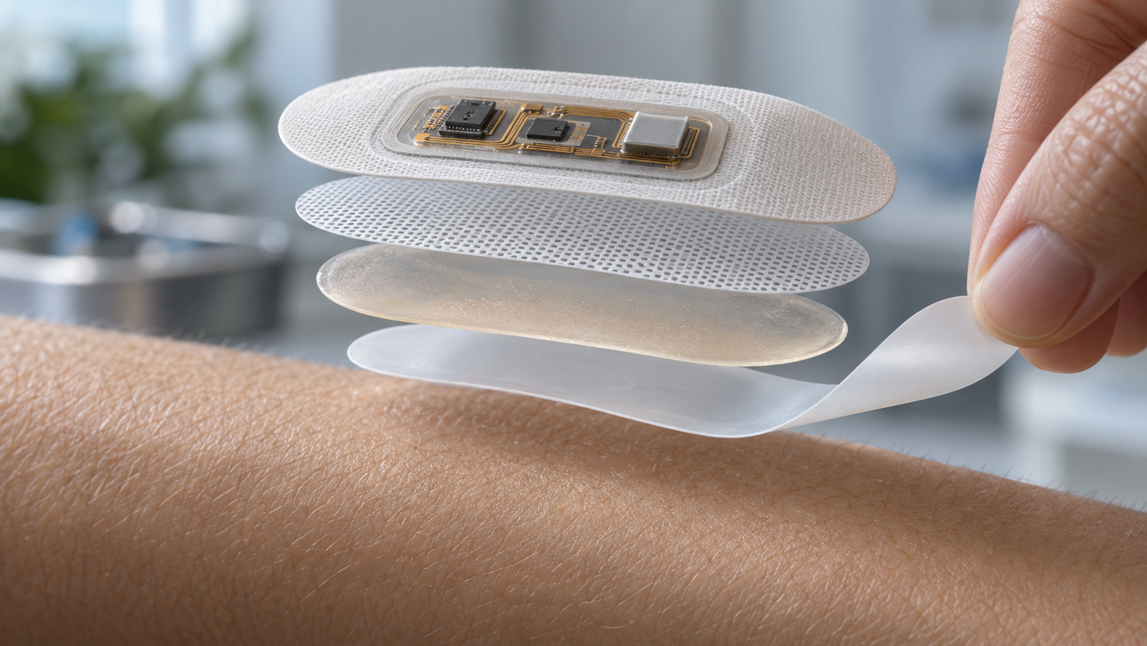
A skin-worn device is not made with one adhesive. 3M's paper divides the jobs adhesives do into three: covering and protecting, bonding to a device, and bonding two substrates together Sourced. In Avery Dennison Medical's wear study, a plastic disc standing in for the sensor body (a sham device) is fixed with tie-layer tape to a skin-adhesive patch, and the patch surrounds the body in a "skirt" arrangement Sourced. The company says this configuration mirrors the wear characteristics and designs of commercial wearables such as continuous glucose monitors (CGMs).
View the original SVG schematic, source notes and numerical details
The three skin-adhesive tapes Avery Dennison Medical tested use the same pressure-sensitive acrylic skin adhesive and differ only in the backing (carrier). MED 5740 has a white thermoplastic polyurethane nonwoven, MED 5741 a white polyester (PET) nonwoven, and MED 5742 a clear 1 mil (about 25 µm) polyurethane film Sourced. The company notes that the three products differ in breathability (moisture vapor transmission), adhesion level and the elasticity of the carrier Sourced. The adhesive family and the finished tape, backing included, have to be considered separately.
The general character of acrylic, rubber and silicone adhesives is covered in this series' articles on tapes and silicones, and the principle that wetting comes first and strength later in the article on adhesives. This article concentrates on what changes when those ideas meet a very particular adherend: skin.
3. Skin adhesive families: acrylic, silicone, hydrocolloid and others
3M's technical paper names three main families of pressure-sensitive adhesive (PSA) for stick-to-skin wearables: acrylic, silicone and synthetic rubber. It adds specialty formulations such as conductive hydrogels and hydrocolloids, which are designed to wick moisture Sourced. The paper also says synthetic rubber adhesives bond strongly to low-surface-energy materials and, for that reason, are often the first choice for bonding the construction layers inside a device rather than for the layer that touches skin Sourced.
Acrylic versus silicone: how they flow, and how stiff they are
3M explains the difference between the two families in terms of how well they wet the skin and how they deform at removal Sourced.
- Acrylic: A traditional acrylic adhesive is slow to wet out and conform to the contours of the skin, and at first touches only the high points. Over time it flows into skin texture and around hair, so adhesion rises, and so does the chance of pulling hair and stratum corneum off at removal. Acrylics are stiffer and deform less, so the skin is pulled harder at removal, the paper says
- Silicone: Silicones have low surface energy and, more importantly, low modulus, so they wet out quickly and form a maximum bond. They are more elastic than traditional acrylics, especially over long timescales, which avoids cold flow around hair and skin cells. At removal the adhesive deforms, causing less trauma to the stratum corneum, according to the paper
3M's paper also shows confocal microscope images of the adhesive faces of medical tapes after removal from a healthy volunteer, with a qualitative difference in the amount of skin cells transferred between acrylic and silicone (the paper adds that results vary with the application) Sourced. This is the series' idea of the "adhesive as a slowly flowing solid" (tape article) and of the low surface energy and low modulus of silicone (silicone article), applied to skin (Commentary).
The structure of a hydrocolloid
According to the abstract of a patent publication [Reference 17], a hydrocolloid adhesive is a composition with hydrocolloid particles dispersed in a rubbery elastomeric base (US 4,952,618, assigned to 3M; we make no reading of claim scope) Sourced. When the material takes up water, the hydrocolloid swells. A review by Thomas (2008) says the impermeable nature of hydrocolloid wound dressings allows washing or showering while helping to prevent the spread of pathogenic microorganisms Sourced. That one material both takes up water and blocks water vapor becomes important in the chapter on sweat and moisture.
4. A materials engineer's view 1: skin is not a steel plate
The strength of a pressure-sensitive tape is normally given as the force to peel it from a stainless steel plate at an angle such as 180° and a constant speed (see this series' article on tapes). The adherend is uniform and the result reproducible. On skin, the same yardstick does not hold.
- Adhesion is lower on skin than on steel. Klode and colleagues tested 56 wound dressings (23 acrylic, 9 silicone, 17 hydrocolloid, 7 polyurethane) on steel and on healthy human skin, following EN 1939:2003. Adhesion on steel was significantly higher than on skin (P < 0.0001), and the relationship differed between families Sourced
- Different steel numbers, same wear result. The three Avery Dennison Medical tapes differed by up to about 2.5 times in peel adhesion to stainless steel (5.1 ÷ 2.0) Our calculation as the table shows. Yet in the 14-day wear study on 20 healthy adults, there was no statistically significant difference among the three, and none came off before day 10 Sourced
- The manufacturer says so itself. The company states that tests on stainless steel and polyethylene are useful because the surfaces are consistent and the tests reproducible, but that skin is complex, dynamic and different from person to person; sweat, oil and hair affect adhesion, and the adhesive must follow an elastic, extensible skin. It concludes that skin is very difficult to duplicate in laboratory testing Sourced
The steel test is best read as a quality-control tool for the material, and performance on skin as something to confirm with a different test (Commentary). The point that ASTM D3330 itself says peel adhesion usually has no direct relationship to functional requirements and gives no design information was covered in the tape article.
| Item (Avery Dennison Medical test-method code) | MED 5740 | MED 5741 | MED 5742 |
|---|---|---|---|
| Backing | white thermoplastic polyurethane nonwoven | white polyester (PET) nonwoven | clear polyurethane film (1 mil) |
| Peel adhesion on polyethylene (TDS-01) | 0.7 lbf/in | 0.9 lbf/in | 0.8 lbf/in |
| Peel adhesion on stainless steel (TDS-04) | 2.1 lbf/in | 2.0 lbf/in | 5.1 lbf/in |
| Quick-stick adhesion on stainless steel (TDS-05) | 1.6 lbf/in | 0.7 lbf/in | 0.2 lbf/in |
| Liner release, 90° (TDS-06) | 190 gf/2in | 200 gf/2in | 150 gf/2in |
| Static shear (TDS-12) | 600 min | 260 min | 1120 min |
| Moisture vapor transmission rate, MVTR (TDS-16) | 400 g/m²·24h | 1000 g/m²·24h | 270 g/m²·24h |
All Sourced (Table 1 of the Avery Dennison Medical white paper; typical values, i.e. the manufacturer's stated values). TDS-01 and the rest are the company's own test-method codes; temperature, speed, dwell time and MVTR test conditions are not given in the paper. All three use the same pressure-sensitive acrylic skin adhesive and differ only in backing.
The skin's side of the interface
3M's paper explains that the outer stratum corneum is continually shed and replaced, and that on average the skin renews itself about every 14 days Sourced (the company's statement; the renewal period varies with site and age). Renewal slows with age, moisture falls, and the skin becomes drier, thinner and more fragile. The paper divides "fragile skin" into four groups Sourced.
- Physiological: infant skin, elderly skin, and fragile zones such as the face and eyelids
- Pathological: acne, atopic dermatitis, rosacea and the like
- Circumstantial: environmental (heat, cold, chemicals) and mechanical (rubbing) stress
- Iatrogenic: medicinal treatment and aesthetic procedures
So the surface being bonded to varies widely with age, body site and health, and cannot be controlled like a material. From the designer's side, no single adhesive is optimal for everyone. That is why 3M suggests that a device meant for the whole population might use an adhesive portfolio covering a range of adhesion levels Sourced.
5. Wear time and removal: a 14-day wear study as an example
"How many days will it stay on" is answered not with a steel-plate number but by sticking the product on people and measuring the time until it comes off. Avery Dennison Medical's white paper is one of the few that publishes the design and results of such a test. It is, however, the company's own technical paper (not peer-reviewed), it tested the company's products, and it used sham devices rather than real sensors Sourced.
| Item | Study design (Avery Dennison Medical white paper) |
|---|---|
| Subjects | Single center; 20 healthy adults, all of whom completed. Exclusions included immunocompromise, use of anti-inflammatory, immunosuppressive or antihistamine drugs, allergy to adhesives or bandages, and damaged skin at the test sites |
| Sham device | A plastic disc of 25 mm × 35 mm × 3.2 mm fixed to the skin-adhesive patch with tie-layer tape (the skirt arrangement of Fig. 1) |
| Site and preparation | Back of the upper arm and lower abdomen (placement randomized). Hair clipped; skin cleaned with 70% isopropanol wipes and left to dry for at least 2 minutes |
| Conditions during wear | No showering or strenuous activity for 3 hours after application; afterwards showering and exercise as normal, with moisture exposure not restricted |
| Assessment | Visits on days 0, 7 and 14. Percentage of the patch still adhered, appearance (visual check for adhesive residue) and comfort (itch, irritation). Detachment time reported by the subject. Time to detachment analyzed with the Kaplan-Meier method |
| Results | No statistically significant difference among the three tapes (same acrylic adhesive, different backing) (p > 0.05). No detachment until day 10. No significant difference by body site. No subject reported itching, irritation or discomfort, and none removed a sham device on purpose |
All Sourced (Avery Dennison Medical white paper [Reference 2]; the manufacturer's own results; products MED 5740, 5741 and 5742).
In the white paper's Fig. 3 (survival curves), all three tapes begin to lose devices after day 10, and the curves fall by day 14. Reading off the figure, the share still attached on day 14 looks like roughly the 60-80% range (our visual reading of the figure; the paper states no numbers). In its discussion the company also says that wear time could extend beyond two weeks depending on conditions, user environment and product design Sourced, but that is the company's view, not a result of this study.
What it shows: a way to measure time-to-detachment on human skin (visit-based assessment plus survival analysis), and that with the same adhesive family, differences in backing did not produce a difference under these conditions. What it does not show: the result is for 20 healthy adults, sham devices, the company's acrylic adhesive and one test site. It tells us nothing about real sensors, people who sweat heavily, infants or older people, or sports use (Commentary).
6. What happens at removal: skin injury (MARSI) and the force numbers
Many of the problems after application arise at removal. A consensus statement by McNichol and colleagues (2013) called skin injury from medical adhesives a prevalent but under-recognized complication, and named it medical adhesive-related skin injury (MARSI) (based on a consensus meeting of 23 experts; the meeting was funded by an unrestricted educational grant from 3M) Sourced. A later consensus paper (LeBlanc and colleagues, 2019) describes the definition as erythema or other skin abnormality (vesicle, erosion, tear and so on) that persists 30 minutes or more after removal of a device containing adhesive, and the types as mechanical, dermatitis and other Sourced.
Peel force versus pain and skin burden: two studies
Is it true, then, that the weaker the peel force, the gentler the adhesive on skin? Two studies show that the answer is not simple.
(1) Klode et al. (2011): force and pain did line up
In the study of 56 wound dressings mentioned above, the median energy needed to remove a dressing from skin was hydrocolloid 2.25 N, acrylic 1.14 N, polyurethane 0.9 N and silicone 0.7 N (as written in the abstract, which gives the unit as N). Pain on removal (visual analogue scale, 0-10) was 6.8 for hydrocolloid, 4.9 for acrylic, 3.1 for polyurethane and 2.5 for silicone, with a significant correlation between adhesion and pain (correlation coefficient 0.806, P = 0.01) Sourced. The authors point out that no standardized test procedure exists for investigating the adhesion of wound dressings Sourced.
(2) Turnbull et al. (2024): force and stratum corneum removal did not line up
A study in Cureus by researchers at 3M compared four commercial medical tapes (two with silicone adhesive and two with acrylic; one of them a product of the authors' employer) on 88 healthy volunteers (32 on the back, 56 on the forearm) Sourced.
- Adhesion: 24 hours after application, each tape was pressed with a 4.5 lb roller and removed on a peel tester at 180° and 90 inches/min. One silicone tape adhered most strongly and one acrylic tape least, so there were differences within a family as well
- Stratum corneum removal: adhesion force did not correlate with the total protein or corneocytes removed. Silicone tapes removed less than acrylic tapes (the most strongly adhering silicone tape carried 31% to 74% less total protein than the other three)
- Skin barrier: after ten successive applications and removals on the forearm, transepidermal water loss (TEWL) showed no significant difference from untaped skin for the silicone tapes, while it rose significantly for the acrylic tapes
- Inflammation marker (IL-1α): results were confounded by position on the forearm and no overall conclusion could be drawn
The authors list as limitations the small number of tapes and the fact that the subjects were healthy volunteers rather than fragile skin. They also offer, as a conjecture, that for the acrylic tape with a plastic backing, which adhered least and disrupted the barrier most, a plastic backing may trap moisture and interfere with adhesion.
Klode et al. measured force versus pain; Turnbull et al. measured force versus stratum corneum removal. They are different quantities, so there is no contradiction. Still, "lower peel force means lighter burden on the skin" cannot be said simply. Turnbull et al. note that earlier work (on wound dressings, and on acrylic tapes under different environmental conditions) likewise found that peel force and damage were not clearly related Sourced. Note also that the Turnbull study was run by researchers at an adhesives supplier, on commercial tapes with paper and similar backings, in 24-hour and repeated-removal tests. It is not a result for long-wear sensor adhesives.
In a separate study, Waring and colleagues applied six wound dressings to the backs of healthy volunteers over 15 days, with removal and reapplication, and reported that the change in transepidermal water loss (TEWL) ranged from almost none to a rise of about 28 g/m²/h depending on the product (abstract) Sourced. Both point the same way: even under the single name "adhesive", the effect on skin changes greatly with the product's design (the combination of adhesive and backing).
7. Our calculation: unit conversions and the order of magnitude of moisture transmission
These conversions, from published numbers, are meant to give a sense of scale Our calculation.
- Peel adhesion unit conversion: with 1 lbf = 4.448 N and 1 in = 25.4 mm, the Avery Dennison Medical peel adhesion on stainless steel of 2.1, 2.0 and 5.1 lbf/in becomes about 9.2, 8.8 and 22.3 N/25 mm (value × 4.448 × 25 ÷ 25.4).
- Order of magnitude of moisture transmission: assume a skin-adhesive tape that is a circle 4 cm in diameter (area 12.6 cm² = 0.00126 m²) and passes water vapor over its whole area. Multiplying MVTR values of 270, 400 and 1000 g/m²·24h by the area gives about 0.34, 0.50 and 1.26 g per day, or about 4.8, 7.0 and 17.6 g over 14 days.
- Ratio of removal energy and pain between families: from the medians of Klode et al., hydrocolloid ÷ silicone = 2.25 ÷ 0.7 = about 3.2 times (energy) and 6.8 ÷ 2.5 = about 2.7 times (pain).
- Thickness conversion: 1 mil = 1/1000 in = about 25.4 µm. The backing of Avery Dennison Medical's MED 5742 is a 1 mil polyurethane film.
Assumptions and limits: the white paper gives no test conditions for peel adhesion (temperature, peel speed, dwell time, width), so the N/25 mm figures cannot be set beside another company's N/25 mm to argue which is better. The moisture calculation ignores that the sensor body blocks most of the area and that the white paper does not give the MVTR test conditions (temperature and humidity difference); it is an order-of-magnitude estimate only. The Klode values are family medians for wound dressings, may differ widely between products of one family, and do not apply directly to sensor adhesives.
8. Sweat and moisture: between breathability and adhesion
Skin continuously gives off water vapor, and exercise and heat add sweat. A patch sits on top of that for days. Moisture acts in two directions.
- It weakens adhesion: Avery Dennison Medical says skin produces sweat, oil and hair, all of which affect adhesion Sourced. 3M likewise says that adhesive selection should consider the user's activity, sweating and climate Sourced
- It can macerate the skin: moisture trapped between skin and tape can lead to a different type of skin injury, maceration (Fig. 4; Commentary)
In patch design the backing's moisture vapor transmission rate (MVTR) is an important variable. Even with the same adhesive, Avery Dennison Medical's three tapes differ by about 3.7 times: 1000 g/m²·24h with the PET nonwoven, 400 with the thermoplastic polyurethane nonwoven and 270 with the polyurethane film Sourced Our calculation (typical values stated by the manufacturer; test conditions not given). Static shear (resistance to slipping), on the other hand, was longest for the film backing with the lowest MVTR, at 1120 minutes, about 4.3 times the 260 minutes of the PET nonwoven Sourced Our calculation. With the same adhesive, the choice of backing moved breathability and resistance to slipping in opposite directions (Commentary).
Yet in the 14-day wear study the three came out without significant difference, as described above. That is, differences in MVTR and shear measured in the laboratory do not necessarily appear in whether a device stays on. The white paper's data do not tell us which property decides the wear result (Commentary).
A hydrocolloid is a rubbery base with water-absorbing particles dispersed in it, and it takes up water and swells (chapter 3). 3M describes hydrocolloids as gentle, cushioning and absorbent, and lists water resistance among the features of silicone Sourced. Even under the one phrase "good with water" there are families that soak it up and families that repel or pass it, and the design thinking differs (Commentary).
9. A materials engineer's view 2: four demands that work against each other
Designing a skin adhesive means meeting four demands at once.
- 1. It must stick: conform to skin texture and hair (wet out). In 3M's account, acrylics wet slowly and gain adhesion over time, while silicones, with a low modulus, wet quickly
- 2. It must stay stuck: withstand sweat, movement and washing, and not slip for the required number of days. 3M's figure suggests raising the adhesion level as days increase
- 3. It must come off gently: not strip the stratum corneum, and cause little pain. Klode et al. found adhesion and pain correlated; Turnbull et al. found force and stratum corneum removal did not line up
- 4. The skin must not react: little irritation or sensitization. This depends on the design of the components and on evaluation under ISO 10993 (chapter 10)
Raising demands 1 and 2 tends to worsen demand 3. Higher adhesion, adhesion that builds over time, and wrapping around hair all increase the force at removal (Commentary). Even so, 3M's paper points to designs such as silicone, with its low modulus, that can wet quickly and yet dissipate force through deformation at removal. The search for compatibility is along a different axis from "strength": modulus and flow (viscoelasticity).
On the other hand, 3M's summary lists silicone as "gentle to moderate bonding" and "short to moderate wear time", which implies limits for long wear Sourced. Combining gentleness with long wear (7-14 days) therefore looks like a design problem that remains open (Commentary). Backing breathability, stretch and thickness, the tie layer, body stiffness and patch area all matter, so design beyond the adhesive itself counts too.
10. Evaluating irritation and sensitization: ISO 10993 and FDA guidance
An adhesive that touches skin for a long time is evaluated for three biological effects: cytotoxicity, sensitization and irritation. The framework is the ISO 10993 series (biological evaluation of medical devices). The series is not a list of tests to pass. It is a framework in which the kind and duration of contact determine which effects to evaluate, and the result is judged as risk.
| Standard (edition) | Title (as shown on the standards body's official page) | Relevance to skin adhesives |
|---|---|---|
| ISO 10993-1:2025 (November 2025) | Biological evaluation of medical devices — Part 1: Requirements and general principles for the evaluation of biological safety within a risk management process | The overall framework, aligned with ISO 14971 risk management. The title of the previous edition (2018) was "Evaluation and testing within a risk management process" |
| ISO 10993-5:2009 | Biological evaluation of medical devices — Part 5: Tests for in vitro cytotoxicity | Tests cytotoxicity by incubating cultured cells in contact with a device or its extracts |
| ISO 10993-10:2021 (November 2021) | Biological evaluation of medical devices — Part 10: Tests for skin sensitization | Assessment of the potential to induce skin sensitization. Its scope includes details of in vivo test procedures and key factors in interpreting results |
| ISO 10993-23:2021 (January 2021) | Biological evaluation of medical devices — Part 23: Tests for irritation | Assessment of irritation potential. Includes pre-test considerations (with in silico and in vitro methods), in vitro and in vivo procedures, and key factors in interpretation. Amendment 1 in 2025 |
| ISO 10993-12:2021 (January 2021) | Biological evaluation of medical devices — Part 12: Sample preparation and reference materials | Selection and preparation of samples, controls, and preparation of extracts. Amendment 1 in 2025 |
All Sourced (the official pages of the standards body EVS [References 10-14]. The ISO site itself could not be fetched automatically, so numbers, titles and scope were confirmed on the page of an ISO member body. The full text of the standards is paid and was not checked).
Kinds and durations of contact: FDA guidance
The FDA guidance "Use of International Standard ISO 10993-1" (issued 8 September 2023; nonbinding recommendations) categorizes devices by kind of contact (surface device on intact skin, mucosal membrane and so on) and by contact duration. The durations are limited (24 hours or less), prolonged (more than 24 hours to 30 days) and long-term (more than 30 days) Sourced. Its Attachment A (Table A.1) lists, for a surface device on intact skin, cytotoxicity, sensitization, and irritation or intracutaneous reactivity as endpoints to consider in all three duration categories Sourced. The guidance says the table is a framework and not a checklist for testing, and that different endpoints may warrant evaluation for particular devices.
Sensitization tests: mostly animal methods
The guidance says the sensitization tests most often submitted are the guinea pig maximization test (GPMT) and the local lymph node assay (LLNA). For devices that only touch the skin (topical devices) the Buehler method may also be used, per ISO 10993-10. For novel materials it is not known whether chemicals will penetrate the skin in an LLNA, so the LLNA is not recommended and the GPMT is Sourced. These are animal tests; they do not directly examine whether a given person will react (Commentary).
The revision to ISO 10993-1:2025: FDA's explanation as of 2026
ISO 10993-1 was revised in 2025 (the sixth edition). According to the record of a town hall held by FDA's CDRH on 9 September 2026 Sourced:
- Devices are now categorized by tissue type (intact skin, intact mucosal membrane and so on), and the "externally communicating device" category was removed. For FDA submissions, however, that term may still be used
- FDA partially recognized the new edition in May 2026. Declarations of conformity to the 2018 edition will be accepted until 1 July 2029. FDA does not plan to update its guidance now and says that its expectations for biocompatibility testing are not raised by the 2025 edition
This article did not check how the revision changes the evaluation of skin-contact adhesives in detail, because the text of the standard is paid.
Standard testing does not rule out individual reactions: case reports
Biocompatibility testing assesses hazard for a population. It does not guarantee that no individual will react. In fact, there are case reports of allergic contact dermatitis caused by isobornyl acrylate (IBOA, an acrylate) at the application site of a continuous glucose monitor. Mine and colleagues (2019) report an 8-year-old girl with type 1 diabetes who had a positive patch test to the adhesive part of the sensor and to IBOA at 0.1%, 0.05% and 0.01% Sourced. Similar case reports exist (Corazza et al. 2018; Kamann and Oppel 2019), and a report (Oppel et al. 2020; by its title) describes a successor model as an IBOA-free generation Sourced.
Case reports do not give an incidence. They do not evaluate the safety of any product, and they are not advice to any individual reader. From a materials engineer's viewpoint, they show that small components of the adhesive, backing or body (monomers and additives) can become the cause of sensitization in long skin contact, and that components can be revised when a product generation changes (Commentary).
11. A materials engineer's view 3: FDA treats adhesives as a separate case
Attachment G of the FDA guidance sets out a simplified policy for devices that touch only intact skin: if the materials are on a list and a history of safe use in legally marketed devices can be documented, biocompatibility testing can be omitted. The list includes ABS, PET, polyethylene, polypropylene, polyurethane, PMMA, silicone, and fabrics such as cotton and silk Sourced.
But Table G.1 of the same guidance lists the following as excluded from this policy Sourced:
- Adhesives used to attach a device directly to the skin (examples given: electrode pads and on-body pump attachment systems). The stated reason: "Adhesives can cause adverse biological responses (e.g., irritation)"
- Hydrogels, in-situ polymerizing materials and absorbable materials, because polymerization or degradation products can change over time
- Materials not on the list (novel materials, bulk metals and so on)
In other words, FDA appears to treat a skin adhesive as something that cannot be represented by the track record of a similarly named material, and is evaluated with its formulation and manufacturing process (Commentary). For skin adhesives, even within "acrylic" or "silicone", differences in residual monomer, additives and tackifying components can matter for the evaluation. The silicone and polyurethane on the list refer to materials as device components, not to skin adhesives (note the exclusion above; Commentary).
The policy also presumes that the manufacturer analyzes post-market complaints (erythema, edema, irritation, sensitization, allergy) on a continuing basis Sourced. Omitting tests does not mean stopping observation.
12. Adhesion test methods: steel, skin and wear studies
The test methods that have appeared so far, arranged by what they look at.
| What is examined | Method (examples) | Source | Notes for reading |
|---|---|---|---|
| Peel adhesion to steel | ISO 29862:2024 (180° from stainless steel and others), ASTM D3330/D3330M-04(2025), JIS Z 0237:2022 | Standards bodies' pages (checked for the tape article) | A quality-control tool. ASTM D3330 says it cannot compare tapes from different manufacturers and gives no design information |
| Holding power (shear) | Static shear (ISO 29863:2018 and others). Avery Dennison Medical reports its own method TDS-12 | Standards bodies; manufacturer white paper | Conditions may differ between manufacturers |
| Adhesion to skin | Remove after 24 h at 180° and 90 inches/min (about 38 mm/s), or with a modified testing machine following EN 1939:2003 | Turnbull et al.; Klode et al. | Klode et al. noted in 2011 that no standardized method existed for the adhesion of wound dressings. The present situation could not be confirmed |
| Stratum corneum removal | Total protein on the removed tape (BCA assay); corneocyte staining (phloxine B) with image analysis; repeated at the same site ten times | Turnbull et al. | The amount removed falls as the number of strips rises (the authors explain that cell cohesion is stronger in deeper layers) |
| Skin barrier | Transepidermal water loss (TEWL), judged by the change from before application | Turnbull et al.; Waring et al. | Interpreted as a difference from an untaped control site, not as an absolute value |
| Wear time | Visit-based assessment and survival analysis of time to detachment (Kaplan-Meier method) | Avery Dennison Medical | Site, preparation, activity and showering conditions must be stated |
| Skin reactions | Tests under ISO 10993-5, -10 and -23, and observation in use | FDA guidance; standards bodies | An assessment of hazard for a population; it does not exclude individual reactions |
"90 inches/min = about 38 mm/s" is our conversion Our calculation (90 × 25.4 ÷ 60 = 38.1). Everything else is Sourced (see each reference).
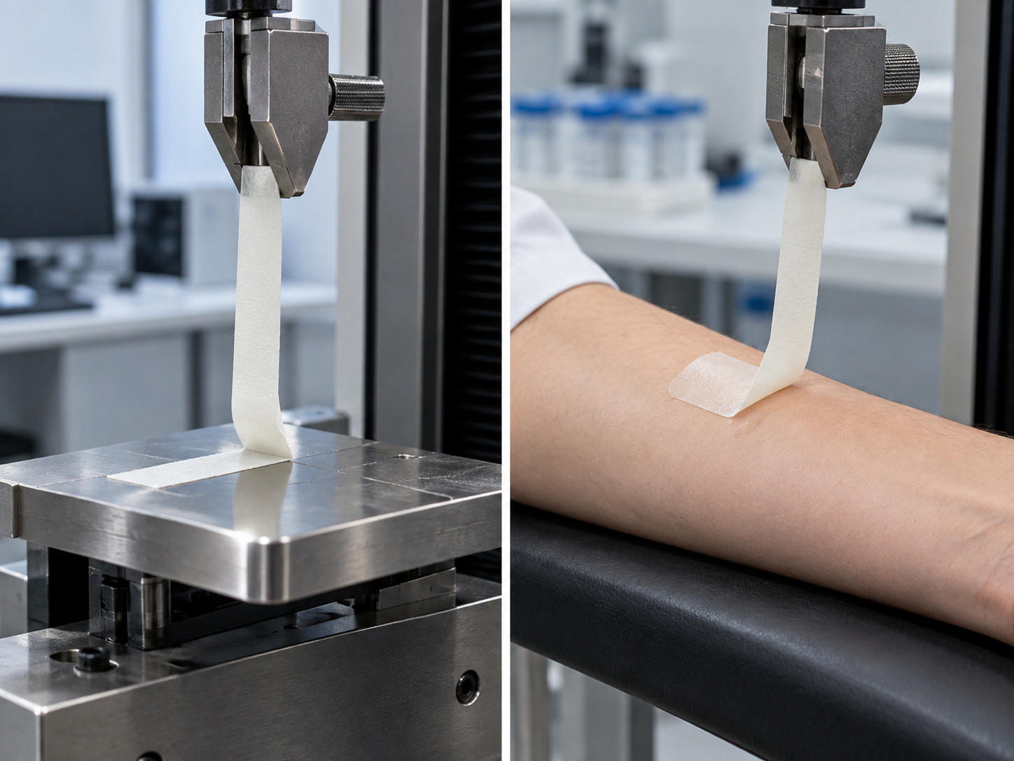
A steel test has a uniform adherend and high reproducibility. A skin test differs between people, between body sites and between days. Avery Dennison Medical's white paper takes the position that both kinds of test are needed and neither replaces the other. For a materials engineer, that means designing and reporting tests that control the material's quality (steel) separately from tests that reproduce how the product is used (skin, wear), and always attaching the adherend, site, preparation, time and speed to the result (Commentary).
13. Open issues and what could not be confirmed
(1) No independent source comparing the families under the same conditions
Among the sources this article consulted, we could not confirm any study that compares acrylic, silicone, hydrocolloid and other families under the same conditions and is independent of adhesive suppliers. 3M and Avery Dennison Medical supply adhesive materials, and the authors of the Turnbull study work at 3M. The Klode and Waring studies concern wound dressings. This article introduces the character of each family but does not declare one better than another.
(2) Avery Dennison Medical's white paper is not peer-reviewed
The 14-day wear study is the company's technical paper: 20 healthy adults, sham devices, one center, and no justification of statistical power is given. Apart from being pressure-sensitive acrylic with different backings, the composition of MED 5740 to 5742 could not be confirmed.
(3) The text of the standards was not checked
The text of the ISO 10993 series and of ISO 29862 and others is paid. Because the ISO site refused automated access, titles and scope were confirmed on the standards body's (EVS) official pages and in the FDA guidance. We did not check how ISO 10993-1:2025 changes the evaluation of devices that touch skin in concrete terms. We also did not check the related Japanese standards (the JIS T 0993 series) or the treatment under Japan's Pharmaceuticals and Medical Devices Act.
(4) The adhesives of actual wearable-sensor products are out of reach
The adhesive composition, layer structure and wear-test data of commercial continuous glucose monitors and similar products could not be confirmed in the public official material this article consulted. The numbers here come from studies of sham devices, commercial medical tapes and wound dressings. For a product's wear time and the frequency of skin reactions, one must consult each product's instructions for use and the regulator's documents.
(5) The skin renewal period is the manufacturer's statement
"Renews every 14 days on average" is 3M's statement, with no source given. The period varies with age, body site and health.
(6) No source quantified the effect of sweat volume or exercise
The studies consulted did not restrict showering or exercise, but we could not confirm any result that quantified the relationship between sweat volume and detachment.
- Skin is not a steel plate. Adhesion on steel was significantly higher than on skin, and tapes whose steel numbers differed by about 2.5 times showed no significant difference in a 14-day wear study Sourced
- Peel force is an incomplete yardstick of skin burden. One study found adhesion and pain correlated, while another found force and stratum corneum removal did not line up Sourced
- Backing and adhesive are designed together. With the same adhesive, MVTR differed by about 3.7 times depending on the backing Sourced Our calculation
- FDA excludes adhesives that attach directly to skin from its simplified evaluation policy, because adhesives can cause adverse biological responses such as irritation Sourced
14. Glossary
- Pressure-sensitive adhesive (PSA)
- An adhesive that sticks under light finger pressure without changing from liquid to solid.
- MARSI
- Medical adhesive-related skin injury: erythema or another skin abnormality (vesicle, erosion, tear) persisting 30 minutes or more after removal of an adhesive device.
- Stratum corneum
- The outermost skin layer, made of dead keratinized cells. It is shed and renewed continually.
- Corneocyte
- A cell of the stratum corneum. Corneocytes cling to the adhesive face when a tape is removed.
- TEWL (transepidermal water loss)
- The amount of water lost from skin as vapor. An indicator of skin barrier condition.
- MVTR (moisture vapor transmission rate)
- How much water vapor a film or tape lets through, in g/m²·24h.
- Maceration
- Softening of skin caused by prolonged retention of moisture.
- Hydrocolloid
- An adhesive with water-swellable particles (gelling agents) dispersed in a rubbery base.
- Sham device
- A test device with no sensing function that imitates only shape and weight.
- Kaplan-Meier method
- A way to estimate, as a survival curve, the time until an event such as detachment occurs.
- Sensitization
- Establishment of an immune response, after which a delayed allergic reaction occurs on later contact.
- GPMT / LLNA
- Animal tests for sensitization: the guinea pig maximization test and the local lymph node assay.
- Static shear
- A holding-power test in which a load is hung from a tape and resistance to slipping is expressed, for example, as time to failure.
- IBOA
- Isobornyl acrylate, an acrylate reported as a contact allergen in case reports.
15. References
- 3M "Designing Wearable Devices for Fragile Skin" (Medical Materials & Technologies technical paper, © 2021) — multimedia.3m.com
- Avery Dennison Medical "Adhesives Enabling Reliable, Multi-Week Wearable Device Attachment" (white paper, © 2020) — medical.averydennison.com
- Turnbull MJ et al. (3M) Comparison of Medical Tape Performance Using Skin Response Quantitative Measurements on Healthy Volunteers. Cureus 16(3): e56548 (2024). DOI 10.7759/cureus.56548 (CC BY 4.0, PMC11027027) — doi.org
- Klode J et al. Investigation of adhesion of modern wound dressings: a comparative analysis of 56 different wound dressings. J Eur Acad Dermatol Venereol 25(8): 933-939 (2011). DOI 10.1111/j.1468-3083.2010.03886.x (abstract checked on PubMed, PMID 21062365) — doi.org
- McNichol L et al. Medical adhesives and patient safety: state of the science: consensus statements for the assessment, prevention, and treatment of adhesive-related skin injuries. J Wound Ostomy Continence Nurs 40(4): 365-380 (2013). DOI 10.1097/WON.0b013e3182995516 (abstract checked on PubMed, PMID 23759927) — doi.org
- LeBlanc K et al. Peristomal Medical Adhesive-Related Skin Injury: Results of an International Consensus Meeting. J Wound Ostomy Continence Nurs 46(2): 125-136 (2019). DOI 10.1097/WON.0000000000000513 (PMC6519893) — doi.org
- Waring M et al. An evaluation of the skin stripping of wound dressing adhesives. J Wound Care 20(9): 412-422 (2011). DOI 10.12968/jowc.2011.20.9.412 (abstract checked on PubMed, PMID 22068140) — doi.org
- U.S. Food and Drug Administration (FDA) Guidance "Use of International Standard ISO 10993-1, 'Biological evaluation of medical devices - Part 1: Evaluation and testing within a risk management process'", 8 September 2023 (nonbinding recommendations; Attachments A and G) — fda.gov
- FDA (CDRH) "Biocompatibility Town Hall #1: Risk Assessment Approaches for Addressing Biocompatibility Endpoints", transcript, 9 September 2026 — fda.gov
- EVS (Estonian Centre for Standardisation and Accreditation) ISO 10993-1:2025: title, scope and edition history — evs.ee
- EVS ISO 10993-5:2009 Tests for in vitro cytotoxicity — evs.ee
- EVS ISO 10993-10:2021 Tests for skin sensitization — evs.ee
- EVS ISO 10993-23:2021 Tests for irritation — evs.ee
- EVS ISO 10993-12:2021 Sample preparation and reference materials — evs.ee
- Heikenfeld J et al. Wearable sensors: modalities, challenges, and prospects. Lab Chip 18(2): 217-248 (2018). DOI 10.1039/C7LC00914C (abstract checked on PubMed, PMID 29182185) — doi.org
- Thomas S Hydrocolloid dressings in the management of acute wounds: a review of the literature. Int Wound J 5(5): 602-613 (2008). DOI 10.1111/j.1742-481X.2008.00541.x (abstract checked on PubMed, PMID 19134061) — doi.org
- U.S. patent US 4,952,618 "Hydrocolloid/adhesive composition" (Minnesota Mining and Manufacturing Co; inventor Roger A. Olsen; granted 28 August 1990; abstract only) — patents.google.com
- Mine A et al. Allergic contact dermatitis caused by isobornyl acrylate when using the FreeStyle Libre. J Diabetes Investig 10(5): 1382-1384 (2019). DOI 10.1111/jdi.13023 (abstract checked on PubMed, PMID 30758915) — doi.org
- Corazza M et al. Allergic contact dermatitis caused by isobornyl acrylate in a young diabetic patient using a continuous glucose monitoring system (Freestyle Libre). Contact Dermatitis 79(5): 320-321 (2018). DOI 10.1111/cod.13075 (title only checked, PMID 30039529) — doi.org
- Kamann S, Oppel E Hydrocolloid blister plaster decreases allergic contact dermatitis caused by Freestyle Libre and isobornyl acrylate. Contact Dermatitis 81(5): 380-381 (2019). DOI 10.1111/cod.13329 (title only checked, PMID 31148185) — doi.org
- Oppel E et al. Freestyle libre 2: The new isobornyl acrylate free generation. Contact Dermatitis 83(5): 429-431 (2020). DOI 10.1111/cod.13638 (title only checked, PMID 32542740) — doi.org
- ISO ISO 29862:2024 Self adhesive tapes — Determination of peel adhesion properties (checked for this series' tape article) — iso.org
- ASTM International ASTM D3330/D3330M-04(2025) Standard Test Method for Peel Adhesion of Pressure-Sensitive Tape — store.astm.org
- Japanese Standards Association (JSA) JIS Z 0237:2022 Testing methods of pressure-sensitive adhesive tapes and sheets (title translated from Japanese) — webdesk.jsa.or.jp
- ISO ISO 29863:2018 Self adhesive tapes — Measurement of static shear adhesion (checked for this series' tape article) — iso.org
16. Claim-to-source audit
| Claim in the text | Basis | Type |
|---|---|---|
| Success of stick-to-skin wearables depends heavily on secure attachment for the required wear time. The three jobs of an adhesive (cover and protect, bond to a device, bond two substrates). The main families are acrylic, silicone and synthetic rubber, with hydrogel (conductive) and hydrocolloid (wicking moisture) as specialties. Synthetic rubber is often first choice for bonding construction layers inside a device | Statements in 3M's technical paper Reference 1 https://multimedia.3m.com/mws/media/2027247O/designing-wearable-devices-for-fragile-skin-hr-pdf-file.pdf | Sourced |
| Acrylic wets out slowly, adhesion builds over time, and removal pulls on the skin harder; silicone has low modulus and low surface energy, wets quickly and deforms at removal with less trauma. Qualitative confocal images. The stratum corneum renews on average in about 14 days. The four groups of fragile skin. Adhesive strength should match wear time. Adhesion-level guidance (0-3, 3-7, 7-14 days). Consider a portfolio of adhesion levels. The seven MARSI names | Statements in 3M's technical paper (the manufacturer's account, no conditions given) Reference 1 https://multimedia.3m.com/mws/media/2027247O/designing-wearable-devices-for-fragile-skin-hr-pdf-file.pdf | Sourced |
| Sham device (25 × 35 × 3.2 mm) fixed to a skin-adhesive patch with tie-layer tape. Three tapes (MED 5740, 5741, 5742) with the same pressure-sensitive acrylic and different backings. Property table (peel adhesion on polyethylene and steel, quick stick, liner release, static shear, MVTR). 20 healthy adults, upper arm and lower abdomen, preparation, showering and exercise allowed, assessment on days 0, 7 and 14, Kaplan-Meier analysis, no significant difference, no detachment until day 10, no reported discomfort. The company's views that skin is hard to reproduce in the laboratory and that wear could exceed two weeks | Avery Dennison Medical white paper (the manufacturer's test results and stated typical values; not peer-reviewed) Reference 2 https://medical.averydennison.com/content/dam/medical/medicalproducts/Documents/Brochures/ADM19001_A4_WP_WEARABLES_061620_WEB.pdf | Sourced |
| Four commercial tapes; 88 healthy volunteers (32 on the back, 56 on the forearm). Removal 24 h after application at 180° and 90 inches/min (4.5 lb roller). One silicone tape adhered most and one acrylic tape least; force did not correlate with total protein or corneocytes removed; total protein 31-74% lower. TEWL not significantly raised for silicone, raised for acrylic. IL-1α: no conclusion. Limitations and the authors' conjecture (plastic backing may trap moisture). Authors are employed at 3M | Full text of Turnbull et al., Cureus 2024 (open access) Reference 3 https://doi.org/10.7759/cureus.56548 | Sourced |
| 56 wound dressings (23 acrylic, 9 silicone, 17 hydrocolloid, 7 polyurethane). Measured on steel and skin following EN 1939:2003. Median energy to remove from skin (hydrocolloid 2.25 N, acrylic 1.14 N, polyurethane 0.9 N, silicone 0.7 N), pain VAS (6.8, 4.9, 3.1, 2.5), adhesion on steel significantly higher than on skin (P < 0.0001), correlation of adhesion with pain 0.806 (P = 0.01). No standardized test procedure | Abstract of Klode et al. (2011) Reference 4 https://doi.org/10.1111/j.1468-3083.2010.03886.x | Sourced |
| Skin injury from adhesives is prevalent but under-recognized. A 23-expert consensus meeting (December 2012; unrestricted educational grant from 3M) | Abstract of McNichol et al. (2013) Reference 5 https://doi.org/10.1097/WON.0b013e3182995516 | Sourced |
| The MARSI definition (erythema or other abnormality persisting 30 minutes or more after removal) and the three types: mechanical, dermatitis, other | LeBlanc et al. (2019), citing the definition of McNichol et al. Reference 6 https://doi.org/10.1097/WON.0000000000000513 | Sourced |
| In a 15-day study of six wound dressings, the change in TEWL ranged from almost none to a rise of about 28 g/m²/h depending on the product | Abstract of Waring et al. (2011) Reference 7 https://doi.org/10.12968/jowc.2011.20.9.412 | Sourced |
| Contact-duration categories (limited 24 h or less; prolonged over 24 h to 30 d; long-term over 30 d). In Attachment A, cytotoxicity, sensitization and irritation (intracutaneous reactivity) are considered for a surface device on intact skin in all three durations. Sensitization tests: GPMT and LLNA submitted, Buehler also for topical devices; LLNA not recommended for novel materials. The simplified policy of Attachment G, its material list, the exclusions of Table G.1 (adhesives attached directly to skin, hydrogels and others), and the premise of post-market complaint analysis. Issued 8 September 2023 | FDA guidance (nonbinding recommendations) Reference 8 https://www.fda.gov/media/142959/download | Sourced |
| The main points of the ISO 10993-1:2025 revision (categorization by tissue type; removal of the "externally communicating device" category). FDA partially recognized it in May 2026. Declarations of conformity to the 2018 edition accepted until 1 July 2029. No near-term guidance update, and FDA's expectations are not raised | Transcript of the FDA (CDRH) town hall of 9 September 2026 Reference 9 https://www.fda.gov/media/194889/download | Sourced |
| Title and scope of ISO 10993-1:2025; effective 18 November 2025; previous edition 17 August 2018 | EVS standard page Reference 10 https://www.evs.ee/en/iso-10993-1-2025 | Sourced |
| Title and scope of ISO 10993-5:2009 (cytotoxicity by incubating cultured cells with a device or extracts) | EVS standard page Reference 11 https://www.evs.ee/en/iso-10993-5-2009 | Sourced |
| Title and scope of ISO 10993-10:2021 (skin sensitization; in vivo test procedures and interpretation), 16 November 2021 | EVS standard page Reference 12 https://www.evs.ee/en/iso-10993-10-2021 | Sourced |
| Title and scope of ISO 10993-23:2021 (irritation; pre-test considerations including in silico and in vitro methods), 20 January 2021, 2025 amendment | EVS standard page Reference 13 https://www.evs.ee/en/iso-10993-23-2021 | Sourced |
| Title and scope of ISO 10993-12:2021 (sample preparation, controls, extracts), 2025 amendment | EVS standard page Reference 14 https://www.evs.ee/en/iso-10993-12-2021 | Sourced |
| The difficulties of wearable sensors become understandable when skin is seen as an information barrier | Abstract of Heikenfeld et al. (2018) Reference 15 https://doi.org/10.1039/C7LC00914C | Sourced |
| The impermeable nature of hydrocolloid wound dressings permits washing and showering and helps prevent the spread of pathogens | Abstract of Thomas (2008) Reference 16 https://doi.org/10.1111/j.1742-481X.2008.00541.x | Sourced |
| A hydrocolloid adhesive is a composition with hydrocolloid particles dispersed in a rubbery elastomeric base | Abstract of US 4,952,618 Reference 17 https://patents.google.com/patent/US4952618A/en | Sourced |
| An 8-year-old girl: positive patch test to the adhesive part of a sensor and to IBOA (0.1, 0.05 and 0.01%) | Abstract of Mine et al. (2019) Reference 18 https://doi.org/10.1111/jdi.13023 | Sourced |
| Similar case reports exist (Corazza et al. 2018; Kamann and Oppel 2019), and a report describing a successor model as an IBOA-free generation (Oppel et al. 2020). All by title | Titles of each paper (checked on Crossref and PubMed) Reference 19 https://doi.org/10.1111/cod.13075, Reference 20 https://doi.org/10.1111/cod.13329, Reference 21 https://doi.org/10.1111/cod.13638 | Sourced |
| Standards for peel adhesion to steel (ISO 29862:2024, ASTM D3330/D3330M-04(2025), JIS Z 0237:2022), ASTM D3330's statement that it cannot compare tapes from different manufacturers and gives no design information, and ISO 29863:2018 (static shear) | Standards bodies' pages confirmed for this series' tape article Reference 22 https://www.iso.org/standard/88050.html, Reference 23 https://store.astm.org/d3330_d3330m-04r25.html, Reference 24 https://webdesk.jsa.or.jp/books/W11M0090/index/?bunsyo_id=JIS+Z+0237%3A2022, Reference 25 https://www.iso.org/standard/70312.html | Sourced |
| Unit conversion of peel adhesion (2.1, 2.0 and 5.1 lbf/in = about 9.2, 8.8 and 22.3 N/25 mm), a maximum ratio of about 2.5 (5.1 ÷ 2.0), the moisture estimate (a 4 cm round patch: about 0.34, 0.50 and 1.26 g/day; about 4.8, 7.0 and 17.6 g over 14 days), the MVTR ratio of about 3.7 and the static shear ratio of about 4.3, the Klode median ratios (about 3.2 and 2.7), 1 mil = 25.4 µm, 90 inches/min = about 38 mm/s | Our calculation. Assumptions: 1 lbf = 4.448 N, 1 in = 25.4 mm, patch area 12.6 cm², water vapor passing over the whole area. Source numbers are the published values of the Avery Dennison Medical white paper and the Klode abstract | Our calculation |
| Assigning the seven names in Fig. 4 to the three types. Treating steel tests as quality control and performance on skin as something to confirm by other tests. The four mutually opposed demands (stick, stay, come off gently, no reaction). Reading FDA's exclusion of adhesives from the simplified policy. Distinguishing silicone as a device component from skin adhesives. The example of backing moving MVTR and static shear in opposite directions. Long wear with gentleness remaining a design problem. The visual reading of the day-14 survival curves (roughly 60-80%) in Fig. 3 | Commentary by this article, built on the sourced facts above (the survival-curve figures are not stated in the paper) | Commentary |
| Adhesive composition, layer structure and test data of actual wearable-sensor products; a same-conditions comparison of families from independent sources; the relationship between sweat volume and detachment; the concrete changes ISO 10993-1:2025 brings for skin-contact devices; Japan's JIS T 0993 series and treatment under the Pharmaceuticals and Medical Devices Act; the adhesive composition of the three Avery Dennison Medical products | Could not be confirmed in the public official sources consulted for this article, and so are not stated | Commentary |
| That Figs. 1 to 7 are explanatory drawings, and that the hero image and Fig. 8 are AI-generated images that do not show real products, testing machines or dimensions | Note by this article | Commentary |
Last updated 3 October 2026. Sources are limited to primary material (technical papers from adhesive-materials manufacturers, peer-reviewed studies, FDA guidance and records, and official pages of standards bodies). Values from manufacturers' documents are treated as the manufacturer's stated values or the company's own test results, and no family or product is declared better than another. Because the article includes structural readings and materials-design interpretations, those are marked as Commentary and kept separate from sourced fact. The adhesive composition and wear-test data of actual wearable-sensor products, independent same-condition comparisons of families, the relationship between sweat volume and detachment, the concrete changes in ISO 10993-1:2025, and the related Japanese standards and legal treatment are not stated, because no published primary source could be confirmed. All figures are explanatory concept graphics. Figs. 1 to 7 are vector drawings; the hero image and Fig. 8 are AI-generated images, and none of them shows a real product, testing machine or cross-section. This article explains materials, evaluation and regulation and is not medical advice.