TECHNOLOGY EXPLAINER
Materials for the Enzyme–Electrode Interface
— enzyme, mediator, immobilisation and membrane set a sensor's performance and life
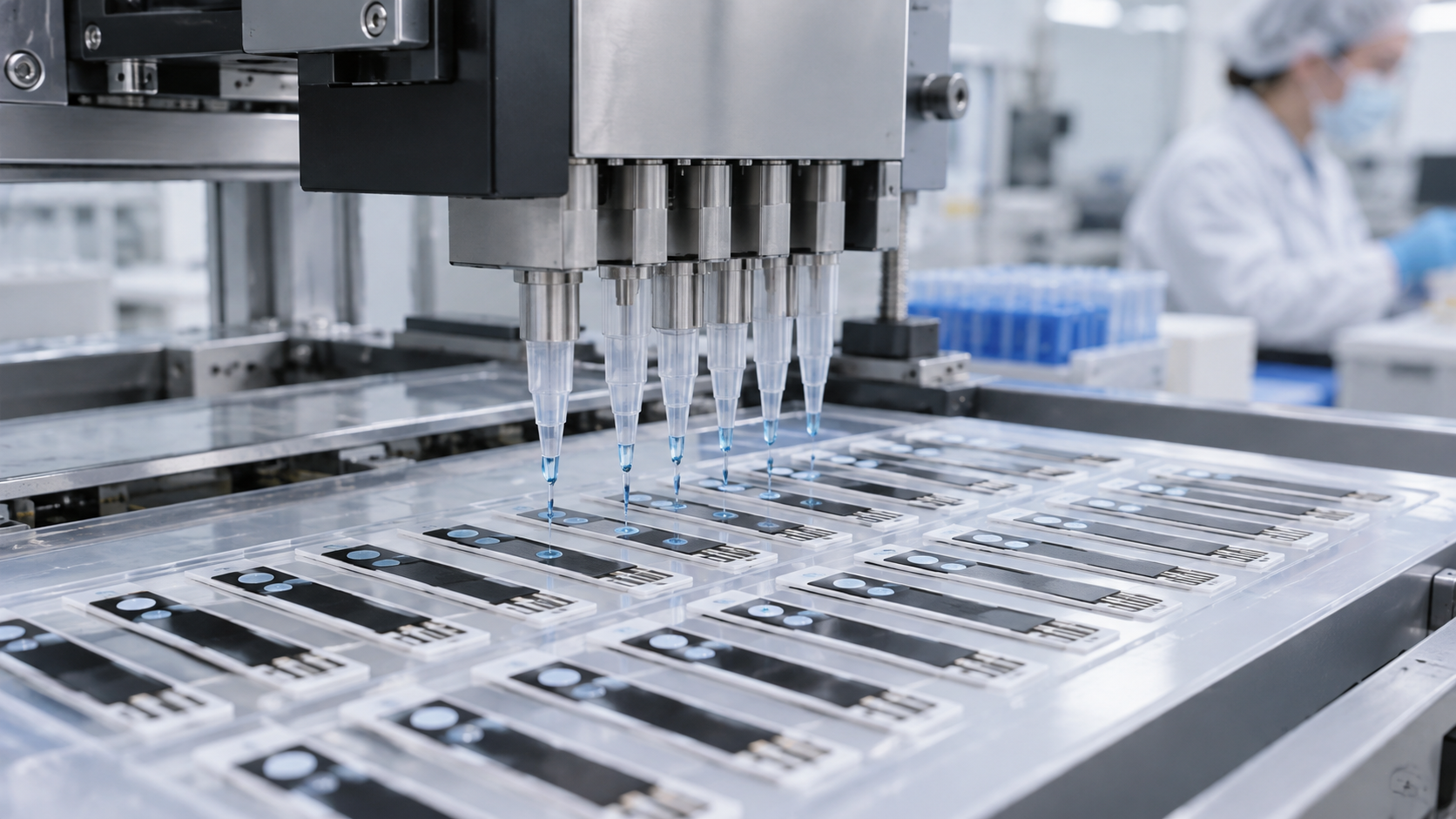
The brain of a biosensor is the enzyme, but what makes it work as a sensor is the materials around the enzyme. The mediator that passes electrons to the electrode, the immobilisation that keeps the enzyme in place and alive, the diffusion-limiting membrane that admits just enough glucose, the coating that resists protein deposition (biofouling), and the stability to survive drying, sterilisation and storage. Using glucose sensors as the example, this explainer sets out the enzyme–electrode interface through a materials lens.
- What the enzyme–electrode interface is, in three points
- The layer structure: electrode, enzyme and membrane
- Choosing the enzyme: glucose oxidase, FAD-GDH and others
- Carrying electrons: mediators and “wired” enzymes
- A materials engineer's view (1): can the enzyme survive drying, heat, sterilisation and storage as a material?
- Immobilising the enzyme: adsorption, entrapment, crosslinking and redox hydrogels
- The diffusion-limiting membrane: polyurethane, Nafion and their roles
- Our calculation: membrane thickness and response time, and the amount of glucose that 1 nA represents
- Biofouling and antifouling
- A materials engineer's view (2): what is being optimised against what
- A company note: Toyobo (diagnostic enzymes)
- Open problems, and what this article could not confirm
- Glossary / References / Claim-to-source audit
Sourced = stated in an official document, a paper or a published patent (link given)
Our calculation = a value this article derived from assumptions it states
Not yet confirmed = research-stage work or a plan with no confirmed track record
Structural readings and materials-design interpretations are marked separately as Commentary.
This article explains technology. It gives no advice on diagnosis, treatment or how to read a measured value, and it neither recommends nor ranks any product or company.
1. What the enzyme–electrode interface is, in three points
An enzyme electrode is an electrode that converts the action of an enzyme that reacts with glucose into an electric current. Its performance is set not by the enzyme alone but by how the interfaces where enzyme, electrode, membrane and body fluid meet are built (our commentary).
- Which enzyme: a review states that current electrochemical CGM sensors are all based on glucose oxidase (GOx)Sourced [Reference 1]. FDA documents likewise say the Dexcom G6 reads a current from hydrogen peroxide produced by GOx, and FreeStyle Libre a current from GOx passing electrons to an electrodeSourced [References 15, 16].
- How electrons are carried: there are three approaches: oxygen takes the electrons (first generation), an artificial electron carrier (a mediator) is used (second generation), and the enzyme passes electrons directly to the electrode (third generation)Sourced [Reference 1].
- What is hard: enzymes are fragile proteins. They have to survive drying and heat in manufacture, sterilisation, storage, and days to weeks in the body, and resist protein deposition (biofouling), while a path for glucose stays open (our commentary).
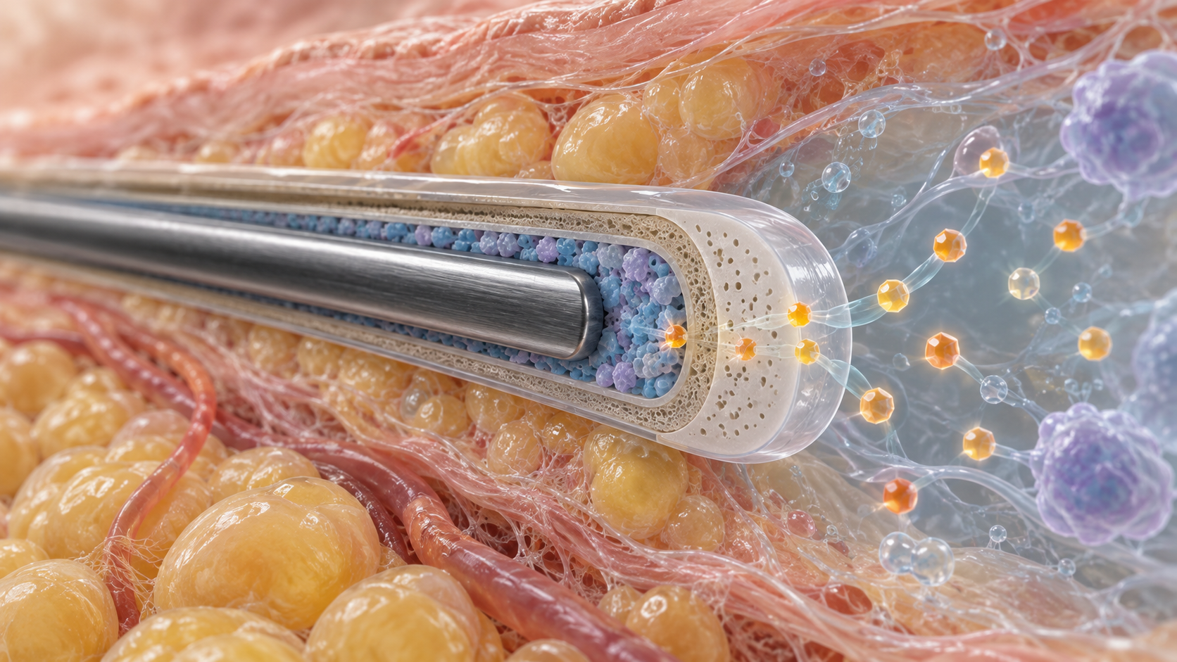
In an enzyme electrode, more of the materials go into “keeping things other than the enzyme away and trimming the amount of glucose” than into “making the enzyme work”. The membrane above the electrode does several jobs at once: it keeps out interferents, throttles glucose so it is not over-supplied, and protects the enzyme from protein deposition (our commentary).
2. The layer structure: electrode, enzyme and membrane
View the original SVG schematic, source notes and numerical details
For in vivo enzyme electrodes, the review by Weltin and colleagues sets out the followingSourced [Reference 5].
- Enzyme-based amperometric microbiosensors are typically made by stacking a set of immobilised membranes on a noble-metal or carbon-based electrode.
- In a first-generation sensor, the enzyme converts the analyte to hydrogen peroxide (consuming oxygen), which is oxidised at a noble-metal electrode to give the current. Because other biogenic substances can also be oxidised at the electrode, it is often covered with a permselective membrane.
- A diffusion-limiting membrane can limit the transport of the analyte relative to oxygen transport, which extends the linear range.
3. Choosing the enzyme: glucose oxidase, FAD-GDH and others
| Enzyme | Main characteristics (as described in reviews, a patent and an FDA label) |
|---|---|
| Glucose oxidase (GOx) | An FAD-dependent glycoprotein. Uses oxygen as the electron acceptor and produces hydrogen peroxide. Very high specificity for β-D-glucose, with minimal activity toward maltose, galactose or xylose. Highly stable and dominant in current CGM. Its cofactor FAD is buried deep in the protein, so it cannot pass electrons directly to an electrode (not a DET type). Because it competes with oxygen, accuracy can fall at low concentrations in mediator-based designs |
| Fungal FAD-dependent GDH | Does not react with oxygen and produces no hydrogen peroxide. No activity toward maltose. Reactivity toward xylose depends on origin (about 10 to 20% of the catalytic efficiency for glucose for A. flavus and A. terreus enzymes; low or negligible for A. niger, A. phoenicis and Mucor). Less stable over the long term than GOx, which has limited its use in CGM. The mainstream enzyme in self-testing strips for blood |
| Bacterial FAD-dependent GDH | Burkholderia cepacia enzyme is the representative. A complex of a catalytic subunit, an electron-transfer subunit with cytochrome and a small subunit; capable of direct electron transfer (DET) to an electrode. Thermally stable. Regarded as a leading candidate for long-term implantable sensors, but at research stage |
| PQQ-dependent GDH | A patent document describes a substrate-specificity problem: it is as reactive with maltose as with glucose |
| NAD(P)-dependent GDH | Strict substrate specificity, but the coenzyme NAD(P) must be added separately, which makes reagents and sensors costly to produce and quality control complex, according to the patent |
All Sourced (Hatada et al. [Reference 1] and Toyobo's patent publication US9657325B2 [Reference 13]). The choice of enzyme is a balance of use (strip or CGM), resistance to interferents and long-term stability (our commentary).
Ferri and colleagues' 2011 review organised the diversity of enzymes, cofactors and final electron acceptors; Hatada and colleagues' 2026 review updates itSourced [References 1, 2].
The FDA drug label for the peritoneal dialysis solution Extraneal (icodextrin) carries a boxed warning that, to avoid falsely high readings caused by maltose, blood glucose monitors based on GDH-PQQ, GDO (glucose-dye-oxidoreductase) and some GDH-FAD methods must not be usedSourced [Reference 11]. Even among “FAD-GDH” enzymes, sugar reactivity differs by origin, which is one way to read why the label says “some” (our commentary). This reports the content of a drug label and is not advice on choosing a meter.
Glucose test systems, including strips for self-testing of blood glucose, are classed as Class II (special controls) in FDA regulation (21 CFR 862.1345)Sourced [Reference 17].
4. Carrying electrons: mediators and “wired” enzymes
- Requirements of a mediator: it reacts rapidly with the reduced enzyme, undergoes reversible electron transfer at low overpotential at the electrode, has stable oxidised and reduced forms, and does not react with oxygenSourced [Reference 3].
- Ferricyanide: one of the mediators most used in blood glucose strips. It is stable in dry, dark conditions, water soluble and a small inorganic complex, but needs special dry, dark packagingSourced [Reference 3].
- Hexaammineruthenium(III) chloride: attracting attention in recent strips. Its redox potential is lower than that of ferricyanide, so it can be used at a lower applied potential, and in general a lower potential means fewer interferences. Its packaging requirements are also less stringentSourced [Reference 3].
- Modifying the enzyme: GOx variants have been reported with higher reactivity toward positively charged mediators such as osmium complexes and ferrocenemethanol, and with mediators covalently tethered to the enzyme surface to aim at “quasi-direct electron transfer (generation 2.5)”Sourced [Reference 1].
What matters for CGM is that the mediator does not leak into the body. The review by Kuznetsova and colleagues says that early second-generation biosensors using mediators such as ferrocene still had the problem of mediator leaching from the electrode, that in 1987 Adam Heller's group addressed it by covalently attaching the mediator to the enzyme to make a “wired” enzyme, and that in the early 1990s he developed osmium-containing redox hydrogels, three-dimensional conductive networks that prevented enzyme leaching and denaturationSourced [Reference 4]. The same review says FreeStyle Libre, launched in 2014, was the first CGM needing no blood calibration and used “wired” enzymes based on redox hydrogelsSourced [Reference 4]. However, the FDA review documents we checked say only that Libre passes electrons to an electrode and do not name the mediator. This point rests on the review's statement alone.
5. A materials engineer's view (1): can the enzyme survive drying, heat, sterilisation and storage as a material?
- Heat in the drying step. Toyobo's patent publication (US9657325B2) says sensor manufacture includes a step of evaporating a GDH solution to dryness on the reaction layer, and that heat treatment at 50 °C or more is often tried to raise production efficiency. Proteins are generally denatured by heat, so the risk rises for enzymes with low thermal stability. It says the upper limit of heat resistance of conventional FAD-GDH in aqueous solution is about 50 to 55 °C, which is insufficient, and sets as its problem an FAD-GDH with high stability in manufacture and in storage after productionSourced [Reference 13]. This is from the publication's background and problem statement, not an interpretation of its claims.
- Modifying the enzyme. The review reports, as improvements in FAD-GDH stability, introduction of a disulfide bond (AfGDH V149C/G190C) that raised the half-life at 45 °C from 8.3 to 110 minutes, and a chimeric enzyme with 100% residual activity at 55 °CSourced [Reference 1].
- Sterilisation and packaging. Sterilisation (radiation, ethylene oxide) and dry, dark packaging act on both the enzyme and low-molecular-weight species such as the mediator (our commentary). That packaging requirements change with the mediator (strict for ferricyanide, lax for hexaammineruthenium) [Reference 3] is an example of how a materials choice even determines a product's logistics.
6. Immobilising the enzyme: adsorption, entrapment, crosslinking and redox hydrogels
An enzyme must stay near the electrode and keep its activity. From the reviews by Weltin et al. and Kuznetsova et al., the methods of immobilisation are as followsSourced [References 4, 5].
| Method | What it involves | Materials points (commentary) |
|---|---|---|
| Crosslinking (BSA + glutaraldehyde) | The most common. The enzyme is dissolved with bovine serum albumin (BSA) and glutaraldehyde is added last, so crosslinking occurs as the droplet dries. It is applied by dip coating, drop coating or dispensing of microdroplets, and further layers can be added later to increase enzyme loading | The low viscosity of the precursor solution allows a high enzyme concentration. The degree of crosslinking affects activity and diffusion |
| Entrapment (inside a polymer membrane) | The enzyme is held inside a polymer membrane. Hydrogels such as PHEMA can also be photopatterned | By varying the thickness of the enzyme membrane and of the diffusion-limiting membrane, sensitivity and linear range can be adjusted over a wide range. Response time is affected |
| Redox hydrogels | A polymer gel carrying redox groups. It carries electrons between enzyme and electrode. Osmium-containing hydrogels are described as having prevented enzyme leaching and denaturation | It combines immobilising the enzyme and immobilising the mediator. The speed of electron transport depends on the degree of crosslinking of the gel and the density of redox centres |
| Crosslinking with PEG diglycidyl ether (PEGDGE) | For implantable devices it is often chosen over glutaraldehyde as a more flexible, more biocompatible crosslinker that better preserves enzyme activity, a review says | In a material used in the body, even the choice of crosslinker is a safety factor |
The “What it involves” column is Sourced (reviews by Weltin et al. [Reference 5] and Kuznetsova et al. [Reference 4]); the “Materials points” column is commentary (this article's framing).
7. The diffusion-limiting membrane: polyurethane, Nafion and their roles
The membrane above the enzyme layer does at least three jobs at once (this article's framing).
- Limits diffusion. It throttles glucose inflow relative to oxygen. Because GOx uses oxygen as a co-substrate, the reaction can be capped by oxygen if glucose stays in excess over oxygen; this membrane widens that linear range (our commentary). The review by Weltin et al. says that dip- or drop-coating thin polyurethane layers over the enzyme membrane is a common way of increasing the linear rangeSourced [Reference 5].
- Stops interferents. A permselective membrane keeps away other biological substances that can be oxidised at the electrode. Nafion (a negatively charged sulfonated fluoropolymer) is a standard example of suppressing anionic species such as ascorbate and urate, and negatively charged proteinsSourced [Reference 8].
- Protects from the environment. In the body, protein deposits on the membrane surface and a capsule eventually forms (next chapters).
As an example of a membrane to raise durability in the body, Yu and colleagues reported using for the first time, as the outer protective membrane of an implantable sensor, an “epoxy-enhanced polyurethane membrane” containing about 30 to 40% epoxy resin adhesive and 50 to 70% polyurethane. The membrane was porous and durable; a sensor lasted 4 to 8 months in solution at room temperature, and eight of nine sensors implanted in three rats functioned for 10 to 56 daysSourced [Reference 6]. This is a research-stage animal study and not the membrane of a commercial product. For polyurethane as a material, see our explainer on polyurethanes and elastomers.
There are also designs that avoid the oxygen limit not by a membrane but by the way of measuring. Gough and colleagues used an enzyme electrode based on differential electrochemical oxygen detection, described as designed to reduce sensitivity to encapsulation, local microvascular perfusion, limited tissue oxygen and enzyme inactivation, and reported that it operated in the subcutaneous tissue of pigs for 222 and 520 daysSourced [Reference 18] (a study of two animals).
8. Biofouling and antifouling
The first thing to deposit on a material that enters the body is protein. Ward's review calls the binding of proteins such as fibrinogen to the surface of a device soon after implantation biofouling, and says it leads to macrophage adhesion and then capsule formationSourced [Reference 9]. Anderson and colleagues say the window in which a material's surface properties modulate the reaction is the first two to four weeks after implantationSourced [Reference 10].
- Membrane fouling and sensitivity: the review by Wisniewski and colleagues addresses the effect of membrane biofouling on sensor sensitivity from the standpoint of limits on glucose transport, and covers methods of evaluation (in vivo and in vitro) and techniques to improve biocompatibilitySourced [Reference 7].
- Antifouling by hydration: a recent review says hydrated interfaces such as PEGylated layers, zwitterionic peptides and conductive hydrogels suppress nonspecific adsorption in protein-rich media while keeping the target accessible, and so improve reliabilitySourced [Reference 8].
- Electrostatic selection: charged polymers such as Nafion are used to exclude anionic interferents [Reference 8].
- It cannot be avoided entirely: Ward's review says that, as of writing, it had not been possible to create a stealth material that evades the mammalian ability to distinguish foreign from selfSourced [Reference 9].
Foreign body reaction, capsule formation and the effect on lifetime are covered in more detail in our explainer on implantable sensors.
9. Our calculation: the amount of glucose that 1 nA represents
For the GOx reaction, the review states that β-D-glucose is oxidised to gluconolactone and the cofactor FAD is reduced to FADH2Sourced [Reference 1]. We calculate on the basis that this corresponds to two electrons per molecule (this article's reading). Taking the Faraday constant as 96,485 C/mol, we find the amount of glucose reacting for a current of 1 nA (1 × 10⁻⁹ A)Our calculation.
- Per second: 1 × 10⁻⁹ / (2 × 96,485) = about 5.2 × 10⁻¹⁵ mol = about 5.2 fmol per second (about 3.1 × 10⁹ molecules per second)
- Per day: 5.2 × 10−15 × 86,400 = about 4.5 × 10⁻¹⁰ mol = about 0.45 nmol. With a molar mass of glucose of 180 g/mol, about 81 ng per day
Assumptions and limits: an idealisation in which all of the current comes from glucose oxidation at two electrons per glucose. The size of a real sensor's current is not given in the documents we checked (1 nA is a number for the calculation). What the calculation conveys is a sense of scale: the amount of glucose the sensor consumes in order to measure is tiny and does not change the glucose on the body's side. It also means the sensor is measuring such a small quantity that small changes in oxygen or in transport through the membrane show up directly in the current (our commentary).
10. A materials engineer's view (2): what is being optimised against what
| Design choice | What it gives | What it costs (commentary) |
|---|---|---|
| A thicker membrane | A wider linear range, better exclusion of interferents, protection of the enzyme | Slower response (diffusion time scales with thickness squared). Lower current (sensitivity) |
| More enzyme loaded | Sensitivity, spare activity for long life | Limits of viscosity and solubility of the precursor, uniformity of immobilisation, manufacturing cost |
| A mediator-based design | Operation at a lower applied potential, with fewer interferences | Mediator leaching and toxicity concerns, competition with oxygen (GOx) |
| FAD-GDH | Not affected by oxygen; suits second generation | Long-term stability lower than GOx, which has limited its use in CGM |
| A hydrophilic outer layer | Less protein deposition | The layer can swell and change diffusion properties; more manufacturing and adhesion steps |
| Heating during drying | Manufacturing efficiency | The risk of the enzyme denaturing from heat |
Parts of the “What it gives” column rest on Sourced material (reviews and a patent publication [References 1, 3, 5, 13]), but the table as a whole is this article's framing (commentary) and not a design theory stated by the authors of those documents.
- “Fast response” is a materials statement: a thin membrane and a large diffusion coefficient (Fig. 4).
- “Resistant to interference” is about the combination of an enzyme and mediator that work at a low potential and a membrane that selects by charge. Indeed, the Dexcom G6 review document says the effect of acetaminophen, a problem in earlier generations, was reduced by sensor designSourced [Reference 15]. The material makeup used is not described in official documents.
- “Long-lasting” is about the whole of enzyme stability, immobilisation, membrane and outer layer, and is not set by any one of them (our commentary).
11. A company note: Toyobo (diagnostic enzymes)
Toyobo is one Japanese company that handles enzymes for diagnostics (a full company profile is covered separately in our series on companies). What this article could confirm in official sources is as follows.
- Enzyme products: Toyobo's English biotechnology page describes its enzymes as “enzymes for in vitro diagnostics, including test items such as blood glucose, cholesterol, and triglycerides”Sourced [Reference 12]. Its enzyme list shows, for glucose, glucose dehydrogenase (NAD(P)-dependent, product number GLD-311), glucose dehydrogenase (PQQ-dependent, GLD-321) and glucose oxidase (from Aspergillus sp., GLO-101)Sourced [Reference 12].
- Patents on FAD-dependent GDH: Google Patents lists FAD-dependent GDH patent publications with Toyobo as applicant (US9657325B2 “Glucose dehydrogenase” and US7662600B2 “Modified flavin adenine dinucleotide dependent glucose dehydrogenase”)Sourced [References 13, 14]. The background of US9657325B2 states that FAD-GDH is an enzyme mainly used for measuring blood glucose, that it came to be used as the main enzyme in glucose sensors because it is not affected by oxygen, that PQQ-dependent GDH has the problem of being as reactive with maltose as with glucose, and that FAD-GDH from the genus Aspergillus is a coenzyme-bound type with high substrate specificity [Reference 13].
- What could not be confirmed: the enzyme list page did not show an FAD-dependent GDH product. Toyobo's current FAD-GDH products, and the companies and products it supplies, could not be confirmed here. The patent descriptions introduce technical background; they do not interpret claims or show adoption in any specific product.
A company that supplies the enzyme itself is positioned as a “materials supplier” to sensor makers. Because improvements in thermal stability and substrate specificity (previous chapters) bear directly on the sensor maker's process and the product's performance, modifying the enzyme determines its value as a material (our commentary).
12. Open problems, and what this article could not confirm
(1) The specific makeup of membrane, immobilisation and mediator by product
The FDA documents checked for this article do not give each product's diffusion-limiting membrane composition, enzyme immobilisation method or type of mediator. Libre's “wired enzyme” and “redox hydrogel” rest on a review's statement [Reference 4].
(2) How fast the enzyme is inactivated in the body
No primary source showing, product by product, in how many days and to what extent the enzyme loses activity in the body could be confirmed. For an implantable example (Eversense's indicator hydrogel), see our explainer on implantable sensors.
(3) Physical properties of membranes such as diffusion coefficients
The diffusion coefficient used in Fig. 4 in Chapter 7 is an assumed value for illustration and not that of any real membrane. Measured values for polyurethane or Nafion are not covered here.
(4) Toyobo's current products and customers
As in Chapter 11, we did not confirm the current state of Toyobo's FAD-GDH products, its customers or comparisons with other companies. No judgment is made on which company is better.
(5) Non-enzymatic sensors, aptamers and so on
Glucose sensors that do not use an enzyme, and other recognition elements such as aptamers and molecularly imprinted polymers, are covered in other articles in this series.
- An enzyme electrode is made of layers. An electrode, an enzyme layer, a diffusion-limiting and permselective membrane and an outer layerSourced
- Current CGM is based on GOx. FAD-GDH is not affected by oxygen but is described as less stable than GOx in the long termSourced
- There are three generations of ways to carry electrons. Mediators allow a lower operating potential and fewer interferences, but leaching is a problemSourced
- The enzyme must survive the process. Heating at 50 °C or more when drying, sterilisation and storage are described as challengesSourced
- Doubling the membrane thickness quadruples the diffusion time. A worked example with an assumed diffusion coefficientOur calculation
- 1 nA corresponds to about 5.2 fmol of glucose per second. The amount of glucose a sensor consumes is tinyOur calculation
13. Glossary
- Enzyme electrode
- An electrode that converts an enzyme's reaction into a current to measure an analyte.
- Glucose oxidase (GOx)
- An FAD-dependent oxidase. Oxidises glucose and, using oxygen as the electron acceptor, produces hydrogen peroxide.
- FAD-GDH
- FAD-dependent glucose dehydrogenase. Does not use oxygen and passes electrons to a mediator.
- PQQ-GDH
- Pyrroloquinoline quinone-dependent glucose dehydrogenase. Described as readily reacting with other sugars such as maltose.
- Mediator
- An artificial electron carrier between enzyme and electrode: ferricyanide, hexaammineruthenium, ferrocene, osmium complexes and others.
- DET
- Direct electron transfer. The enzyme passes electrons straight to the electrode.
- Redox hydrogel
- A polymer gel carrying redox groups. It immobilises the enzyme and carries electrons.
- Wired enzyme
- An enzyme electrically connected to the electrode by tethering a mediator to it covalently or similarly.
- Diffusion-limiting membrane
- A membrane that throttles analyte inflow to widen the linear range. Polyurethane and others.
- Permselective membrane
- A membrane that passes only certain species. Nafion keeps anionic interferents away.
- Biofouling
- Proteins and cells depositing on and covering a material's surface.
- BSA / glutaraldehyde
- Bovine serum albumin and a crosslinker. The most common combination for immobilising an enzyme in a film.
- PEGDGE
- Poly(ethylene glycol) diglycidyl ether. A crosslinker described as flexible and highly biocompatible.
- Faraday constant
- The electric charge carried by one mole of electrons: 96,485 C/mol.
14. References (primary sources)
- Hatada M, Okuda-Shimazaki J, Sode K. “Glucose Oxidoreductases for Glucose Monitoring: A Bird's Eye View of Glucose-Sensing Enzymes II”, J Diabetes Sci Technol 20:1543 (2026) (PMC) — pmc.ncbi.nlm.nih.gov
- Ferri S, Kojima K, Sode K. “Review of glucose oxidases and glucose dehydrogenases: a bird's eye view of glucose sensing enzymes”, J Diabetes Sci Technol 5:1068 (2011) — doi.org
- Loew N et al. “Mediator Preference of Two Different FAD-Dependent Glucose Dehydrogenases Employed in Disposable Enzyme Sensors”, Sensors 17:2636 (2017) (PMC) — pmc.ncbi.nlm.nih.gov
- Kuznetsova LS et al. “Redox Polymers in Electrochemical Biosensing: Molecular Design, Electron Transfer, and Hybrid Nanocomposites”, Biosensors 16 (2026) (PMC) — pmc.ncbi.nlm.nih.gov
- Weltin A, Kieninger J, Urban GA. “Microfabricated, amperometric, enzyme-based biosensors for in vivo applications”, Anal Bioanal Chem 408:4503 (2016) (PMC) — pmc.ncbi.nlm.nih.gov
- Yu B, Long N, Moussy Y, Moussy F. “A long-term flexible minimally-invasive implantable glucose biosensor based on an epoxy-enhanced polyurethane membrane”, Biosens Bioelectron 21:2275 (2006) — doi.org
- Wisniewski N, Moussy F, Reichert WM. “Characterization of implantable biosensor membrane biofouling”, Fresenius J Anal Chem 366:611 (2000) — doi.org
- Review (Int J Mol Sci 27, 2026) “Why Sensors Fail in Biological Samples: Fouling, Blocking, Matrix Effects and Prevention Solutions” (PMC; authors and DOI not present in the bibliographic record we retrieved) — pmc.ncbi.nlm.nih.gov
- Ward WK. “A review of the foreign-body response to subcutaneously-implanted devices: the role of macrophages and cytokines in biofouling and fibrosis”, J Diabetes Sci Technol 2:768 (2008) — doi.org
- Anderson JM, Rodriguez A, Chang DT. “Foreign body reaction to biomaterials”, Semin Immunol 20:86 (2008) — doi.org
- U.S. Food and Drug Administration (FDA) “EXTRANEAL (icodextrin) peritoneal dialysis solution” prescribing information (including the boxed warning), 2016 — www.accessdata.fda.gov
- Toyobo Biotechnology business (English), “Enzyme List” page — www.toyobo-global.com
- Toyobo (patent publication) US9657325B2 “Glucose dehydrogenase” (Google Patents; applicant: Toyobo Co Ltd) — patents.google.com
- Toyobo (patent publication) US7662600B2 “Modified flavin adenine dinucleotide dependent glucose dehydrogenase” (Google Patents; applicant: Toyobo Co Ltd) — patents.google.com
- FDA “DEN170088 Dexcom G6 Decision Summary”, 27 March 2018 — www.accessdata.fda.gov
- FDA “K233537 510(k) summary and clearance letter (FreeStyle Libre 2 / Libre 3)”, 23 April 2024 — www.accessdata.fda.gov
- FDA (eCFR) “21 CFR 862.1345 Glucose test system” — www.ecfr.gov
- Gough DA et al. “Function of an implanted tissue glucose sensor for more than 1 year in animals”, Sci Transl Med 2:42ra53 (2010) — doi.org
- FDA “DEN180058 t:slim X2 insulin pump with interoperable technology: Decision Summary” — www.accessdata.fda.gov
15. Claim-to-source audit
| Claim in the text | Basis | Type |
|---|---|---|
| That current electrochemical CGM sensors are all based on GOx. The three generations of enzyme electrodes (oxygen and hydrogen peroxide / mediator / direct electron transfer). The properties of GOx (FAD-dependent, oxygen as electron acceptor, specificity, stability, non-DET, competition with oxygen). The properties of fungal and bacterial FAD-GDH (inert to oxygen, no maltose reactivity, origin-dependent xylose reactivity, lower long-term stability than GOx, DET and thermal stability of the bacterial enzyme). GOx variants and mediator reactivity / quasi-DET. Examples of FAD-GDH stabilisation (a disulfide bond raising the half-life at 45 °C from 8.3 to 110 minutes; a chimeric enzyme with 100% residual activity at 55 °C). That second-generation designs work at a lower applied potential and reduce interference. The GOx reaction (gluconolactone and FADH2) | Review by Hatada et al. Reference 1 https://pmc.ncbi.nlm.nih.gov/articles/PMC13518614/ | Sourced |
| The existence of the 2011 review of the diversity of GOx and GDH enzymes, and that Hatada et al. update it | Reference 2 https://doi.org/10.1177/193229681100500507 | Sourced |
| The requirements of a mediator; ferricyanide (needs special dry, dark packaging); hexaammineruthenium(III) chloride (lower potential, less stringent packaging); how they are used in blood glucose strips | Paper by Loew et al. (PMC) Reference 3 https://pmc.ncbi.nlm.nih.gov/articles/PMC5712826/ | Sourced |
| The problem of mediator leaching, the “wired enzyme” of 1987, osmium hydrogels of the early 1990s, and the review's statement that the 2014 FreeStyle Libre used wired enzymes (redox hydrogels). The choice between glutaraldehyde and PEGDGE. Electron transport speed in relation to crosslinking and redox-centre density | Review by Kuznetsova et al.; Libre materials rest on the review only Reference 4 https://pmc.ncbi.nlm.nih.gov/articles/PMC13604726/ | Sourced |
| The layer structure of in vivo enzyme electrodes, BSA + glutaraldehyde crosslinking, hydrogels such as PHEMA and photopatterning, that a diffusion-limiting membrane limits analyte transport relative to oxygen and extends the linear range, thin coatings of polyurethane or Nafion, permselective membranes | Review by Weltin et al. (PMC) Reference 5 https://pmc.ncbi.nlm.nih.gov/articles/PMC4909808/ | Sourced |
| The epoxy-enhanced polyurethane membrane (about 30 to 40% epoxy resin adhesive, 50 to 70% polyurethane); porous and durable; 4 to 8 months in solution at room temperature; eight of nine sensors in three rats functioned for 10 to 56 days | Animal (rat) study Reference 6 https://doi.org/10.1016/j.bios.2005.11.002 | Sourced |
| A review of the effect of membrane biofouling on sensor sensitivity in terms of limits on glucose transport, covering evaluation methods and techniques to improve biocompatibility | Only the abstract was checked Reference 7 https://doi.org/10.1007/s002160051556 | Sourced |
| Hydrated antifouling interfaces (PEGylated, zwitterionic, conductive hydrogels), and that Nafion suppresses anionic interferents (ascorbate, urate) and negatively charged proteins | Review (PMC) Reference 8 https://pmc.ncbi.nlm.nih.gov/articles/PMC13300016/ | Sourced |
| The sequence from biofouling (adsorption of fibrinogen and others) to capsule formation, and that a stealth material has not been created | Reference 9 https://doi.org/10.1177/193229680800200504 | Sourced |
| That the period in which surface properties modulate the reaction is the first two to four weeks after implantation | Reference 10 https://doi.org/10.1016/j.smim.2007.11.004 | Sourced |
| The Extraneal label's boxed warning: to avoid falsely high readings due to maltose, glucose monitors based on GDH-PQQ, GDO and some GDH-FAD methods must not be used | FDA-approved drug label Reference 11 https://www.accessdata.fda.gov/drugsatfda_docs/label/2016/021321s034lbl.pdf | Sourced |
| That Toyobo's biotechnology page describes its enzymes as being for in vitro diagnostics (blood glucose, cholesterol, triglycerides). That its enzyme list shows GLD-311 (NAD(P)-dependent), GLD-321 (PQQ-dependent) and GLO-101 (Aspergillus sp.). That no FAD-dependent GDH product appears in the list | Page checked in October 2026 Reference 12 https://www.toyobo-global.com/seihin/xr/enzyme/enzyme_list/ | Sourced |
| US9657325B2: that the applicant is Toyobo. The background (uses of FAD-GDH, no effect from oxygen, maltose reactivity of PQQ-GDH, the coenzyme of NAD(P)-GDH, heat treatment of 50 °C or more during drying, the heat-resistance limit of conventional FAD-GDH of about 50 to 55 °C, and the problem of stability in manufacture and storage) | Background and problem statement of the patent publication; claims are not interpreted Reference 13 https://patents.google.com/patent/US9657325B2/en | Sourced |
| US7662600B2: that the applicant is Toyobo and the title is “Modified flavin adenine dinucleotide dependent glucose dehydrogenase” | Reference 14 https://patents.google.com/patent/US7662600B2/en | Sourced |
| Dexcom G6: GOx produces hydrogen peroxide and a current; the effect of acetaminophen, a problem in earlier generations, was reduced by sensor design; shelf-life testing supporting up to 3 months for the sensor | Reference 15 https://www.accessdata.fda.gov/cdrh_docs/reviews/DEN170088.pdf | Sourced |
| FreeStyle Libre: GOx passes electrons to an electrode and produces a current; electron-beam sterilisation of the sensor pack | Reference 16 https://www.accessdata.fda.gov/cdrh_docs/pdf23/K233537.pdf | Sourced |
| That glucose test systems are Class II (special controls) under 21 CFR 862.1345 | Reference 17 https://www.ecfr.gov/current/title-21/chapter-I/subchapter-H/part-862/subpart-B/section-862.1345 | Sourced |
| That an enzyme electrode based on differential electrochemical oxygen detection operated for 222 and 520 days in the subcutaneous tissue of two pigs | Animal (pig) study Reference 18 https://doi.org/10.1126/scitranslmed.3001148 | Sourced |
| Gamma sterilisation of the t:slim X2 cartridge | Reference 19 https://www.accessdata.fda.gov/cdrh_docs/reviews/DEN180058.pdf | Sourced |
| 1 nA = about 5.2 fmol/s (about 3.1 × 10⁹ molecules/s), about 0.45 nmol (about 81 ng) per day, and membrane diffusion time t = L²/(2D) (with D = 1 × 10⁻⁸ cm²/s: 5 µm → 12.5 s, 10 → 50, 20 → 200, 40 → 800 s) | Our calculation. Assumes a Faraday constant of 96,485 C/mol, two electrons per glucose, an idealisation in which all current comes from glucose oxidation, a molar mass of glucose of 180 g/mol and an assumed diffusion coefficient. The 1 nA and the diffusion coefficient are illustrations and not values for any real sensor or membrane | Our calculation |
| The makeup of membranes, immobilisation and mediators by product; the rate of enzyme inactivation in the body; measured diffusion coefficients and other membrane properties; Toyobo's current FAD-GDH products and customers; non-enzymatic sensors | Not stated in the official documents and papers checked for this article, so not given here | Commentary |
| The framing that a membrane does three jobs at once; the table of what is optimised against what; the reading that an enzyme's thermal stability is a condition for whether the process works; the way of translating a specification sheet into the language of materials; the reading that modifying the enzyme determines its value as a material | This article’s own framing and commentary based on published content. Not views expressed by the authors or regulators of those documents | Commentary |
| That Figs. 1, 2 and 3 are concept diagrams (vector drawings), Fig. 4 is a drawing that includes our calculation, and that the hero image and Fig. 5 are AI-generated images | A note by this article | Commentary |
Last updated 3 October 2026. Sources are limited to primary material (peer-reviewed papers and reviews, FDA review documents, regulation text and a drug label, companies' official pages, and published patent documents). No market-size or market-share estimates are used. Because the article includes structural readings and materials-design interpretations, those are marked as Commentary and kept separate from sourced fact. The makeup of membranes, immobilisation and mediators by product, the rate of enzyme inactivation in the body, measured diffusion coefficients of membranes, and Toyobo's current FAD-GDH products and customers are not covered, because they could not be confirmed in published primary sources. Figs. 1, 2 and 3 are concept diagrams, Fig. 4 includes our calculation, and the hero image and Fig. 5 are AI-generated images; none of them shows a real product, cross-section or measured data. This article explains technology. It gives no advice on diagnosis, treatment or how to read a measured value, and it neither recommends nor ranks any product or company.