LNPs combine four lipid components. A comparison of 56 aminolipids linked apparent pKa to potency in mice; formulation ratios and mixing then influence particle quality. Molecular design and processing are both engineering variables.
Lipid Nanoparticles — Ionizable-Lipid Design and Mixing
Based on regulatory documents and peer-reviewed primary sources / text-source review: September 2026 / illustration and English editorial revision: October 2026.
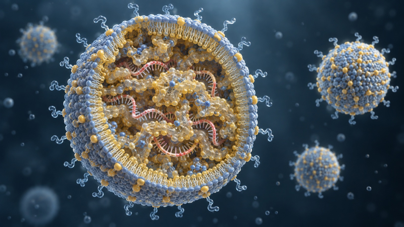
The mRNA article covers LNPs in vaccines, including component roles, per-dose composition and the cold chain. This article focuses on ionizable-lipid design, lipid ratios, manufacture and delivery beyond the liver. Nucleic-acid chemistry and contract manufacturing are covered in the nucleic-acid medicines and CDMO articles.
- LNPs at a glance
- Ionizable lipids: pKa and potency
- Materials viewpoint: pKa requires molecular design
- Four-component molar ratios
- Author calculation from an approved composition
- PEG lipids: designed to dissociate
- Mixing: T-junctions and microfluidics
- Manufacture and specifications in the EPAR
- Materials viewpoint: specifications as requirements
- Delivery: apoE and endosomal escape
- Extrahepatic delivery research
- Challenges and unresolved questions
- Glossary / references / source audit
Facts are statements in cited publications. Author calculations use explicitly stated assumptions. Unresolved/future items have not been confirmed as established outcomes. Interpretations are this article’s organization and reading of materials and process design, not statements by the cited authors. Source fact Author calculation Unresolved / future
This is an explanation of materials and formulation technology, not an assessment or comparison of treatment efficacy or safety, and not medical advice. Product composition and approval dates are taken from regulatory documents. Animal-study findings must not be treated as human outcomes.
1. LNPs at a glance
- LNPs package siRNA or mRNA to protect it from degradation and assist cellular delivery. The Onpattro assessment describes lipids associating with siRNA to protect it in circulation and support delivery to the liver.
- The four components are an ionizable lipid, a structural phospholipid such as DSPC, cholesterol, and a PEG lipid. Ionizable and PEG lipids in this approved formulation were assessed as novel excipients.
- The assessment’s cryo-TEM description is of spherical particles with electron-dense cores, without the conventional liposomal bilayer structure. This is not simply a hollow liposome bag. Source fact
Jayaraman et al. compared 56 aminolipids and found a strong relationship between apparent pKa and gene-silencing potency, with an optimum of 6.2–6.5 in the mouse FVII model. The engineering point is molecular design and measurement in the particle state, not only blending ratios. Source fact
2. Ionizable lipids: pKa and potency
Ionizable lipids are approximately neutral near physiological pH and become positively charged under acidic conditions. The Onpattro assessment explains why this behavior matters. Source fact
The assessment attributes particle formation, siRNA association, cellular uptake, membrane fusion and endosomal release to the physicochemical properties of DLin-MC3-DMA. This lipid was selected after extensive evaluation of related cationic lipids. Source fact
The assessment describes near-neutral charge at about pH 7.4 during circulation and acidic endosomal conditions near pH 5 that favor particle disruption and release. This is a formulation mechanism, not a universal guarantee of efficient delivery. Source fact
DLin-MC3-DMA has formula C43H79NO2 and molecular weight 642.09, with two linoleyl (C18:2) chains. The assessment describes a colorless to pale-yellow liquid, soluble in organic solvents and practically insoluble in water. Source fact
In 2012, Jayaraman et al. synthesized 56 aminolipids and measured apparent pKa and the dose required for 50% silencing of mouse factor VII (FVII ED50). Source fact
| Item | Description in Jayaraman et al. (2012) |
|---|---|
| Main finding | Apparent lipid pKa strongly correlated with mouse FVII-silencing ED50; optimum pKa 6.2–6.5. |
| pKa measurement | TNS fluorescence titration from pH 3 to 10 in 0.5-unit steps. TNS fluoresces when associated with positively charged membrane surfaces, not when free in solution. |
| Structure and pKa | Changing ester–amine spacing by 1–5 methylene groups gave pKa values of 4.17–7.16. |
| Most potent lipid | DLin-MC3-DMA (lipid 16): pKa 6.44, ED50 0.03 mg/kg. Both acidic and basic shifts reduced potency. |
| Comparison conditions | Up to three extrusions through an 80 nm membrane; diameter 70–90 nm; dispersity 0.11±0.04 (n=56); siRNA/total-lipid mass ratio approximately 0.05. |
All findings in the comparison are from Jayaraman et al. (2012) [Reference 1] and mouse experiments, not human efficacy measurements. Source fact
3. Materials viewpoint: pKa requires molecular design
This is not simply a formulation variable adjusted by the mixing ratio. Jayaraman et al. changed nitrogen substituents and the methylene spacing between ester and amine groups; the resulting pKa values spanned 4.17–7.16. Molecular structure is the control variable. Source fact
A desired pKa shift of only 0.3 can therefore require changing molecular structure. The 56-lipid comparison is an experimental structure–property study, not a universal predictive recipe.
The TNS assay measures positive membrane-surface charge by pH titration after particle formation. It yields an apparent particle-associated pKa rather than merely a free-molecule dissociation constant. The analogy with powder zeta potential or resin acid-value measurements is this article’s interpretation. Source fact
The comparison controlled other particle properties: up to three passes through an 80 nm membrane, 70–90 nm particle diameter, and dispersity 0.11±0.04. Holding size reasonably constant helps isolate the relationship with pKa. Source fact
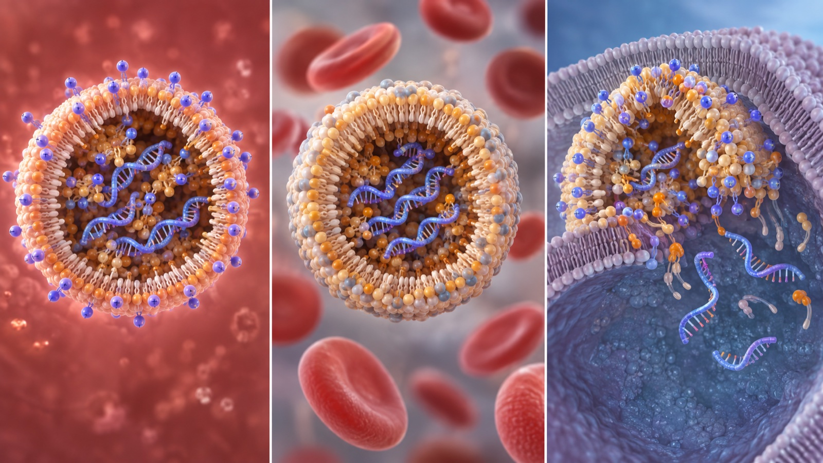
View the original schematic and source annotations
Figure 2. Original vector schematic. Stage descriptions: EMA Onpattro assessment [7]; optimum pKa range: Jayaraman (2012) [1]. Acidic aqueous-phase mixing is a general preparation concept. The drawing is not an actual molecular structure or particle cross-section.
4. Four-component molar ratios
After choosing the lipid’s pKa, the next variable is the ratio of the four components. Jayaraman et al. explicitly describe this subsequent optimization.
The initial molar ratio of aminolipid/DSPC/cholesterol/PEG lipid was 40/10/40/10. Source fact
Further optimization gave 50/10/38.5/1.5. In the mouse FVII model the ED50 reached 0.005 mg/kg, a sixfold improvement over the 40/10/40/10 composition. The same lipid composition was also tested in cynomolgus monkeys. Source fact
The ionizable-lipid fraction rose from 40 to 50 mol%, while PEG lipid fell from 10 to 1.5 mol%, less than one-sixth of its initial fraction. PEG-lipid behavior is discussed below.
5. Author calculation from an approved composition
Does the optimized molar ratio correspond to an approved formulation? The next calculation works backward from the label’s mass composition.
- Patisiran (siRNA): 2 mg, equivalent to 2.1 mg patisiran sodium.
- DLin-MC3-DMA: 13.0 mg.
- Cholesterol: 6.2 mg.
- DSPC: 3.3 mg.
- PEG2000-C-DMG: 1.6 mg.
- Potassium dihydrogen phosphate 0.2 mg; sodium chloride 8.8 mg; disodium hydrogen phosphate heptahydrate 2.3 mg; water for injection. pH approximately 7.0.
- Patisiran sodium: C412H480N148Na40O290P40; molecular weight 14,304 Da.
Masses come from the FDA label. Molecular weights are 642.09 for DLin-MC3-DMA and 2,650±300 Da for PEG2000-C-DMG from the EMA assessment, and 386.7 for cholesterol and 790.1 for DSPC from PubChem. Source fact
Divide each mass by its molecular weight, then normalize the molar amounts. The percentages are author calculations. Author calculation
- DLin-MC3-DMA: 13.0 ÷ 642.09 = 20.2 µmol.
- Cholesterol: 6.2 ÷ 386.7 = 16.0 µmol.
- DSPC: 3.3 ÷ 790.1 = 4.18 µmol.
- PEG2000-C-DMG: 1.6 ÷ 2,650 = 0.60 µmol.
- Total 40.99 µmol; molar percentages: ionizable lipid 49.3%, DSPC 10.2%, cholesterol 39.0%, PEG lipid 1.5%.
The calculated 49.3/10.2/39.0/1.5 ratio is close to the paper’s optimized 50/10/38.5/1.5 composition. Author calculation
Further conversions below are author calculations, not published manufacturing feed ratios. Author calculation
- Lipid/siRNA mass ratio: 6.2 + 13.0 + 3.3 + 1.6 = 24.1 mg; 24.1 ÷ 2 ≈ 12.
- siRNA/total-lipid mass ratio: 2 ÷ 24.1 = 0.083; the paper’s comparison used approximately 0.05.
- Ionizable lipid per siRNA: 2 mg ÷ 14,304 = 0.140 µmol siRNA; 20.2 ÷ 0.140 ≈ 145 molecules.
- Amine/phosphate comparison: 40 phosphorus atoms per siRNA; 145 ÷ 40 ≈ 3.6, assuming one fully charged amine per lipid.
Limitations: the PEG-lipid molecular-weight range changes its calculated fraction to approximately 1.7% at 2,350 Da and 1.4% at 2,950 Da. Cholesterol and DSPC weights are from PubChem. The amine/phosphate estimate assumes one amine per ionizable lipid and complete charge, giving an upper-bound comparison; actual protonation depends on pH and pKa. It is not a manufacturing input ratio.
6. PEG lipids: designed to dissociate
Why reduce PEG lipid from 10 to 1.5 mol%? The assessment describes a deliberate balance between particle formation and subsequent uptake.
PEG-lipid chain length was selected so that PEG lipids dissociate from circulating particles over time, enabling uptake of the less-PEG-covered particles by hepatocytes and siRNA release. Source fact
PEG2000-C-DMG is described as (C2H4O)n C36H73NO5, with n approximately 47 and molecular weight 2,650±300 Da. It is a white to gray-white powder soluble in water, chloroform and acetonitrile. Source fact
Dissociation is part of the design. Hydrophobic-chain length affects how quickly PEG lipid leaves the particle. Mui et al. examined this quantitatively. Source fact
Mui et al. compared PEG lipids with dialkyl chains containing 14, 16 or 18 carbons and measured transfer from LNPs to plasma lipoproteins in vivo. Source fact
The paper describes 1.5 mol% as a threshold minimizing the chain-length effect on liver gene silencing, while still allowing changes in pharmacokinetics and distribution. Source fact
At 2.5 and 3.5 mol%, C18 PEG lipids markedly impaired hepatocyte gene silencing, whereas the shorter C14 dimyristyl chains had little effect in that study. Source fact
PEG lipids are needed to control self-assembly during particle formation but can inhibit hepatocyte delivery and gene silencing. Their useful manufacturing role and their later biological effect can conflict. Source fact
PEG lipid therefore helps arrest assembly and control particle size during preparation, but persistent PEG coverage can hinder uptake after administration. Source fact
The response is to reduce the fraction and facilitate dissociation. PEG2000-C-DMG uses dimyristyl (C14) chains. Mui et al. reported less inhibition for these shorter chains even at higher PEG fractions. Source fact
Materials engineers know additives needed during processing but undesirable during use. Unlike a volatile additive removed by drying or baking, PEG-lipid dissociation occurs in the body. Encoding removal behavior in chain length is this article’s interpretation.
7. Mixing: T-junctions and microfluidics
LNP manufacture centers on mixing an ethanol phase containing lipids with an aqueous phase containing nucleic acid. The mixing technology has evolved over time.
| Study | Method | Reported outcome |
|---|---|---|
| Jeffs et al. (2005) | T-junction merging DNA buffer and ethanol–lipid feeds, rapidly lowering ethanol concentration; scalable preparation without extrusion. | Monodisperse vesicles below 200 nm; DNA encapsulation above 80%. Cryo-TEM showed a mixture of uni-, bi- and multilamellar vesicles. |
| Belliveau et al. (2012) | Microfluidic mixing on nanoliter scales within milliseconds. | Reproducible limiting-size LNPs of 20 nm or larger with near-complete siRNA encapsulation; PDI as low as 0.02. An optimized system achieved 50% mouse-hepatocyte silencing at 10 µg/kg siRNA. |
Sources: Jeffs et al. (2005) [Reference 2] and Belliveau et al. (2012) [3]. Belliveau et al. discuss variability in encapsulation, uniformity and reproducibility with older macroscopic mixing approaches producing particles of 70 nm or larger. These publications concern preparation and preclinical evaluation. Source fact
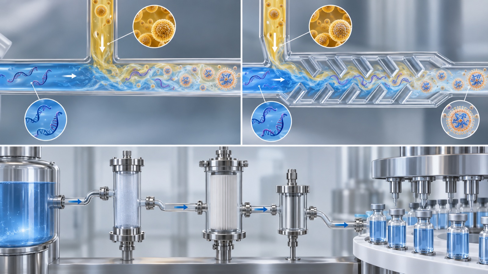
View the original schematic and source annotations
Figure 4. Original vector schematic. Mixing studies: Jeffs (2005) [2] and Belliveau (2012) [3]; five formulation stages: EMA Onpattro assessment [7]. Channel geometry and dimensions are illustrative. The historical mixers are not identified here as the equipment used to manufacture the approved product.
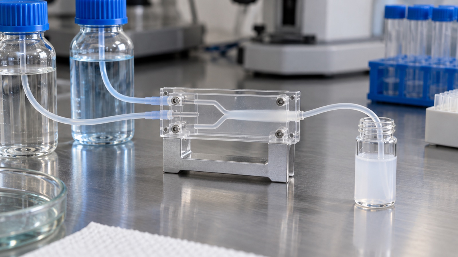
8. Manufacture and specifications in the EPAR
Drugs@FDA records approval of Onpattro (patisiran) on August 10, 2018. The EMA public assessment describes formulation manufacture and quality control. Source fact
The assessment states that LNPs form by mixing aqueous patisiran drug-substance solution with an ethanol solution containing the four lipids. Source fact
The five main formulation stages are solution preparation; mixing to form LNPs; ultrafiltration with buffer exchange and initial concentration; dilution and bioburden filtration; and sterilizing filtration followed by vial filling. Specific proprietary operating conditions are not disclosed here. Source fact
Development evaluated different ionizable lipids and the relative molar ratios of the four lipids iteratively, rather than optimizing only one variable. Source fact
The assessment links formulation particle size to pharmacokinetics and describes control during the LNP-forming manufacturing step. The size was chosen mainly for liver delivery; the stated range is approximately 60–100 nm. Source fact
Release and shelf-life specifications include the following quality attributes: Source fact
Identity by IPRP-HPLC/MS and AX-HPLC/UV; purity and impurities; assay by AX-HPLC/UV; individual lipid identity and content by RP-HPLC/ELSD; duplex-siRNA encapsulation by fluorescence assay; pH; osmolality; particle size by DLS; copper by ICP-MS; residual ethanol by GC; residual EDTA; particulate matter; endotoxins; sterility; content uniformity; extractable volume; container-closure integrity; and in vitro siRNA release using a kinetic assay.
The assessment also records specification-setting discussions. CHMP did not accept the initially proposed statistical widths and requested limits within ±3 SD or narrower; some specifications were tightened. Source fact
A white film after transport is another materials issue. The assessment attributes it to the LNP formulation’s response to mechanical stress. The label describes withdrawing through a filter needle and gently inverting the infusion bag rather than shaking or stirring. This is a description of the cited documents, not instructions for clinical preparation. Source fact
9. Materials viewpoint: specifications as requirements
From a materials viewpoint, the highlighted specification items are quantities that can change with processing. This linkage is an interpretation by the article.
- Individual lipid content: whether all four components meet the intended composition.
- Encapsulation: whether nucleic acid is inside particles; it can depend on mixing. Source fact
- Particle size (DLS): controlled through the LNP-forming step during manufacture. Source fact
- Residual ethanol: a quality attribute arising from the solvent-based process.
- In vitro release: measures release as well as loading; the assessment records addition of this specification during review. Source fact
Even with the same ingredient list, mixing can change size and encapsulation. Specifications therefore address structure and behavior as well as composition. A similar issue arises in the DDS article’s discussion of liposomal formulations.
The assessment describes DLin-MC3-DMA and PEG2000-C-DMG as chemically synthesized lipids made from well-defined starting materials to acceptable specifications. It discusses defined critical quality attributes and process parameters, along with organic, residual-solvent, inorganic/elemental and genotoxic impurities. No animal- or human-derived materials are used in PEG2000-C-DMG manufacture. Source fact
The interpretation here is that lipid excipients require drug-substance-like quality discipline. For a materials supplier, auxiliaries and dispersants may require impurity-profile control as rigorous as that of the principal ingredient.
10. Delivery: apoE and endosomal escape
Why do ionizable LNPs reach hepatocytes without an attached liver ligand? Akinc et al. investigated the role of adsorbed apolipoprotein E. Source fact
Their study identified apoE, involved in physiological lipoprotein clearance and hepatocyte uptake, as an endogenous targeting ligand for ionizable LNPs, but not for cationic LNPs. Source fact
Experiments used recombinant apoE in vitro, wild-type and apoE-deficient mice in vivo, and LDL-receptor-deficient mice to examine receptor dependence. Source fact
The authors also developed multivalent GalNAc clusters as exogenous ligands for the asialoglycoprotein receptor (ASGPR). Endogenous apoE and exogenous GalNAc were both promising liver-delivery strategies in those experiments. Source fact
After uptake, another barrier remains. Gilleron et al. tracked delivery using imaging and electron microscopy. Source fact
Depending on cell type, LNPs entered through constitutive and induced pathways involving clathrin-dependent endocytosis and macropinocytosis. Source fact
Direct detection of colloidal-gold-labeled siRNA gave an estimated 1–2% escape from endosomes to cytosol. Escape occurred during a limited interval in a compartment with both early- and late-endosome features. This is a result in particular experimental systems, not a universal efficiency. Source fact
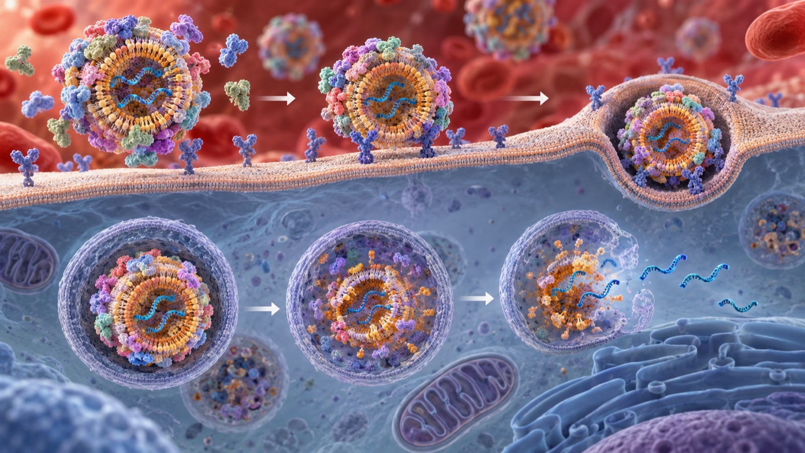
View the original schematic and source annotations
Figure 6. Original vector schematic. PEG-lipid dissociation: EMA assessment [7]; apoE-mediated uptake: Akinc (2010) [4]; uptake and escape: Gilleron (2013) [5]. Four stages are organized here for explanation. These cell and animal findings do not establish the same delivery or escape values in humans.
The 1–2% estimate highlights a potential materials-development bottleneck: circulation and uptake alone do not ensure cytosolic delivery. Improving the final membrane-crossing step could reduce dose requirements, but that is an engineering interpretation, not a clinical conclusion.
Sabnis et al. reported new aminolipids for mRNA delivery in rodent and primate models. Mechanistic studies attributed improved delivery partly to increased endosomal escape. Their one-month toxicity evaluations in rats and nonhuman primates found no adverse events with the studied LNP system; this does not establish human safety. Source fact
Membrane interaction and formulation stability can conflict. Using pH-dependent ionization to respond differently in circulation and endosomes is the design interpretation here. The analogy with cleavable ADC linkers concerns environment-sensitive material behavior, not identical mechanisms.
11. Extrahepatic delivery research
Liver delivery can involve the apoE–receptor pathway. Delivery elsewhere requires changing this distribution behavior. The following examples are research reports, not approved treatments.
| Study | Approach | Reported result (animal studies) |
|---|---|---|
| Cheng et al. (2020) | SORT: add an auxiliary component to systematically change the tissue distribution of different LNP formulations. | Lung-, spleen- and liver-targeted formulations; editing of epithelial/endothelial cells, B/T cells and hepatocytes. Compatible with mRNA, Cas9 mRNA/guide RNA and Cas9 ribonucleoprotein. |
| Rurik et al. (2022) | CD5-targeted LNPs deliver modified CAR-encoding mRNA to T lymphocytes in a mouse heart-failure model. | Transient functional CAR-T cells formed in vivo; fibrosis decreased and cardiac function recovered after injury. |
Sources: Cheng et al. (2020) [Reference 8] and Rurik et al. (2022) [9]. Both report animal experiments. The CAR-T article separately explains the cells themselves. Source fact
12. Challenges and unresolved questions
(1) Extrahepatic delivery remains a research question
The extrahepatic examples above are animal studies. This article’s September 2026 survey did not comprehensively establish FDA or EMA approval status for LNP formulations targeting organs beyond the liver. Approval status remains unresolved here. Unresolved / future
(2) Public sources do not identify the proprietary mixer
The assessment says aqueous and ethanol solutions are mixed but does not identify the particular mixer design, flow rate, temperature or lipid concentration in the public material reviewed here. It states that critical process parameters and acceptable ranges were defined. Absence of public details is not evidence that the process is uncontrolled. Source fact
(3) How far can endosomal escape improve?
Gilleron et al.’s 1–2% estimate applies to specific cells and conditions. This survey did not establish a general later value showing how far endosomal escape had improved. Source fact Unresolved / future
- The assessed LNP has four components and an electron-dense core, not a conventional hollow liposome structure. Source fact
- The 56-lipid mouse comparison found an optimum apparent pKa of 6.2–6.5; DLin-MC3-DMA was 6.44. Source fact
- Optimization changed 40/10/40/10 to 50/10/38.5/1.5; author calculation from the label gives approximately 49.3/10.2/39.0/1.5. Author calculation
- PEG-lipid dissociation is part of the design; chain length changes its behavior, and 1.5 mol% was a threshold in the cited study. Source fact
- Mixing two solutions is central; quality specifications address size, encapsulation and release as well as composition. Source fact
- Endosomal escape was estimated at 1–2% in the cited cellular systems, not universally in humans. Source fact
13. Glossary
- LNP
- Lipid nanoparticle carrying nucleic acid; the formulation discussed uses four lipid components.
- Ionizable lipid
- Lipid that becomes positively charged in acid and is approximately neutral near physiological pH.
- pKa
- pH at half ionization; here an apparent particle-associated value measured by TNS.
- TNS assay
- Fluorescence-based pH titration of positive membrane-surface charge using 2-(p-toluidino)-6-naphthalenesulfonic acid.
- DLin-MC3-DMA
- Onpattro ionizable lipid: molecular weight 642.09; reported apparent pKa 6.44.
- DSPC
- Distearoylphosphatidylcholine, a structural phospholipid.
- PEG lipid
- PEG-conjugated lipid controlling particle assembly and dissociating over time.
- Molar ratio
- Ratio of component amounts, such as 50/10/38.5/1.5.
- Encapsulation efficiency
- Fraction of added nucleic acid contained in particles.
- PDI
- Polydispersity index, an indication of particle-size dispersity.
- Microfluidic mixing
- Rapid mixing of two feeds in small channels.
- Ultrafiltration
- Membrane operation for concentration and buffer exchange.
- apoE
- Apolipoprotein E; can associate with ionizable LNPs and mediate hepatocyte uptake.
- Endosomal escape
- Nucleic-acid transfer from endosomes to cytosol; estimated at 1–2% in the cited study.
- cryo-TEM
- Transmission electron microscopy of frozen specimens for structural observation.
- Novel excipient
- Excipient without prior approved-formulation use requiring additional assessment.
- SORT
- Selective organ targeting, a research strategy adding components to change organ distribution.
14. Primary references
- Jayaraman, M. et al.“Maximizing the potency of siRNA lipid nanoparticles for hepatic gene silencing in vivo”Angewandte Chemie International Edition 51(34), 8529–8533 (2012). doi:10.1002/anie.201203263 — pmc.ncbi.nlm.nih.gov
- Jeffs, L.B. et al.“A scalable, extrusion-free method for efficient liposomal encapsulation of plasmid DNA”Pharmaceutical Research 22(3), 362–372 (2005). doi:10.1007/s11095-004-1873-z — pubmed.ncbi.nlm.nih.gov
- Belliveau, N.M. et al.“Microfluidic Synthesis of Highly Potent Limit-size Lipid Nanoparticles for In Vivo Delivery of siRNA”Molecular Therapy — Nucleic Acids 1, e37 (2012). doi:10.1038/mtna.2012.28 — pmc.ncbi.nlm.nih.gov
- Akinc, A. et al.“Targeted delivery of RNAi therapeutics with endogenous and exogenous ligand-based mechanisms”Molecular Therapy 18(7), 1357–1364 (2010). doi:10.1038/mt.2010.85 — pmc.ncbi.nlm.nih.gov
- Gilleron, J. et al.“Image-based analysis of lipid nanoparticle-mediated siRNA delivery, intracellular trafficking and endosomal escape”Nature Biotechnology 31(7), 638–646 (2013). doi:10.1038/nbt.2612 — pubmed.ncbi.nlm.nih.gov
- FDA/NLM DailyMed“ONPATTRO (patisiran) prescribing information” — dailymed.nlm.nih.gov
- European Medicines Agency (CHMP)“Onpattro — Assessment report”EMA/554262/2018(PDF) — ema.europa.eu
- Cheng, Q. et al.“Selective organ targeting (SORT) nanoparticles for tissue-specific mRNA delivery and CRISPR-Cas gene editing”Nature Nanotechnology 15(4), 313–320 (2020). doi:10.1038/s41565-020-0669-6 — pmc.ncbi.nlm.nih.gov
- Rurik, J.G. et al.“CAR T cells produced in vivo to treat cardiac injury”Science 375(6576), 91–96 (2022). doi:10.1126/science.abm0594 — pubmed.ncbi.nlm.nih.gov
- Mui, B.L. et al.“Influence of Polyethylene Glycol Lipid Desorption Rates on Pharmacokinetics and Pharmacodynamics of siRNA Lipid Nanoparticles”Molecular Therapy — Nucleic Acids 2, e139 (2013). doi:10.1038/mtna.2013.66 — pmc.ncbi.nlm.nih.gov
- Sabnis, S. et al.“A Novel Amino Lipid Series for mRNA Delivery: Improved Endosomal Escape and Sustained Pharmacology and Safety in Non-human Primates”Molecular Therapy 26(6), 1509–1519 (2018). doi:10.1016/j.ymthe.2018.03.010 — pmc.ncbi.nlm.nih.gov
- NIH/NCBI PubChem“Cholesterol (CID 5997)”molecular formula and molecular weight — pubchem.ncbi.nlm.nih.gov
- NIH/NCBI PubChem“1,2-Distearoyl-sn-glycero-3-phosphocholine (CID 94190)”molecular formula and molecular weight — pubchem.ncbi.nlm.nih.gov
- FDA Drugs@FDA“NDA 210922 ONPATTRO”approval history — accessdata.fda.gov
15. Claim–source correspondence (audit)
| Claim in this article | Evidence / limitations | Classification |
|---|---|---|
| In 2012, Jayaraman et al. synthesized 56 aminolipids and measured apparent pKa and the dose required for 50% silencing of mouse factor VII (FVII ED50). This is not simply a formulation variable adjusted by the mixing ratio. Jayaraman et al. changed nitrogen substituents and the methylene spacing between ester and amine groups; the resulting pKa values spanned 4.17–7.16. Molecular structure is the control variable. The comparison controlled other particle properties: up to three passes through an 80 nm membrane, 70–90 nm particle diameter, and dispersity 0.11±0.04. Holding size reasonably constant helps isolate the relationship with pKa. The initial molar ratio of aminolipid/DSPC/cholesterol/PEG lipid was 40/10/40/10. Further optimization gave 50/10/38.5/1.5. In the mouse FVII model the ED50 reached 0.005 mg/kg, a sixfold improvement over the 40/10/40/10 composition. The same lipid composition was also tested in cynomolgus monkeys. TNS titration pH 3–10 in 0.5-unit steps; lipid 16 pKa 6.44 and ED50 0.03 mg/kg; optimum 6.2–6.5; siRNA/lipid mass ratio approximately 0.05. | Jayaraman et al. (2012) [Reference 1] https://pmc.ncbi.nlm.nih.gov/articles/PMC3470698/ | Source fact Source fact |
| T-junction merging DNA buffer and ethanol–lipid feeds, rapidly lowering ethanol concentration; scalable preparation without extrusion. Monodisperse vesicles below 200 nm; DNA encapsulation above 80%. Cryo-TEM showed a mixture of uni-, bi- and multilamellar vesicles. | Jeffs et al. (2005) [Reference 2] https://pubmed.ncbi.nlm.nih.gov/15835741/ | Source fact Source fact |
| Microfluidic mixing on nanoliter scales within milliseconds. Reproducible limiting-size LNPs of 20 nm or larger with near-complete siRNA encapsulation; PDI as low as 0.02. An optimized system achieved 50% mouse-hepatocyte silencing at 10 µg/kg siRNA. Older macroscopic preparations showed variability at 70 nm or larger. | Belliveau et al. (2012) [Reference 3] https://pmc.ncbi.nlm.nih.gov/articles/PMC3442367/ | Source fact Source fact |
| Their study identified apoE, involved in physiological lipoprotein clearance and hepatocyte uptake, as an endogenous targeting ligand for ionizable LNPs, but not for cationic LNPs. Experiments used recombinant apoE in vitro, wild-type and apoE-deficient mice in vivo, and LDL-receptor-deficient mice to examine receptor dependence. The authors also developed multivalent GalNAc clusters as exogenous ligands for the asialoglycoprotein receptor (ASGPR). Endogenous apoE and exogenous GalNAc were both promising liver-delivery strategies in those experiments. | Akinc et al. (2010) [Reference 4] https://pmc.ncbi.nlm.nih.gov/articles/PMC2911264/ | Source fact Source fact |
| Depending on cell type, LNPs entered through constitutive and induced pathways involving clathrin-dependent endocytosis and macropinocytosis. Direct detection of colloidal-gold-labeled siRNA gave an estimated 1–2% escape from endosomes to cytosol. Escape occurred during a limited interval in a compartment with both early- and late-endosome features. This is a result in particular experimental systems, not a universal efficiency. | Gilleron et al. (2013) [Reference 5] https://pubmed.ncbi.nlm.nih.gov/23792630/ | Source fact Source fact |
| ONPATTRO per-mL composition: Patisiran (siRNA): 2 mg, equivalent to 2.1 mg patisiran sodium. DLin-MC3-DMA: 13.0 mg. Cholesterol: 6.2 mg. DSPC: 3.3 mg. PEG2000-C-DMG: 1.6 mg. Potassium dihydrogen phosphate 0.2 mg; sodium chloride 8.8 mg; disodium hydrogen phosphate heptahydrate 2.3 mg; water for injection. pH approximately 7.0. Patisiran sodium: C412H480N148Na40O290P40; molecular weight 14,304 Da. Formulated for hepatocyte delivery. | ONPATTRO label [Reference 6] https://dailymed.nlm.nih.gov/dailymed/drugInfo.cfm?setid=e87ec36f-b4b4-49d4-aea4-d4ffb09b0970 | Source fact Source fact |
| The assessment attributes particle formation, siRNA association, cellular uptake, membrane fusion and endosomal release to the physicochemical properties of DLin-MC3-DMA. This lipid was selected after extensive evaluation of related cationic lipids. The assessment describes near-neutral charge at about pH 7.4 during circulation and acidic endosomal conditions near pH 5 that favor particle disruption and release. This is a formulation mechanism, not a universal guarantee of efficient delivery. DLin-MC3-DMA has formula C43H79NO2 and molecular weight 642.09, with two linoleyl (C18:2) chains. The assessment describes a colorless to pale-yellow liquid, soluble in organic solvents and practically insoluble in water. PEG-lipid chain length was selected so that PEG lipids dissociate from circulating particles over time, enabling uptake of the less-PEG-covered particles by hepatocytes and siRNA release. PEG2000-C-DMG is described as (C2H4O)n C36H73NO5, with n approximately 47 and molecular weight 2,650±300 Da. It is a white to gray-white powder soluble in water, chloroform and acetonitrile. PEG lipids are needed to control self-assembly during particle formation but can inhibit hepatocyte delivery and gene silencing. Their useful manufacturing role and their later biological effect can conflict. Drugs@FDA records approval of Onpattro (patisiran) on August 10, 2018. The EMA public assessment describes formulation manufacture and quality control. The assessment states that LNPs form by mixing aqueous patisiran drug-substance solution with an ethanol solution containing the four lipids. The five main formulation stages are solution preparation; mixing to form LNPs; ultrafiltration with buffer exchange and initial concentration; dilution and bioburden filtration; and sterilizing filtration followed by vial filling. Specific proprietary operating conditions are not disclosed here. Development evaluated different ionizable lipids and the relative molar ratios of the four lipids iteratively, rather than optimizing only one variable. The assessment links formulation particle size to pharmacokinetics and describes control during the LNP-forming manufacturing step. The size was chosen mainly for liver delivery; the stated range is approximately 60–100 nm. Identity by IPRP-HPLC/MS and AX-HPLC/UV; purity and impurities; assay by AX-HPLC/UV; individual lipid identity and content by RP-HPLC/ELSD; duplex-siRNA encapsulation by fluorescence assay; pH; osmolality; particle size by DLS; copper by ICP-MS; residual ethanol by GC; residual EDTA; particulate matter; endotoxins; sterility; content uniformity; extractable volume; container-closure integrity; and in vitro siRNA release using a kinetic assay. The assessment also records specification-setting discussions. CHMP did not accept the initially proposed statistical widths and requested limits within ±3 SD or narrower; some specifications were tightened. A white film after transport is another materials issue. The assessment attributes it to the LNP formulation’s response to mechanical stress. The label describes withdrawing through a filter needle and gently inverting the infusion bag rather than shaking or stirring. This is a description of the cited documents, not instructions for clinical preparation. The assessment describes DLin-MC3-DMA and PEG2000-C-DMG as chemically synthesized lipids made from well-defined starting materials to acceptable specifications. It discusses defined critical quality attributes and process parameters, along with organic, residual-solvent, inorganic/elemental and genotoxic impurities. No animal- or human-derived materials are used in PEG2000-C-DMG manufacture. DSPC supports structure and stability; cholesterol influences permeability. Cryo-TEM shows an electron-dense core without a conventional liposomal bilayer. Ionizable and PEG lipids were novel excipients. | EMA Onpattro assessment (EMA/554262/2018) [Reference 7] https://www.ema.europa.eu/en/documents/assessment-report/onpattro-epar-public-assessment-report_en.pdf | Source fact Source fact |
| SORT: add an auxiliary component to systematically change the tissue distribution of different LNP formulations. Lung-, spleen- and liver-targeted formulations; editing of epithelial/endothelial cells, B/T cells and hepatocytes. Compatible with mRNA, Cas9 mRNA/guide RNA and Cas9 ribonucleoprotein. | Cheng et al. (2020) [Reference 8] https://pmc.ncbi.nlm.nih.gov/articles/PMC7735425/ | Source fact Source fact |
| CD5-targeted LNPs deliver modified CAR-encoding mRNA to T lymphocytes in a mouse heart-failure model. Transient functional CAR-T cells formed in vivo; fibrosis decreased and cardiac function recovered after injury. | Rurik et al. (2022) [Reference 9] https://pubmed.ncbi.nlm.nih.gov/34990237/ | Source fact Source fact |
| Mui et al. compared PEG lipids with dialkyl chains containing 14, 16 or 18 carbons and measured transfer from LNPs to plasma lipoproteins in vivo. The paper describes 1.5 mol% as a threshold minimizing the chain-length effect on liver gene silencing, while still allowing changes in pharmacokinetics and distribution. At 2.5 and 3.5 mol%, C18 PEG lipids markedly impaired hepatocyte gene silencing, whereas the shorter C14 dimyristyl chains had little effect in that study. PEG lipids are needed to control self-assembly during particle formation but can inhibit hepatocyte delivery and gene silencing. Their useful manufacturing role and their later biological effect can conflict. | Mui et al. (2013) [Reference 10] https://pmc.ncbi.nlm.nih.gov/articles/PMC3894582/ | Source fact Source fact |
| Sabnis et al. reported new aminolipids for mRNA delivery in rodent and primate models. Mechanistic studies attributed improved delivery partly to increased endosomal escape. Their one-month toxicity evaluations in rats and nonhuman primates found no adverse events with the studied LNP system; this does not establish human safety. | Sabnis et al. (2018) [Reference 11] https://pmc.ncbi.nlm.nih.gov/articles/PMC5986714/ | Source fact Source fact |
| Cholesterol: C27H46O; molecular weight 386.7. | PubChem CID 5997 [Reference 12] https://pubchem.ncbi.nlm.nih.gov/compound/5997 | Source fact Source fact |
| DSPC: C44H88NO8P; molecular weight 790.1. | PubChem CID 94190 [Reference 13] https://pubchem.ncbi.nlm.nih.gov/compound/94190 | Source fact Source fact |
| ONPATTRO NDA 210922 approved August 10, 2018. | Drugs@FDA [Reference 14] https://www.accessdata.fda.gov/scripts/cder/daf/index.cfm?event=overview.process&ApplNo=210922 | Source fact Source fact |
| DLin-MC3-DMA: 13.0 ÷ 642.09 = 20.2 µmol. Cholesterol: 6.2 ÷ 386.7 = 16.0 µmol. DSPC: 3.3 ÷ 790.1 = 4.18 µmol. PEG2000-C-DMG: 1.6 ÷ 2,650 = 0.60 µmol. Total 40.99 µmol; molar percentages: ionizable lipid 49.3%, DSPC 10.2%, cholesterol 39.0%, PEG lipid 1.5%. Lipid/siRNA mass ratio: 6.2 + 13.0 + 3.3 + 1.6 = 24.1 mg; 24.1 ÷ 2 ≈ 12. siRNA/total-lipid mass ratio: 2 ÷ 24.1 = 0.083; the paper’s comparison used approximately 0.05. Ionizable lipid per siRNA: 2 mg ÷ 14,304 = 0.140 µmol siRNA; 20.2 ÷ 0.140 ≈ 145 molecules. Amine/phosphate comparison: 40 phosphorus atoms per siRNA; 145 ÷ 40 ≈ 3.6, assuming one fully charged amine per lipid. PEG-lipid molar fraction is approximately 1.7% at 2,350 Da and 1.4% at 2,950 Da. | Limitations: the PEG-lipid molecular-weight range changes its calculated fraction to approximately 1.7% at 2,350 Da and 1.4% at 2,950 Da. Cholesterol and DSPC weights are from PubChem. The amine/phosphate estimate assumes one amine per ionizable lipid and complete charge, giving an upper-bound comparison; actual protonation depends on pH and pKa. It is not a manufacturing input ratio. | Author calculation Author calculation |
| The extrahepatic examples above are animal studies. This article’s September 2026 survey did not comprehensively establish FDA or EMA approval status for LNP formulations targeting organs beyond the liver. Approval status remains unresolved here. Gilleron et al.’s 1–2% estimate applies to specific cells and conditions. This survey did not establish a general later value showing how far endosomal escape had improved. | The extrahepatic examples above are animal studies. This article’s September 2026 survey did not comprehensively establish FDA or EMA approval status for LNP formulations targeting organs beyond the liver. Approval status remains unresolved here. | Unresolved / future Unresolved / future |
| The assessment says aqueous and ethanol solutions are mixed but does not identify the particular mixer design, flow rate, temperature or lipid concentration in the public material reviewed here. It states that critical process parameters and acceptable ranges were defined. Absence of public details is not evidence that the process is uncontrolled. | The assessment says aqueous and ethanol solutions are mixed but does not identify the particular mixer design, flow rate, temperature or lipid concentration in the public material reviewed here. It states that critical process parameters and acceptable ranges were defined. Absence of public details is not evidence that the process is uncontrolled. | Interpretation / scope note |
| Author interpretations of molecular pKa design, TNS measurement analogies, detachable PEG additives, process-sensitive specifications, excipient quality discipline, membrane-crossing bottlenecks, ADC-linker analogy and two extrahepatic-delivery approaches. | Author interpretation or scope note, not a regulatory conclusion. | Interpretation / scope note |
| This is an explanation of materials and formulation technology, not an assessment or comparison of treatment efficacy or safety, and not medical advice. Product composition and approval dates are taken from regulatory documents. Animal-study findings must not be treated as human outcomes. | Author interpretation or scope note, not a regulatory conclusion. | Interpretation / scope note |
| Educational ImageGen illustrations and vector schematics are distinguished from real specimens and measured data. | Author interpretation or scope note, not a regulatory conclusion. | Interpretation / scope note |
Text-source review: September 26, 2026. Sources include FDA labels, EMA assessment reports, Drugs@FDA, PubChem and peer-reviewed papers. Interpretations and calculations are distinct from source facts. The pKa comparisons, escape estimates, SORT and CD5-targeted LNP findings are cellular or animal results, not human clinical outcomes. Proprietary mixing conditions and a comprehensive extrahepatic-approval review were not confirmed. This is an explanation of materials and formulation technology, not an assessment or comparison of treatment efficacy or safety, and not medical advice. Product composition and approval dates are taken from regulatory documents. Animal-study findings must not be treated as human outcomes. Images are educational models, not measured data; quantitative SVGs remain available.