TECHNOLOGY EXPLAINER
Aptamers
— Putting a nucleic-acid "artificial receptor" to work on an electrode
An aptamer is a short single-stranded nucleic acid (DNA or RNA) whose sequence has been picked out by test-tube selection because it binds a specific molecule. It can serve as the recognition element in place of an antibody, and it can be made by chemical synthesis. Sensors that fix aptamers on an electrode and turn the shape change on binding into an electrical signal are under study, and it has become clear that what limits their use in blood over long periods is less the nucleic acid itself than the materials on the electrode surface. Two aptamer drugs have been approved, but this article could not confirm any aptamer-based sensor that a regulator has authorized.
- What an aptamer is (the short version)
- SELEX: picking out nucleic acids that bind
- Our calculation: sequence space is far larger than the number of molecules you can hold
- How detection works: turning a shape change on binding into a signal
- A materials engineer's view (1): the monolayer on the electrode sets the sensor's lifetime
- Compared with antibodies: stability, regeneration, cost
- What happens in biological fluids: degradation, desorption, fouling, drift
- A materials engineer's view (2): how to confirm that it really binds
- What can be confirmed about practical use, and what is still research
- What is still hard, and what could not be confirmed
- Glossary / References / Claim-to-source audit
Sourced = stated in a published source (link given)
Our calculation = a value this article calculated from stated assumptions
Not yet confirmed = a plan, a target, or something for which no result could be confirmed
Interpretation of material design is marked "Commentary" and kept separate.
This article explains materials, principles, evaluation and regulation. It is not advice on diagnosis, treatment or medication, or on how to read a measured value, and it does not rank any product or company.
1. What an aptamer is (the short version)
- What it is: a short single-stranded nucleic acid that binds a specific target. A 1992 paper described aptamers as nucleic acid molecules that bind specific molecular targets, and reported single-stranded DNA that binds human thrombin, an enzyme of blood coagulation Sourced [Reference 3].
- How it is made: by SELEX, which repeats a cycle of choosing, from a mixture of random-sequence nucleic acids, those that bind the target and then amplifying them. Two groups reported it in 1990 Sourced [References 1 and 2].
- Where it is used: in drugs (two products approved by the FDA), in research-use multiplexed protein measurement, and in research on sensors in which aptamers are fixed on electrodes Sourced. This article could not confirm a regulatory authorization for an aptamer-based sensor.
An aptamer is not a material that binds anything you select it for. It is a chain of materials and evaluation work: confirm that it binds, fix it to a surface, and keep it working in a biological fluid (Commentary).
2. SELEX: picking out nucleic acids that bind
SELEX (Systematic Evolution of Ligands by EXponential enrichment) was reported in 1990 by Tuerk and Gold as a way to select RNA that binds bacteriophage T4 DNA polymerase Sourced [Reference 1]. In the same year Ellington and Szostak reported in vitro selection of RNA molecules that bind specific ligands Sourced [Reference 2]. The idea is to start from a population with varied sequences, keep only those that bound the target, amplify them, and select again.
The first single-stranded DNA aptamer: thrombin
In 1992 Bock and colleagues selected single-stranded DNA that binds thrombin from a DNA population containing a random region of 60 nucleotides. The binding strength (dissociation constant) was in the range 25 to 200 nM; 32 sequences shared a conserved region of 14 to 17 bases, and several of them at nanomolar concentrations inhibited fibrin-clot formation in vitro Sourced [Reference 3]. Thrombin is not a protein that interacts with nucleic acids. This was an example showing that nucleic acids that bind even a protein unrelated to nucleic acids can be selected.
Small molecules are hard to select for: Capture-SELEX
Small organic molecules such as drugs and metabolites are often not suitable for immobilization on solid surfaces, which makes them difficult targets for conventional selection. Stoltenburg and colleagues reported Capture-SELEX, in which the library, not the target, is immobilized on magnetic beads, and described the successful selection of DNA aptamers for the aminoglycoside antibiotic kanamycin A Sourced [Reference 4].
Usually the target is fixed to a solid and the nucleic acids are passed over it. With small molecules, fixing the target changes its shape and properties, so the nucleic-acid library was fixed instead. The same issue appears in sensors: an aptamer fixed on a surface does not necessarily behave like a free aptamer in solution (Commentary). How it is attached to the surface (attachment position, chain length, density) affects performance, as Section 5 describes.
3. Our calculation: sequence space is far larger than the number of molecules you can hold
If the random region is N nucleotides long, the number of possible sequences is 4N Our calculation.
- 40 nucleotides: 440 ≈ 1.2×1024 sequences
- 60 nucleotides: 460 ≈ 1.3×1036 sequences (the length of the random region in the library of Bock et al. [Reference 3])
- Molecules in hand: if you use a library of 1 nmol (10-9 mol), the Avogadro constant of 6.022×10²³ /mol gives about 6.0×10¹⁴ molecules. Even assuming every one is a different sequence, you could test only about 4.5×10⁻²² of the 60-nucleotide space (about 5.0×10⁻¹⁰ for 40 nucleotides).
Assumptions and limits: 1 nmol is an assumption made here; actual amounts differ between experiments. Assuming that every molecule is a different sequence gives an upper bound. The calculation only shows how much of the space can be tested; it does not show how well the resulting aptamers perform, or whether the best sequence is in the library.
4. How detection works: turning a shape change on binding into a signal
Many aptamers change their three-dimensional structure when the target binds, and sensor designs turn this change into a signal. Two typical designs follow.
(1) Fluorescence: a "structure switch" in which a duplex comes apart
Nutiu and Li (2003) reported a design in which a DNA aptamer carrying a fluorophore is paired with a short DNA carrying a quencher to form a duplex. Without the target, the two are close and fluorescence is quenched. With the target, the aptamer prefers to form the aptamer–target complex, the short DNA is released, and fluorescence appears. The authors state that the strategy is easy to generalize to any aptamer without prior knowledge of its secondary or tertiary structure Sourced [Reference 5].
(2) Electrochemical: the speed of electron transfer changes (E-AB sensors)
In an electrochemical aptamer-based (E-AB) sensor, a redox-active molecule (the reporter) is attached to the end of an aptamer fixed on an electrode. When the target binds and the aptamer changes shape, the electron-transfer rate between the reporter and the electrode changes, and this can be read as a current Sourced [Reference 11]. In 2005 Xiao and colleagues reported a sensor for label-free electronic detection of thrombin in blood serum [Reference 6] and an architecture whose signal increases through target-induced strand displacement (signal-on) [Reference 7] Sourced.
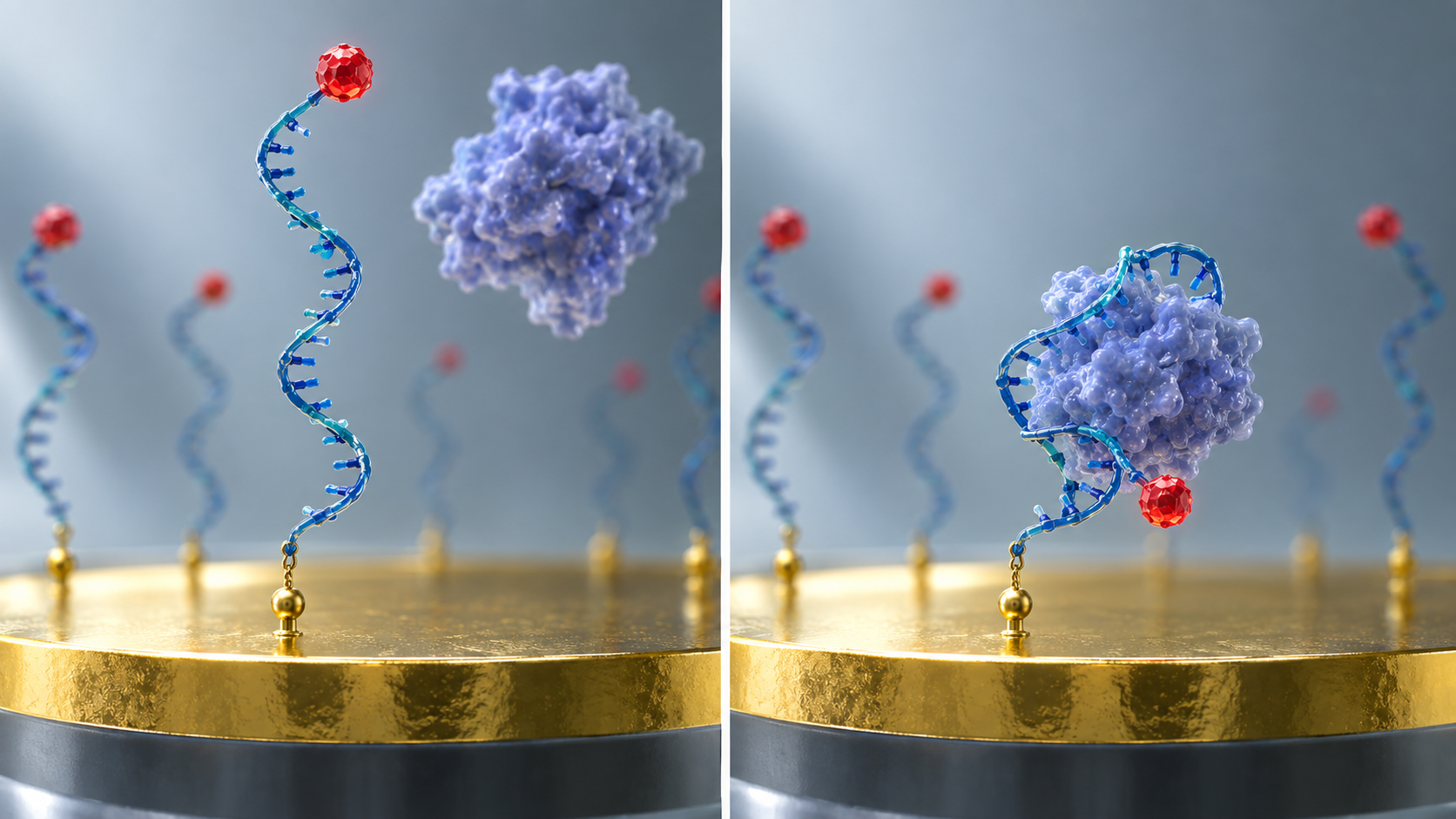
View the original SVG schematic, source notes and numerical details
Demonstrations in animals and blood (research stage)
| Report | Analyte | Subject and conditions (as the authors describe them) |
|---|---|---|
| Ferguson et al. 2013 Sci Transl Med [Reference 8] | Doxorubicin, kanamycin | In live rats and in human whole blood for several hours; subminute temporal resolution; described as needing no exogenous reagents and operating at room temperature |
| Arroyo-Currás et al. 2017 PNAS [Reference 9] | Four drugs | Sensors placed in the bloodstream of awake, ambulatory rats for several hours; 3 s temporal resolution |
| Dauphin-Ducharme et al. 2019 ACS Sensors [Reference 10] | Vancomycin | Measurement in finger-prick-scale samples of human whole blood; subject-specific pharmacokinetics in rats; closed-loop control of plasma levels in rats. "Calibration-free" is the authors' description |
All Sourced (abstracts of the papers). All are studies in animals or in whole blood; this article could not confirm a primary source reporting continuous measurement inside the human body. The numbers belong to each study's conditions, and this table is not a ranking of reports measured under different conditions.
Aptamers can also be combined with field-effect transistors (FETs). To measure cortisol, present in sweat at low nanomolar levels, Wang and colleagues (2022) reported a flexible sensor array that couples an aptamer to nanometer-thin-film In2O3 FETs, and a smartwatch-type system that carries it Sourced [Reference 13]. The FET approach is covered in the "field-effect transistor sensors" explainer in this series, and handling of sweat and other fluids in the explainer on biomarkers in body fluids.
5. A materials engineer's view (1): the monolayer on the electrode sets the sensor's lifetime
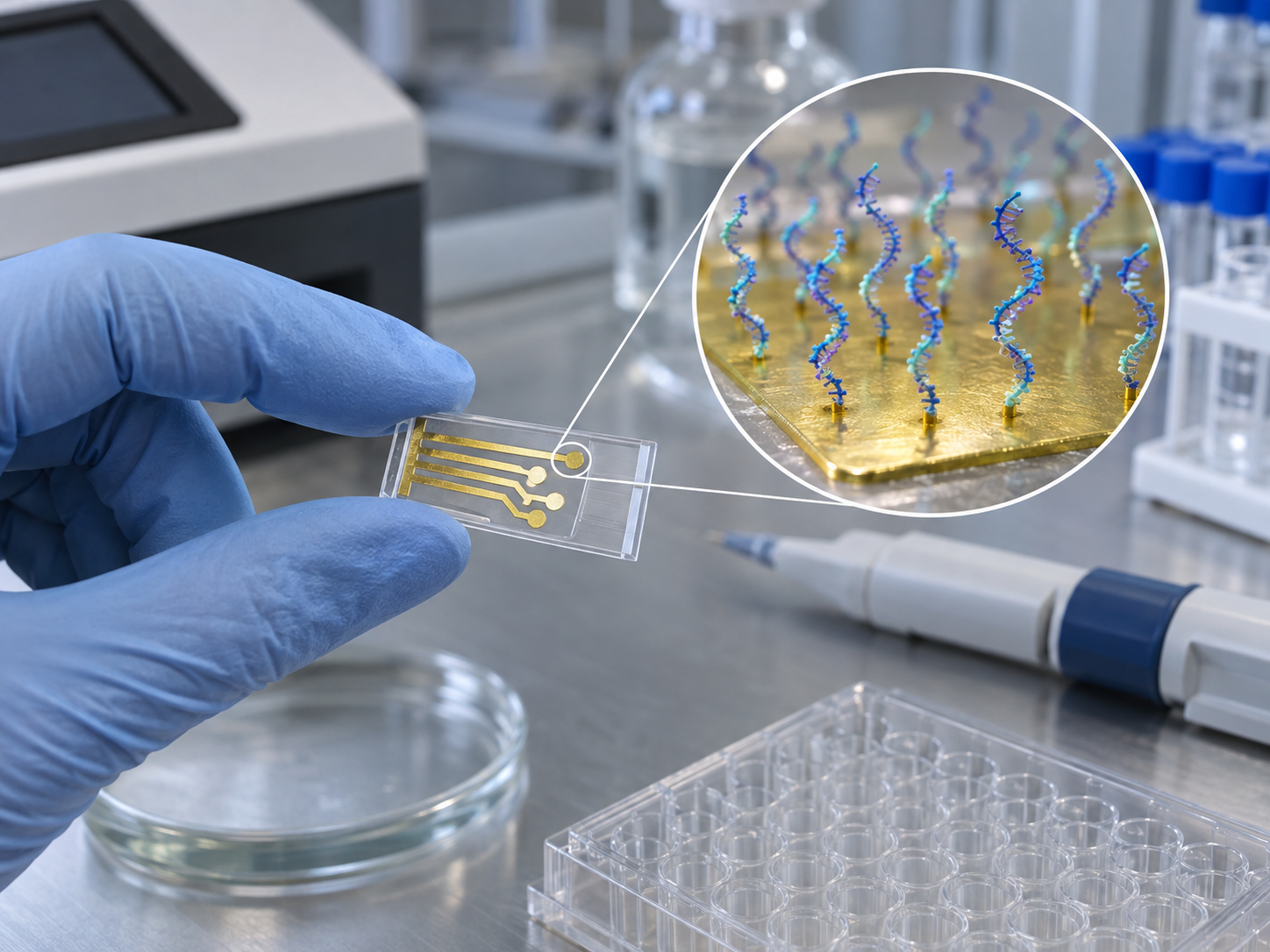
The surface of an E-AB sensor is built of an aptamer monolayer and an alkanethiol monolayer filling the gaps, both on a gold electrode Sourced [Reference 11]. Shaver and colleagues (2021) took up the fact that the in vivo operational life of state-of-the-art E-AB sensors is limited to less than 12 hours, and investigated the cause Sourced [Reference 11].
- They compared natural d-DNA with the nuclease-resistant mirror-image l-DNA in undiluted biological fluids at 37 °C, adding nuclease
- Result: under the conditions tested, nuclease hydrolysis was not the dominant cause of signal loss. The main cause was loss of both the blocking alkanethiol monolayer and the aptamer monolayer from the electrode surface
- The authors say that l-DNA aptamers may extend operational life in the long term, but that the problem of passive monolayer loss must be addressed first
Watkins and colleagues (2023) then reported conditions under which monolayers lasted at least one week in serum at body temperature, against the conventional view that such alkylthiolate monolayers last no more than several days Sourced [Reference 12]. Their means were: (1) increase van der Waals interactions between adjacent monolayer molecules to raise the activation energy for desorption; (2) optimize the electrochemical measurement to reduce alkylthiolate oxidation and electric-field-induced desorption; and (3) mitigate fouling by albumin and other solutes with zwitterionic membranes and zwitterion-based blocking layers. They also report that short-term (hours) improvements to sensor longevity can actually increase degradation in the longer term (days). The authors of that paper disclose patent and business interests in commercializing E-AB sensors [Reference 12].
What this suggests is that lifetime is limited by the interface materials, not by the recognition element (Commentary). The gold–sulfur bond is not permanent, and the potential applied for the measurement itself promotes desorption. In sensor design, materials and measurement conditions cannot be separated. Electrode-interface materials are covered in the explainers on enzymes and electrode-interface materials and on electrochemical sensors in this series, and the hydrophilic gels and membranes that suppress fouling in biological fluids in the explainer on hydrogels.
6. Compared with antibodies: stability, regeneration, cost
Aptamers are often introduced as "chemically synthesized substitutes for antibodies". What can actually be confirmed is organized below.
| Aspect | What can be confirmed about aptamers | How to read the comparison with antibodies (Commentary) |
|---|---|---|
| Manufacture, cost, stability | A review describes aptamers, compared with antibodies, as having minimal immunogenicity, high chemical synthesis production, low cost and high chemical stability Sourced [Reference 15] | Antibodies are generally produced in animals or cultured cells. Price comparisons depend on scale, modification and purification, so this article did not confirm specific prices |
| Weaknesses | The same review says that susceptibility to nuclease degradation, rapid excretion through renal filtration and insufficient binding affinity hindered their development as drug candidates Sourced [Reference 15] | Mirror-image nucleic acids and chemical modifications are ways to address these (Section 7) |
| Regeneration and repeated measurement | Because binding is reversible and no reagent addition is needed, E-AB sensors have been shown to follow changes in blood concentration continuously Sourced [References 8, 9 and 14] | Common antibody-based immunoassays often involve adding reagents or washing steps. But this is a difference in measurement format, not in the recognition element, and research on continuous measurement with antibodies also exists |
| Certainty that it binds | There are aptamers reported in the literature that did not bind their target in an independent test Sourced [Reference 16] (Section 8) | Antibodies also need quality control. For aptamers, confirming binding sequence by sequence is a practical necessity for the user |
The aptamer column follows the review [Reference 15] and the abstracts of the papers (Sourced). The right column is this article's Commentary, not the result of testing the performance of individual antibodies.
7. What happens in biological fluids: degradation, desorption, fouling, drift
In a sensor placed in blood or serum, several things proceed at once. Including the results of Section 5, they are organized here.
Li and colleagues (2016) noted that E-AB sensors often suffer severe baseline drift in undiluted whole blood, and reported a method that puts two redox reporters on the aptamer, one responding to the target and the other, at a distinct potential, serving as a reference, and takes the difference. In flowing, undiluted whole blood, drift of up to 50% fell to less than 2% over many hours of continuous operation Sourced [Reference 14].
How aptamer drugs protect themselves
The review says that the weakness of easy degradation is being addressed by chemical modification and technological advances [Reference 15]. In FDA documents, pegaptanib (Macugen) is described as "an aptamer, a pegylated modified oligonucleotide" Sourced [Reference 18]. Avacincaptad pegol (Izervay) is "a ribonucleic acid (RNA) aptamer, covalently bound to an approximately 43-kiloDalton (kDa) branched polyethylene glycol (PEG) molecule", with a molecular weight of approximately 56 kDa Sourced [Reference 20]. These, however, are drugs injected into the eye, under conditions different from a sensor placed in blood. The protection used in the drugs does not necessarily solve the lifetime problem of a sensor (Commentary).
8. A materials engineer's view (2): how to confirm that it really binds
In 2020 Bottari and colleagues pointed to the lack of a universally accepted and robust quality control protocol for aptamer performance Sourced [Reference 16]. They took up three aptamers for ampicillin (a β-lactam antibiotic) that had been reported in the literature to have nanomolar affinity and had been used in several detection strategies.
- A well-known colorimetric assay based on gold-nanoparticle aggregation gave results conflicting with the original report
- They therefore examined binding in solution by a combination of isothermal titration calorimetry (ITC), native nano-electrospray ionization mass spectrometry and 1H NMR
- Result: none of the three aptamers showed specific binding to its intended target
This is a result for three aptamers; it does not mean that aptamers in general do not bind. What it suggests is that a result from one method alone (especially a method that reads a colour or signal change) can be weak evidence of binding. It is the same idea as in materials evaluation, where one does not rely on a single analytical technique but checks with methods of different principles (Commentary). When a sensor's limit of detection or selectivity (response to interferents) is quoted, it should likewise be read together with what it was compared against and in which fluid, at what temperature and pH it was measured. The idea of the limit of detection is covered in the explainer on label-free detection and limits of detection in this series.
9. What can be confirmed about practical use, and what is still research
"Practical use" covers distinct stages for drugs, research instruments and diagnostic sensors. What can be confirmed from official regulatory documents is set out separately below.
| Category | Content (as stated in the documents) | Source |
|---|---|---|
| Approved as drugs (aptamers) | Pegaptanib sodium (Macugen): NDA 21-756. The approval letter's electronic signature is dated 17 December 2004 (the original-approval entry in Drugs@FDA shows 17 September 2004, a discrepancy). Indicated for neovascular (wet) age-related macular degeneration. Marketing status in Drugs@FDA: Discontinued. Avacincaptad pegol (Izervay): NDA 217225. Approved on 4 August 2023. Indicated for geographic atrophy secondary to age-related macular degeneration. A complement C5 inhibitor, given by intravitreal injection. The most recent label revision is February 2026. | FDA approval letters and labels [References 17 to 22]. These are new drug approvals (NDA), not approvals of a sensor |
| Research-use measurement | A platform that uses aptamers containing modified nucleotides (SOMAmers) was reported in 2010 to measure 813 proteins in 15 µL of serum or plasma (median limit of detection 1 pM, median coefficient of variation 5%). The manufacturer's page states "For Research Use Only. Not for use in diagnostic procedures" and describes measurement of 10,000 unique human proteins | Gold et al. 2010 [Reference 23]; manufacturer's page [Reference 24] (manufacturer's stated value) |
| Sensors (research stage) | E-AB: continuous measurement in the bloodstream of rats and in human whole blood (after collection). FET: a smartwatch-type prototype that measures cortisol in sweat (correlation of saliva and sweat in participants, and on-body validation) | The papers [References 8 to 10 and 13] |
| Authorization as a diagnostic sensor | Could not be confirmed. On 3 October 2026 this article searched the FDA's public database (openFDA 510(k)) for device names containing "aptamer" and found no match. This is a search by name, however, and not an exhaustive check of products whose review documents alone state that the technology is an aptamer | This article's own check (Commentary) |
Clearance or approval is a judgment for a specific intended use and does not mean "safe and accurate however it is used". FDA 510(k) clearance, De Novo authorization and approval (NDA and others) are different systems; the drugs here are approvals (NDA).
10. What is still hard, and what could not be confirmed
(1) No record of continuous use inside the human body could be confirmed
Within the scope of this article, reports of continuous in vivo measurement with E-AB sensors are in rats [References 9 and 10]. Shaver and colleagues state that the in vivo operational life of state-of-the-art sensors, under 12 hours, prohibits long-term continuous monitoring in humans [Reference 11]. The one-week operation of Watkins and colleagues is a result in serum at 37 °C [Reference 12]. Not yet confirmed
(2) Verification of binding is not uniform across reports
Bottari and colleagues point to the lack of a standard procedure for verifying binding [Reference 16]. For any given aptamer, one needs to check by what method and under what conditions binding was confirmed.
(3) Regulatory category
This article could not confirm authorization of a diagnostic sensor that uses an aptamer as its recognition element. Approval as a drug does not vouch for performance as a sensor. Medical-device categories are covered in the explainer on device approval categories and home use in this series.
(4) Price and storage comparison with antibodies
General statements exist in a review [Reference 15], but this article has not confirmed a primary source that sets aptamer and antibody storage stability or price side by side for the same target under the same conditions.
- Aptamers are nucleic acids chosen by SELEX, which began with two reports in 1990 Sourced
- The sequences that can be tested are a tiny part of those possible: about 4.5×10⁻²² for 60 nucleotides (assuming 1 nmol) Our calculation
- E-AB sensors turn a structural change into a change in electron-transfer rate. Demonstrations exist in animals and whole blood Sourced
- Lifetime is limited by the interface materials. Under the conditions tested, monolayer desorption rather than nucleic-acid degradation was the main cause Sourced
- Binding has to be confirmed. There are cases where verification found no binding Sourced
- Two drug approvals exist. Authorization as a sensor could not be confirmed Sourced
11. Glossary
- Aptamer
- A short single-stranded nucleic acid (DNA or RNA) selected to bind a specific molecule.
- SELEX
- A method that finds binding nucleic acids by repeating selection and amplification.
- Library
- The large mixture of nucleic acids with random regions that SELEX starts from.
- Dissociation constant (Kd)
- A measure of binding strength; the smaller, the tighter. Expressed in units such as nM.
- Capture-SELEX
- A selection method that immobilizes the library, not the target, on a solid such as beads. Used for small-molecule targets.
- Structure switching
- A design in which target binding switches the aptamer's three-dimensional structure or binding partner.
- E-AB sensor
- An electrochemical aptamer-based sensor that reads the structural change of an electrode-bound aptamer as a change in current.
- Redox reporter
- A molecule that is alternately oxidized and reduced. Attached to the end of an aptamer, it gives a signal through its electron-transfer rate.
- Self-assembled monolayer (SAM)
- A film of a single layer of molecules on a surface, such as thiols on a gold electrode.
- Nuclease
- An enzyme that cuts nucleic acids. Present in blood and other body fluids.
- l-DNA
- The mirror image of natural d-DNA. Resistant to degradation by nucleases.
- Drift
- A slow shift of a sensor's baseline signal over time when the quantity being measured has not changed.
- PEGylation
- Attaching polyethylene glycol to a molecule, for example to slow its excretion.
- SOMAmer
- An aptamer containing modified nucleotides. Used in a research-use platform for multiplexed protein measurement.
- NDA
- New Drug Application, the submission to the FDA for approval of a new drug. A system separate from the application categories for medical devices.
12. References
- Tuerk C, Gold L. "Systematic evolution of ligands by exponential enrichment: RNA ligands to bacteriophage T4 DNA polymerase", Science 1990;249:505-510 — doi.org
- Ellington AD, Szostak JW. "In vitro selection of RNA molecules that bind specific ligands", Nature 1990;346:818-822 — doi.org
- Bock LC et al. "Selection of single-stranded DNA molecules that bind and inhibit human thrombin", Nature 1992;355:564-566 — doi.org
- Stoltenburg R et al. "Capture-SELEX: Selection of DNA Aptamers for Aminoglycoside Antibiotics", J Anal Methods Chem 2012;2012:415697 — doi.org
- Nutiu R, Li Y. "Structure-switching signaling aptamers", J Am Chem Soc 2003;125:4771-4778 — doi.org
- Xiao Y et al. "Label-free electronic detection of thrombin in blood serum by using an aptamer-based sensor", Angew Chem Int Ed 2005;44:5456-5459 — doi.org
- Xiao Y et al. "A reagentless signal-on architecture for electronic, aptamer-based sensors via target-induced strand displacement", J Am Chem Soc 2005;127:17990-17991 — doi.org
- Ferguson BS et al. "Real-time, aptamer-based tracking of circulating therapeutic agents in living animals", Sci Transl Med 2013;5:213ra165 — doi.org
- Arroyo-Currás N et al. "Real-time measurement of small molecules directly in awake, ambulatory animals", Proc Natl Acad Sci USA 2017;114:645-650 — doi.org
- Dauphin-Ducharme P et al. "Electrochemical aptamer-based sensors for improved therapeutic drug monitoring and high-precision, feedback-controlled drug delivery", ACS Sens 2019;4:2832-2837 — doi.org
- Shaver A et al. "Nuclease hydrolysis does not drive the rapid signaling decay of DNA aptamer-based electrochemical sensors in biological fluids", Langmuir 2021;37:5213-5221 — doi.org
- Watkins Z et al. "Week-long operation of electrochemical aptamer sensors: new insights into self-assembled monolayer degradation mechanisms and solutions for stability in serum at body temperature", ACS Sens 2023;8:1119-1131 — doi.org
- Wang B et al. "Wearable aptamer-field-effect transistor sensing system for noninvasive cortisol monitoring", Sci Adv 2022;8:eabk0967 — doi.org
- Li H et al. "Dual-reporter drift correction to enhance the performance of electrochemical aptamer-based sensors in whole blood", J Am Chem Soc 2016;138:15809-15812 — doi.org
- Ni S et al. "Chemical modifications of nucleic acid aptamers for therapeutic purposes", Int J Mol Sci 2017;18:1683 — doi.org
- Bottari F et al. "Do aptamers always bind? The need for a multifaceted analytical approach when demonstrating binding affinity between aptamer and low molecular weight compounds", J Am Chem Soc 2020;142:19622-19630 — doi.org
- U.S. Food and Drug Administration (FDA) Approval letter, NDA 21-756 (Macugen, pegaptanib sodium injection), 2004 — www.accessdata.fda.gov
- FDA MACUGEN prescribing information (NDA 21-756/S-006, S-007), 2006 (PDF) — www.accessdata.fda.gov
- FDA Approval letter, NDA 217225 (IZERVAY, avacincaptad pegol), 4 August 2023 (PDF) — www.accessdata.fda.gov
- FDA IZERVAY prescribing information (NDA 217225/S-004, revised February 2026) (PDF) — www.accessdata.fda.gov
- FDA Drugs@FDA: NDA 021756 (Macugen), approval history and marketing status — www.accessdata.fda.gov
- FDA Drugs@FDA: NDA 217225 (Izervay), approval history — www.accessdata.fda.gov
- Gold L et al. "Aptamer-based multiplexed proteomic technology for biomarker discovery", PLoS ONE 2010;5:e15004 — doi.org
- SomaLogic SomaScan platform (manufacturer's page, checked 3 October 2026) — somalogic.com
13. Claim-to-source audit
| Claim in the text | Basis | Label |
|---|---|---|
| That SELEX was reported in 1990 by Tuerk and Gold (RNA that binds T4 DNA polymerase) and by Ellington and Szostak (in vitro selection of RNA that binds specific ligands) | Titles of the Science and Nature papers[Reference 1] https://doi.org/10.1126/science.2200121[Reference 2] https://doi.org/10.1038/346818a0 | Sourced |
| That in 1992 Bock et al. selected single-stranded DNA that binds thrombin from DNA with a 60-nucleotide random region, with binding strength of 25 to 200 nM, a conserved region of 14 to 17 bases in 32 sequences, and inhibition of fibrin-clot formation in vitro | Abstract of the Nature 1992 paper[Reference 3] https://doi.org/10.1038/355564a0 | Sourced |
| That small organic molecules are often unsuitable for immobilization on solid surfaces, and that Capture-SELEX immobilizes the library on magnetic beads and succeeded in selecting DNA aptamers for kanamycin A | Abstract of the J Anal Methods Chem 2012 paper[Reference 4] https://doi.org/10.1155/2012/415697 | Sourced |
| The design in which a duplex of a fluorophore-labelled aptamer and a quencher-labelled DNA dissociates in the presence of the target to give fluorescence; the authors' statement that it is easy to generalize without prior knowledge of the aptamer's structure | Abstract of the JACS 2003 paper[Reference 5] https://doi.org/10.1021/ja028962o | Sourced |
| That an E-AB sensor works by a structural change on target binding altering the electron-transfer rate between reporter and electrode, and that a blocking alkanethiol monolayer and an aptamer monolayer are on the electrode surface | Abstract of the Langmuir 2021 paper[Reference 11] https://doi.org/10.1021/acs.langmuir.1c00166 | Sourced |
| That Xiao et al. reported, in 2005, label-free electronic detection of thrombin in blood serum and a signal-on architecture through target-induced strand displacement | Titles of the Angew Chem and JACS papers[Reference 6] https://doi.org/10.1002/anie.200500989[Reference 7] https://doi.org/10.1021/ja056555h | Sourced |
| Demonstrations of E-AB sensors in animals and blood: Ferguson et al. (rats and human whole blood, several hours, subminute resolution, no reagents, room temperature); Arroyo-Currás et al. (four drugs, awake rats, 3 s resolution); Dauphin-Ducharme et al. (vancomycin, finger-prick-scale human whole blood, rat pharmacokinetics and concentration control; "calibration-free" is the authors' description) | Abstracts of each paper[Reference 8] https://doi.org/10.1126/scitranslmed.3007095[Reference 9] https://doi.org/10.1073/pnas.1613458114[Reference 10] https://doi.org/10.1021/acssensors.9b01616 | Sourced |
| That Wang et al. reported measuring cortisol, at low nanomolar levels in sweat, with a flexible array coupling an aptamer to In2O3 thin-film FETs and a smartwatch-type system | Abstract of the Sci Adv 2022 paper[Reference 13] https://doi.org/10.1126/sciadv.abk0967 | Sourced |
| That the in vivo operational life of state-of-the-art E-AB sensors is under 12 hours; that, from the comparison of d-DNA and l-DNA, nuclease hydrolysis was not the dominant cause under the conditions tested and that loss of both the blocking and aptamer monolayers was | Abstract of the Langmuir 2021 paper[Reference 11] https://doi.org/10.1021/acs.langmuir.1c00166 | Sourced |
| The conditions that kept alkylthiolate monolayers for at least one week in serum at 37 °C (stronger van der Waals interactions, adjusted measurement conditions, zwitterionic membranes); that short-term improvements can increase degradation in the longer term; that the authors disclose patent and business interests in commercialization | Abstract of the ACS Sens 2023 paper[Reference 12] https://doi.org/10.1021/acssensors.2c02403 | Sourced |
| That severe baseline drift occurs in undiluted whole blood and that a dual reporter reduced drift of up to 50% to less than 2%; that E-AB operation is reversible and single-step | Abstract of the JACS 2016 paper[Reference 14] https://doi.org/10.1021/jacs.6b08671 | Sourced |
| That a review lists the advantages of aptamers (minimal immunogenicity, high chemical synthesis production, low cost, high chemical stability) and the obstacles (nuclease degradation, rapid renal excretion, insufficient binding affinity) | Abstract of the Int J Mol Sci 2017 review[Reference 15] https://doi.org/10.3390/ijms18081683 | Sourced |
| That three ampicillin aptamers showed no specific binding to the intended target by ITC, native mass spectrometry and 1H NMR; the point about the lack of a universally accepted quality control protocol | Abstract of the JACS 2020 paper[Reference 16] https://doi.org/10.1021/jacs.0c08691 | Sourced |
| Pegaptanib sodium (Macugen): NDA 21-756; approval letter signed 17 December 2004; indicated for neovascular age-related macular degeneration; described as a pegylated modified oligonucleotide aptamer; the discrepancy with the Drugs@FDA original-approval date (17 September 2004); marketing status Discontinued | FDA approval letter, label and Drugs@FDA[Reference 17] https://www.accessdata.fda.gov/drugsatfda_docs/appletter/2004/021756ltr.pdf[Reference 18] https://www.accessdata.fda.gov/drugsatfda_docs/label/2006/021756s006,s007lbl.pdf[Reference 21] https://www.accessdata.fda.gov/scripts/cder/daf/index.cfm?event=overview.process&ApplNo=021756 | Sourced |
| Avacincaptad pegol (Izervay): NDA 217225; approved 4 August 2023; geographic atrophy secondary to age-related macular degeneration; complement C5 inhibitor; an RNA aptamer covalently bound to an approximately 43 kDa branched PEG (about 56 kDa); intravitreal injection; label revised February 2026 | FDA approval letter, label and Drugs@FDA[Reference 19] https://www.accessdata.fda.gov/drugsatfda_docs/appletter/2023/217225Orig1s000ltr.pdf[Reference 20] https://www.accessdata.fda.gov/drugsatfda_docs/label/2026/217225s004lbl.pdf[Reference 22] https://www.accessdata.fda.gov/scripts/cder/daf/index.cfm?event=overview.process&ApplNo=217225 | Sourced |
| That a SOMAmer-based platform was reported in 2010 to measure 813 proteins in 15 µL (median limit of detection 1 pM, median coefficient of variation 5%); that the manufacturer's page says "For Research Use Only. Not for use in diagnostic procedures" and describes 10,000 unique human proteins | Abstract of the PLoS ONE 2010 paper and the manufacturer's page (manufacturer's stated value)[Reference 23] https://doi.org/10.1371/journal.pone.0015004[Reference 24] https://somalogic.com/somascan-platform/ | Sourced |
| The 4N calculation (440 ≈ 1.2×1024, 460 ≈ 1.3×1036), 1 nmol ≈ 6.0×10¹⁴ molecules, and the fraction testable (about 4.5×10⁻²² for 60 nucleotides, about 5.0×10⁻¹⁰ for 40) | Our calculation. 1 nmol and "every molecule is a different sequence" are assumptions made here; the Avogadro constant is 6.022×10²³ /mol. This is not a measure of performance | Our calculation |
| Regulatory authorization of a diagnostic sensor that uses an aptamer as its recognition element | Not confirmed in this article's search (3 October 2026, device names containing "aptamer" in the openFDA 510(k) database; no match). A search by name, not an exhaustive check | Not yet confirmed |
| Reports of continuous measurement inside the human body with E-AB sensors | No primary source could be confirmed within the scope of this article | Not yet confirmed |
| A same-conditions comparison of price and storage stability between aptamers and antibodies | No primary source was confirmed within the scope of this article, so none is stated | Commentary |
| The readings that the monolayer wears out before the nucleic acid, that an immobilized aptamer does not necessarily behave like a free one, that lifetime is limited by the interface materials, the grouping into three factors, that the protection used in drugs does not necessarily solve the sensor problem, and that one method alone can be weak evidence of binding | This article's own organization and interpretation of published content; not views expressed by the authors of the papers | Commentary |
| That Figs. 1, 3, 5 and 6 are explanatory drawings, that Fig. 2 is a drawing that includes our calculation, and that the hero image and Fig. 4 are AI-generated images | Our note | Commentary |
Last updated 3 October 2026. Sources are limited to primary material (peer-reviewed papers, FDA approval documents and the manufacturer's page). Interpretation of material design is marked Commentary and kept separate from sourced fact. Continuous measurement inside the human body with E-AB sensors, regulatory authorization of an aptamer-based diagnostic sensor, and a same-conditions comparison of price and storage stability with antibodies are not stated, because no primary source could be confirmed. The discrepancy between the original approval date of pegaptanib in Drugs@FDA (17 September 2004) and the signature date of the approval letter (17 December 2004) is shown as found. All figures are explanatory concept graphics. Figs. 1, 2, 3, 5 and 6 are vector drawings; the hero image and Fig. 4 are AI-generated images, and none of them shows a real product, micrograph or arrangement of molecules.