TECHNOLOGY EXPLAINER
Implantable Sensors
— materials design for sitting inside the body for three or six months
A sensor that is surgically placed under the skin and powered wirelessly from an outside transmitter to measure glucose has actually been approved. Unlike a disposable thread-like sensor, it is meant to keep working in the same place for months. Meanwhile the body tries to wall off the foreign object. Using official documents and peer-reviewed papers on a confirmable example (a fluorescence-based implantable CGM), this explainer sets out mechanism, biocompatibility, foreign body reaction and capsule formation, lifetime, procedure, and power and communication through a materials lens.
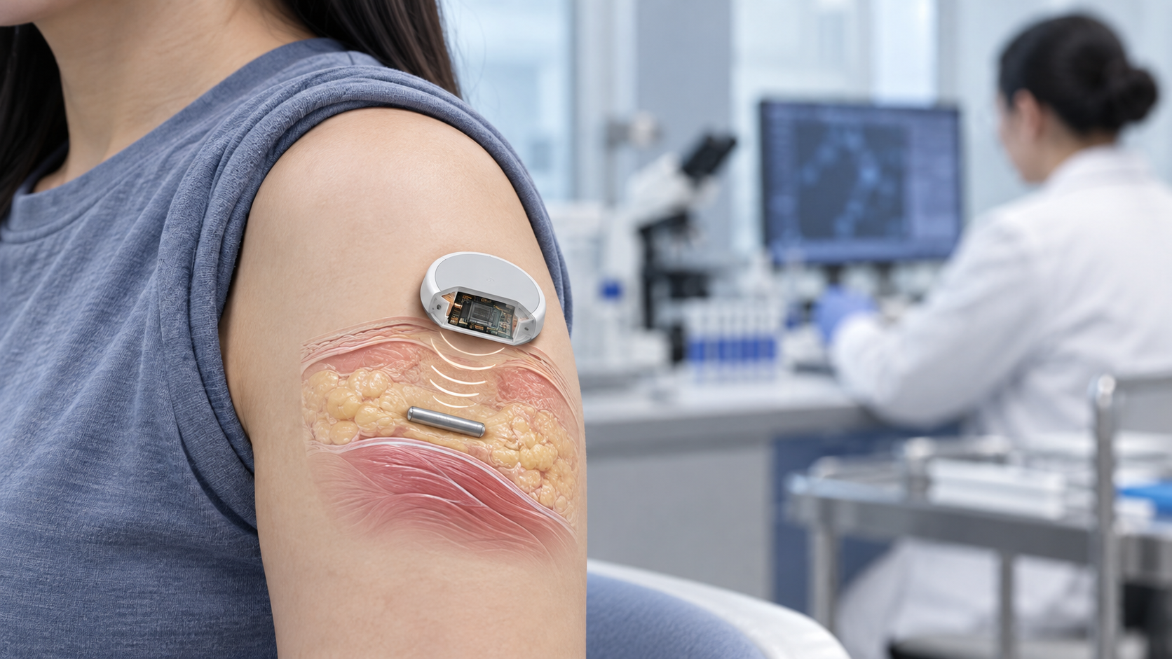
- What an implantable sensor is, in three points
- A confirmable example: the fluorescence-based implantable CGM — structure, power and communication
- A materials engineer's view (1): what is inside a fluorescence sensor
- Foreign body reaction and capsule formation: what the body does
- Lifetime: from 90 to 180 days, and what changed
- Our calculation: the size of the sensor, and how to read the lifetime data
- Surgical procedure: what happened on insertion and removal
- Biocompatibility, sterilisation and storage: what the review document looks at
- Interfering substances and restrictions on use
- FDA classification: implantables go through PMA (approval)
- A materials engineer's view (2): the price of staying a long time
- Open problems, and what this article could not confirm
- Glossary / References / Claim-to-source audit
Sourced = stated in an official document or a paper (link given)
Our calculation = a value this article derived from assumptions it states
Not yet confirmed = research-stage work or a plan with no confirmed track record
Structural readings and materials-design interpretations are marked separately as Commentary.
This article explains technology. It gives no advice on diagnosis, treatment, whether to undergo a procedure, or how to read a measured value, and it does not recommend any product.
Whether an implantation is appropriate is a matter for healthcare professionals and the product's instructions for use.
1. What an implantable sensor is, in three points
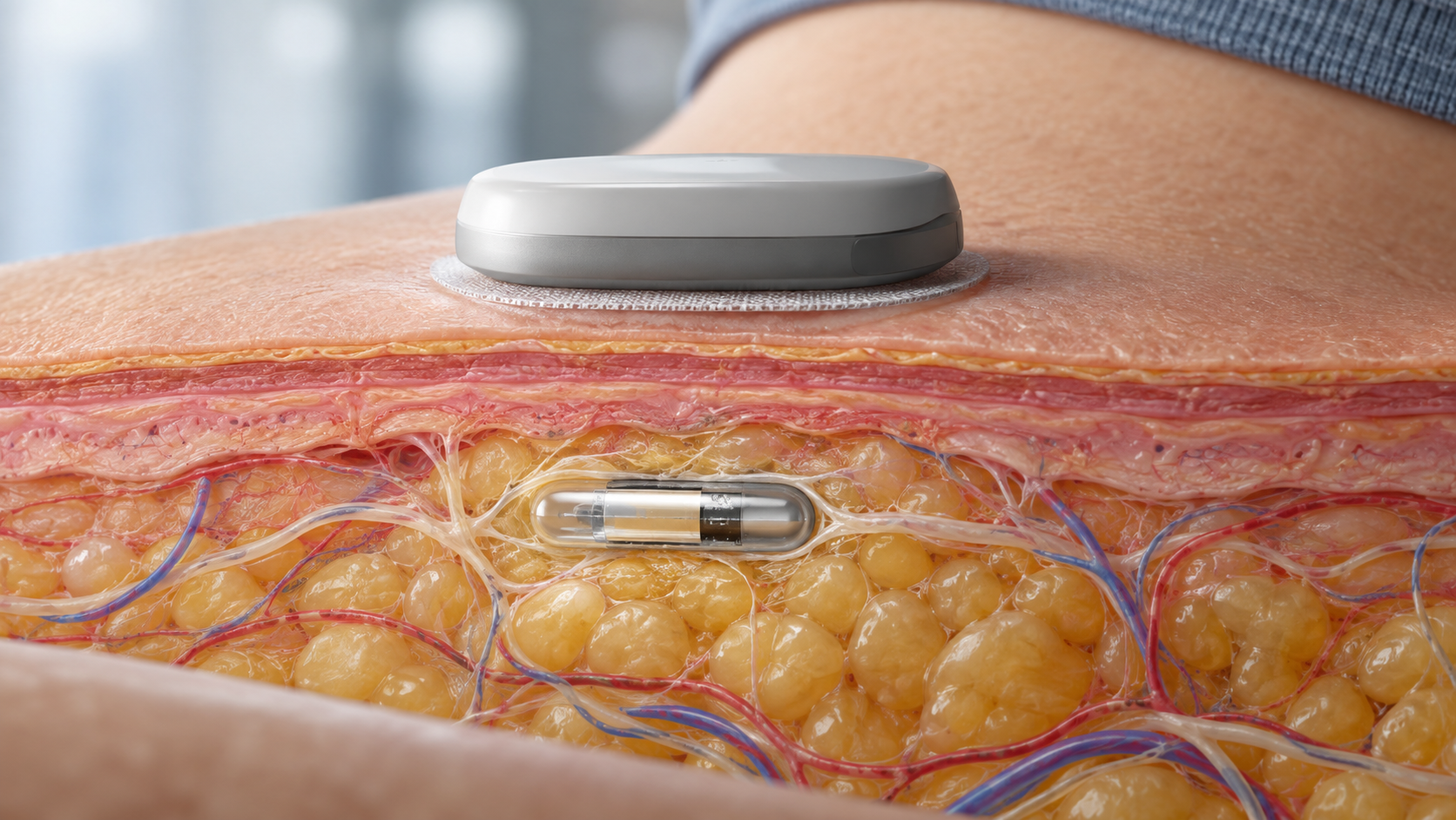
An implantable sensor is a sensor placed entirely beneath the skin that goes on measuring a substance in the body for months (our commentary). This article is limited to a confirmable example in FDA public documents. The approved product we could confirm is a fluorescence-based implantable CGM (Eversense). How CGM works in general is covered in our explainer on continuous glucose monitoring.
- How it differs: a transcutaneous sensor is a thread inserted through the skin and replaced every 10 to 15 days. The Eversense sensor is inserted under the skin by a healthcare provider and left in place. The labelled wear period was up to 90 days at first approval and was later extended to up to 180 daysSourced
- How it measures: not with an enzyme and electrode but by fluorescence. The FDA documents say the sensor detects glucose in interstitial fluid using fluorescence and measures every 5 minutesSourced
- Is there a battery: the FDA documents describe the implanted sensor as powered by radiofrequency (RF) from the transmitterSourced. It is a design that avoids a second operation to change a battery (our commentary)
The hard part of an implantable sensor is not measuring but staying. Over weeks the body coats an incoming foreign object with protein, surrounds it with cells and finally wraps it in a fibrous capsule. Because the capsule makes it harder for substances such as glucose to pass, reviews point out that sensor lifetime can be set not by the principle of measurement but by the interaction with the bodySourced.
2. A confirmable example: the fluorescence-based implantable CGM — structure, power and communication
Eversense is a fluorescence-based implantable CGM approved by the FDA through PMA (premarket approval). The summary of safety and effectiveness (SSED) describes the system as four componentsSourced [Reference 1].
View the original SVG schematic, source notes and numerical details
| Component | What the SSED says |
|---|---|
| Sensor | Detects glucose in interstitial fluid with a fluorescence-sensing mechanism. Inserted subcutaneously by a physician, powered by RF from the transmitter and measuring every 5 minutes; it sends fluorescence readings to the transmitter. A silicone collar contains 1.75 mg of an anti-inflammatory steroid (dexamethasone acetate) that elutes locally. Operating life is the lesser of 90 days or the device's end of life |
| Transmitter | Worn on the skin over the sensor. Powers the sensor, calculates glucose values from the fluorescence readings and sends them to the app over BLE. Held by an adhesive patch. Contains a rechargeable battery and signals alerts by vibration |
| App | Shows values, trends and alerts. The user calibrates by entering blood glucose measurements. Sensor-specific calibration parameters are downloaded once from a server |
| Insertion tools | A blunt dissector (creates the subcutaneous space) and an insertion tool (places the sensor). The sensor is sterilised and shipped in a holder |
All Sourced (PMA P160048 SSED [Reference 1], description at first approval; reportable range 40 to 400 mg/dL).
On power and communication, the SSED's test tables give the following specificationsSourced [Reference 1]. The sensor antenna operates at 13.56 MHz; NFC (near-field communication) reading must work at a distance of up to 12 mm; the sensor's electronics must communicate using the ISO 15693 protocol and be able to excite the fluorescent glucose indicator and detect the fluorescent light it emits; and the transmitter must communicate with the smartphone over BLE within 10 m. In effect, the sensor in the body works like a small wireless tag that draws power from outside and operates only briefly (our commentary).
3. A materials engineer's view (1): what is inside a fluorescence sensor
- 1. The indicator hydrogel (the part that measures). The SSED says the sensor's electronics excite the fluorescent indicator and detect the light it emitsSourced [Reference 1]. The application for the E3 sensor states that the formulation of the indicator hydrogel was changed to extend the in vivo functional lifeSourced [Reference 3]. The glucose-binding part contains boronic acid, and a review also says the synthetic fluorescent receptors used in implantable CGM derive from diboronic acid compoundsSourced [References 4, 9]. The materials design of hydrogels themselves is covered in our explainer on hydrogels.
- 2. The silicone collar (the boundary with the body). It contains an anti-inflammatory steroid that elutes locallySourced [Reference 1]. It is a design that gives the material a drug to dampen the body's reaction (commentary). For silicone as a material, see our explainer on silicones.
- 3. The epoxy-joined end cap (the seal). The SSED says a plastic end cap is attached with epoxy after the electronics assembly is installed, sealing the end and giving a smooth surfaceSourced [Reference 1]. The joint also figures in the root-cause analysis of breakage on removal (Chapter 7).
- 4. The electronics (excitation and detection). They handle optical excitation, detection and NFC communication [Reference 1], and are powered by RF without a battery.
In the thread-like transcutaneous sensor (see the CGM explainer) the enzyme, membrane and electrode were the stars. In an implantable one, optical materials, drug-eluting materials, sealing materials and electronics share one small housing (our commentary).
4. Foreign body reaction and capsule formation: what the body does
Anderson and colleagues call the body's response to a material placed in it the foreign body reaction, and describe it as the end-stage response of inflammation and wound healing, made up of macrophages and foreign body giant cellsSourced [Reference 5]. Ward's review lays out the sequence of the response to a subcutaneous implant from the viewpoint of sensorsSourced [Reference 6].
- The first weeks: Anderson and colleagues state that the period in which a material's surface properties modulate the reaction is the first two to four weeks after implantation, but that the foreign body reaction at the tissue–material interface is present for the whole life of the deviceSourced [Reference 5].
- The capsule: Ward describes how, in response to macrophage-derived signals, fibroblasts become myofibroblasts, and collagen and other extracellular matrix form a dense, hypocellular fibrous capsule. The capsule can become impermeable or hypopermeable to many compoundsSourced [Reference 6].
- Can it be avoided: the same review states that thousands of biomaterials have been tested, but that, as of writing, it had not been possible to create a stealth material that evades the mammalian ability to distinguish foreign from selfSourced [Reference 6].
- The role of an anti-inflammatory drug (a research example): Klueh and colleagues implanted glucose sensors subcutaneously in mice and reported that daily intraperitoneal dexamethasone extended function to as long as 2 weeks, whereas control sensors usually lost function within the first 24 hours. The result indicates an important role for inflammation in loss of sensor functionSourced [Reference 7]. This is a short-term study in mice and does not show effects in humans or the performance of any product.
The Eversense silicone collar is designed to elute dexamethasone acetate locally. The SSED says that risks from local and systemic exposure are “not expected to occur”, while noting that the clinical studies did not include subjects taking dexamethasone or other glucocorticoid medicationsSourced [Reference 1]. The labelling also contraindicates use of the system in people for whom dexamethasone or dexamethasone acetate may be contraindicatedSourced [Reference 1].
5. Lifetime: from 90 to 180 days, and what changed
The value of an implantable is that it needs replacing less often. So how long it keeps working is at the centre of the design.
| FDA procedure | Date | What the SSED says |
|---|---|---|
| PMA P160048 (original approval) | 21 June 2018 | Adults (18 and over), up to 90 days. Adjunctive use. Product code QCD |
| P160048/S006 | 6 June 2019 | Extended to non-adjunctive use. Product code QHJ |
| P160048/S016 (E3) | 10 February 2022 | Up to 180 days. Changes to the sensor's indicator hydrogel formulation, to the algorithm that calculates glucose, and to calibration frequency (one or two per day after day 21). Indicated to replace fingerstick glucose measurement for treatment decisions |
All Sourced (FDA PMA database and SSEDs [References 1 to 3, 10]). Further supplements to the same PMA have followed since 2022; their content was not checked for this article.
The study that supported the extension to 180 days is PROMISE. The FDA summary and a peer-reviewed paper state the followingSourced [References 3, 4].
- Design: sensors were inserted in 181 adults at 8 US sites (85 with one sensor, 96 with one in each arm), from December 2018 to May 2020. 170 (94%) completed day 180. The reference was plasma from centrifuged blood measured on a YSI 2300 bedside glucose analyser.
- Accuracy: the overall MARD of the original sensor was 9.1% (49,613 pairs, 40 to 400 mg/dL), with 85.6% within 15%/15 mg/dL and 92.9% within 20%/20 mg/dL. Accuracy was lowest on day 1; MARD by visit was 11.0% on day 1, 7.7% on day 60 and 10.4% on day 180.
- Sensor survival: the Kaplan–Meier estimate of the share of original sensors still functioning was 98% at day 90, 90% at day 120, 74% at day 150 and 65% at day 180.
- The modified sensor (E3): 43 participants wore the modified sensor as a second sensor. MARD through 180 days was 8.5% (12,034 pairs), and survival was 96% from day 30 to 120, 94% at day 150 and 90% at day 180.
As residual uncertainty, the SSED records that the E3 sensor was evaluated in a relatively small group of 43 subjects, that the effect of repeated insertion and removal over a long 180-day wear has not been evaluated, and that the rate of adverse events in non-adjunctive use was not evaluated because the E3 sensor's output was blinded in the studySourced [Reference 3].
The PROMISE paper describes a modified sensor in which the indicator hydrogel includes the monomer 4-vinylphenylboronic acid (VPBA). The authors' design thinking is that VPBA acts as a target for reactive oxygen species (ROS), a “sacrificial boronic acid” that protects the boronic acid moiety involved in glucose binding, because oxidation can limit the effectiveness and duration of implanted optical sensorsSourced [Reference 4].
There is an idea here that materials people will recognise: place something that oxidises first next to the part you want to protect, the same logic as an antioxidant or a sacrificial anode (our commentary). The SSED, however, describes the formulation change for the “E3 sensor” without giving the specific composition, and nothing in what we read says outright that it is identical to the VPBA of the paper. The link comes from the passage in the FDA document [Reference 3] that calls the modified sensor the “SBA sensor” when discussing the uncertainty of its evaluation (our commentary).
6. Our calculation: the size of the sensor, and how to read the lifetime data
The paper describes the sensor as cylindrical, 3.5 × 18.3 mmSourced [Reference 4]. Assume an ideal cylinder (diameter 3.5 mm, length 18.3 mm)Our calculation.
- Volume: π × (3.5/2)² × 18.3 = about 176 mm³ = about 0.18 mL
- Surface area: side π × 3.5 × 18.3 + two ends 2 × π × (3.5/2)² = about 201 + about 19 = about 220 mm² (about 2.2 cm²)
Assumptions and limits: the real device has a collar and shaped ends and is not an ideal cylinder. This is a rough estimate of the order of magnitude of the tissue-contact area; the volume of about 0.18 mL is close to a few drops of water (our commentary). The body's reaction takes place at the surface, so this surface of about 2 cm² is the boundary between the body and the foreign object (commentary).
- The pattern of MARD: for the original sensor, MARD is high on day 1 (11.0%), lowest at day 60 (7.7%) and rises again at day 180 (10.4%) [Reference 4]. The shape is one of lower accuracy early and late in wear (our commentary).
- Survival probability: for the original sensor it falls steeply in the later part, from 90% at day 120 to 74% at day 150 and 65% at day 180 [Reference 4].
- Caution: the 90% for E3 comes from 43 people and the 65% for the original sensor from a study of 181. The numbers of people differ, and the text we read did not give confidence intervals for the Kaplan–Meier estimates.
7. Surgical procedure: what happened on insertion and removal
For an implantable, the procedure is part of the product. Here are the procedure-related facts from the FDA summariesSourced [References 1, 3].
| Item | What the SSED says |
|---|---|
| Who does it | A qualified physician (a healthcare provider in the later E3 document) inserts it with the supplied insertion tools. A blunt dissector creates the subcutaneous space (pocket) and an insertion tool places the sensor |
| Depth | Intended insertion depth is 3 to 5 mm. In a usability test with simulated tissue, four specimens that were dissected measured 3.3 to 3.9 mm |
| Problems that occurred | In clinical studies, four insertions that were too deep (one in PRECISE II, three in PRECISION). One needed exploratory surgery under general anaesthesia; three needed removal by a surgeon under local anaesthesia. The blunt dissector was redesigned in response, adding two guides and shortening the metal part |
| Removal | Five cases of difficult removal (one a serious adverse event). Two cases of sensor breakage. In some removals the end cap came off or could not be found; the root-cause analysis concluded that the cap was most likely grasped with forceps. The end cap was redesigned to be flush with the sensor body, and manufacturing validation showed it withstood greater removal forces |
| Safety in the E3 study | 558 insertion and removal procedures in 181 people. Two infections at the incision site (0.36% of procedures). All sensors were removed at the first attempt. The most common related adverse events were dermatological (skin irritation from the adhesive patch, skin atrophy, hypopigmentation, infection) at 9.9%, followed by bleeding and bruising at 7.7% |
All Sourced (PMA P160048 SSED [Reference 1] and P160048/S016 SSED [Reference 3]). These events are observations in the studies described in the review documents and are not a basis for judging the safety or suitability of any individual procedure.
The redesign of the end cap was a question of whether the material and structure withstand the forces of removal. The SSED traces the cause to how the cap was grasped, and says the cap was made flush and shown, in tests simulating the forces of removal, to withstand more force than before [Reference 1]. For a material left in the body a long time, the design is set not only by insertion but by the forces of taking it out (our commentary). A broken sensor leaving fragments in the body is also one of the key safety risks the SSED lists [Reference 1].
8. Biocompatibility, sterilisation and storage: what the review document looks at
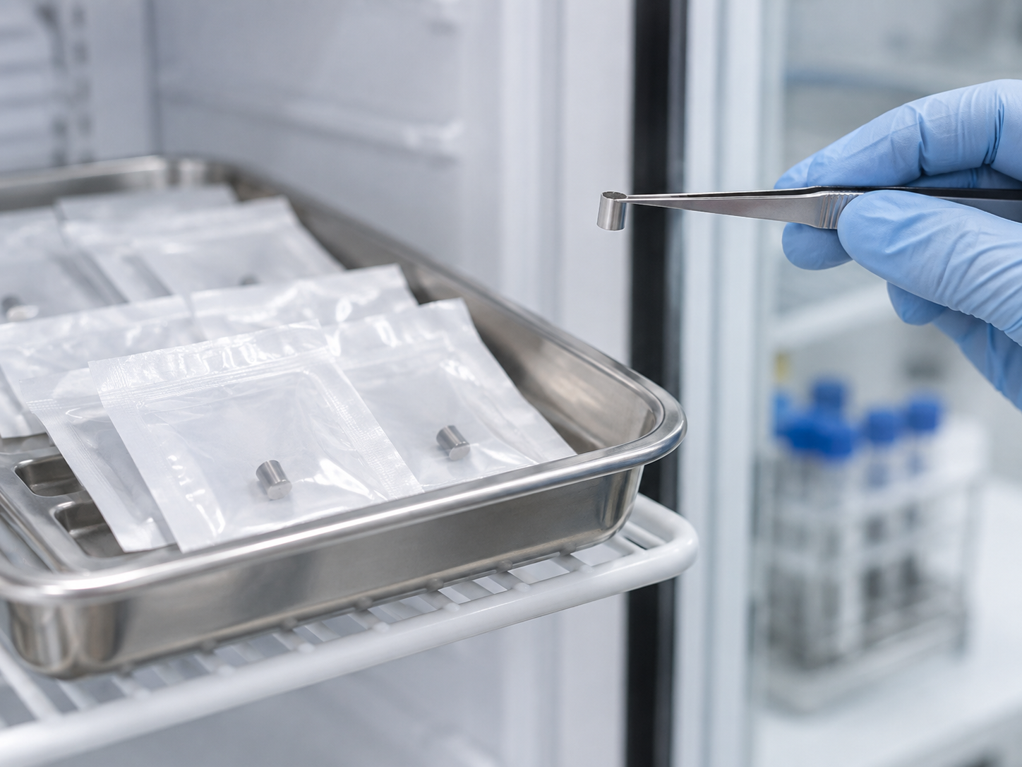
- Biocompatibility: for the sensor, tests were done for cytotoxicity, sensitisation, intracutaneous reactivity, acute systemic toxicity, pyrogenicity, 4-, 13- and 26-week subcutaneous implantation in rats, genotoxicity (three tests), chemical characterisation (ISO 10993-17 and -18) and particulates (ISO 14708-1), all reported as passingSourced [Reference 1].
- Animal study: the sensor with its steroid-eluting collar and an approved pacing lead eluting the same drug from silicone were implanted subcutaneously in rats, and tissue was examined at 30 and 90 days; no adverse tissue reactions were seen with eitherSourced [Reference 1].
- Sterilisation: the sensor is sterilised in its holder with ethylene oxide to a sterility assurance level (SAL) of 10−6. Residuals of ethylene oxide and ethylene chlorohydrin are reported to meet the limits of ISO 10993-7Sourced [Reference 1].
- Storage: the sensor at original approval was to be stored at 2 to 8 °C with a labelled expiry of one month, with shelf-life studies ongoingSourced [Reference 1]. The current storage conditions were not checked for this article.
Here, too, there is a difference from the previous article (continuous glucose monitoring). Some transcutaneous disposable sensors are electron-beam sterilised (Libre). This implantable sensor, by contrast, is reported to be sterilised with ethylene oxide. Different sterilisation methods place different demands on the tolerance of the hydrogel and the indicator (our commentary).
9. Interfering substances and restrictions on use
Even with fluorescence, substances other than glucose can shift readings. The paper explains the mechanism as followsSourced [Reference 4]. Signal is raised by substances that bind to the indicator hydrogel or that emit light within the sensor's operating spectral range, and reduced by substances that absorb the excitation light or absorb or quench the fluorescence. What follows reports what the review documents and the paper state; it is not a basis for any individual's decision about use.
| Item | What the documents say | Source |
|---|---|---|
| Mannitol and sorbitol | When given intravenously or as a component of an irrigation or peritoneal dialysis solution, blood concentrations rise and sensor glucose values can be falsely high. Amounts in a typical diet do not affect results (listed as a contraindication) | [References 1, 3] |
| Tetracycline-class antibiotics | The paper lists tetracycline-class antibiotics, in addition to the above, as interferents of the original sensor | [Reference 4] |
| Dexamethasone | The system is contraindicated in people for whom dexamethasone or dexamethasone acetate may be contraindicated | [References 1, 3] |
| MRI | At original approval, the sensor and transmitter were described as incompatible with MRI, with a statement not to undergo MRI while the sensor is inserted. In the E3 document, the transmitter is MR unsafe and must be removed before MRI, and the sensor is covered by separate MRI safety information | [References 1, 3] |
All Sourced (FDA SSEDs and a peer-reviewed paper). The paper states that the interference from vitamin C and acetaminophen seen with some other systems is not seen with this sensor, but this article has not confirmed that point in a review document. Labelling can change with version and region, so any actual decision rests on the latest instructions for use and a healthcare professional.
Physical conditions were also evaluated. In the study before first approval (PRECISE II), accuracy visits included upper-arm exercise sessions and 30-minute compression sessionsSourced [Reference 1]. They reflect the environment of use, in which a sensor under the skin is subject to external forces (our commentary).
10. FDA classification: implantables go through PMA (approval)
Most transcutaneous CGM, in which a thread-like sensor is inserted from outside the body, is treated as Class II (iCGM) through De Novo or 510(k) (see the CGM explainer). An implantable, by contrast, is classified in the FDA product classification database as Class III and is subject to PMA (premarket approval)Sourced [References 10, 11, 12].
| Product code | Name | Description in the FDA classification database |
|---|---|---|
| QCD | Continuous Glucose Monitor, Implanted, Adjunctive Use | A fully implanted CGM intended to detect trends and track patterns in interstitial glucose values. Adjunctive use: to complement, not replace, information from standard home blood glucose monitoring devices |
| QHJ | Sensor, Glucose, Implanted, Non-Adjunctive Use | An implanted glucose sensor intended to determine glucose levels and the direction and rate of change in people with diabetes. Non-adjunctive use: to replace information from standard blood glucose monitoring devices for diabetes-related treatment decisions |
All Sourced (FDA product classification database [References 11, 12]; both are device class 3, with no individual 21 CFR regulation number assigned).
FDA PMA is an “approval”, a separate procedure from a 510(k) “clearance” or a De Novo “grant”. The original approval of Eversense (P160048) was on 21 June 2018, the extension to non-adjunctive use (S006) on 6 June 2019 and the 180-day E3 (S016) on 10 February 2022Sourced [References 1 to 3, 10]. Each approval is a judgment for a specific intended use; it does not mean the device is safe and accurate in any use.
11. A materials engineer's view (2): the price of staying a long time
- The measuring principle and the capsule. Gough and colleagues reported an implanted sensor that operated for 222 and 520 days in the subcutaneous tissue of two pigs. The sensor was an enzyme electrode based on differential electrochemical oxygen detection, described as designed to reduce sensitivity to encapsulation, to variation in local microvascular perfusion, to limited tissue oxygen and to enzyme inactivation. The lag between blood and tissue glucose was 11.8 ± 5.7 minutes on rising and 6.5 ± 13.3 minutes on falling glucose; it resulted mainly from mass transfer in the tissue rather than the sensor's response and showed no systematic change over the implant periodSourced [Reference 8]. This is an animal study (two pigs), not the performance of a product in people.
- A fork in design. Faced with a capsule that narrows the path for substances, the answers diverge: measure in a way less affected by the capsule (Gough), dampen the reaction with a drug (Eversense's steroid-eluting collar), protect the indicator from oxidation (VPBA) (our commentary).
- Procedure and material interact. As the end-cap redesign shows [Reference 1], the removal procedure sets the demands on the material's joints. The FDA itself lists the effect of repeated insertion and removal as not evaluated [Reference 3].
- Storage as a weak point. The sensor at original approval had a one-month expiry under refrigeration [Reference 1]. It is an example of how a product containing a hydrogel and an indicator can carry storage constraints (our commentary).
12. Open problems, and what this article could not confirm
(1) Implantable sensors other than Eversense
The approved implantable glucose sensor this article could confirm in official documents was Eversense. The approval status of other products, or of implantable sensors that measure something other than glucose, was not confirmed and is not covered.
(2) The specific composition of the indicator, hydrogel and housing materials
The SSED does not specify the chemical structure of the fluorescent indicator, the composition of the hydrogel or the material of the housing. What this article could write is limited to boronic acid, VPBA and the cylindrical dimensions from the paper, and the silicone collar and the epoxy joining of the end cap from the SSED.
(3) Wear beyond 180 days, and supplements since 2022
The FDA PMA database shows further supplements to the same PMA after the E3 approval (2022). This article checked only the SSEDs for the original approval, the extension to non-adjunctive use (S006) and the 180-day E3 (S016), and did not check the content of later changes, including any further extension of wear.
(4) Histological data in people, such as capsule thickness
No primary source in the documents checked shows capsule thickness, tissue structure or the state around the sensor in people. The account of foreign body reaction rests on reviews [References 5, 6] and animal studies [References 7, 8] and is general.
- The confirmable example is a fluorescence-based implantable CGM (Eversense). The sensor has no battery and is powered by RF from the transmitterSourced
- Lifetime went from 90 to 180 days. In the 180-day study, the share of original sensors still functioning was 65% and of the modified sensor (E3) 90%Sourced. The difference is 25 pointsOur calculation
- The body walls off the foreign object. It goes from protein adsorption to a dense fibrous capsule that makes passage of substances harderSourced
- The materials share four jobs. An indicator hydrogel, drug-containing silicone, an epoxy-joined seal and electronicsSourced
- An implantable is Class III and goes through PMA (approval). It is a different procedure from the Class II iCGM of the transcutaneous typeSourced
- Even “taking it out” is part of the design. The end-cap redesign and the lack of evaluation of repeated insertion and removal are examplesSourced
13. Glossary
- Implantable sensor
- A sensor placed entirely beneath the skin that measures a substance in the body for months.
- Fluorescence-based
- A method in which a fluorescent indicator that binds glucose is excited by light and concentration is derived from changes in the emitted fluorescence.
- Boronic acid
- A chemical group that binds sugars such as glucose. Used in the binding site of fluorescent glucose sensors.
- Foreign body reaction
- The end-stage response of inflammation and wound healing to a material in the body, involving macrophages and foreign body giant cells.
- Biofouling
- Proteins and cells adhering to and covering a material's surface.
- Fibrous capsule
- A dense, hypocellular layer of collagen and other matrix formed around a foreign object. It is hard for substances to pass through.
- PMA
- Premarket approval. The premarket route for Class III devices; the FDA's “approval”.
- SSED
- Summary of Safety and Effectiveness Data. A review summary published after PMA approval.
- Non-adjunctive use
- Use that replaces information from a home blood glucose meter and is used for treatment decisions.
- NFC
- Near-field communication. Uses the 13.56 MHz band to supply power and communicate over distances of millimetres to centimetres.
- Kaplan–Meier estimate
- A statistical method that estimates, over time, the share still “surviving” (here, still functioning) during observation.
- End cap
- A component that seals the end of a cylindrical sensor.
- SAL
- Sterility assurance level. The probability that a viable microorganism remains after sterilisation; 10−6 is one in a million or less.
- MARD
- Mean absolute relative difference: the mean of the absolute difference from the reference divided by the reference (%).
14. References (primary sources)
- U.S. Food and Drug Administration (FDA) “PMA P160048 FDA Summary of Safety and Effectiveness Data (Eversense Continuous Glucose Monitoring System)”, approved 21 June 2018 — www.accessdata.fda.gov
- FDA “PMA P160048/S006 FDA Summary of Safety and Effectiveness Data (extension to non-adjunctive use)”, approved 6 June 2019 — www.accessdata.fda.gov
- FDA “PMA P160048/S016 FDA Summary of Safety and Effectiveness Data (Eversense E3 CGM System, up to 180 days)”, approved 10 February 2022 — www.accessdata.fda.gov
- Garg SK et al. “Evaluation of Accuracy and Safety of the Next-Generation Up to 180-Day Long-Term Implantable Eversense Continuous Glucose Monitoring System: The PROMISE Study”, Diabetes Technol Ther 24:84 (2022) (PMC) — pmc.ncbi.nlm.nih.gov
- Anderson JM, Rodriguez A, Chang DT. “Foreign body reaction to biomaterials”, Semin Immunol 20:86 (2008) — doi.org
- Ward WK. “A review of the foreign-body response to subcutaneously-implanted devices: the role of macrophages and cytokines in biofouling and fibrosis”, J Diabetes Sci Technol 2:768 (2008) — doi.org
- Klueh U et al. “Inflammation and glucose sensors: use of dexamethasone to extend glucose sensor function and life span in vivo”, J Diabetes Sci Technol 1:496 (2007) — doi.org
- Gough DA et al. “Function of an implanted tissue glucose sensor for more than 1 year in animals”, Sci Transl Med 2:42ra53 (2010) — doi.org
- Hatada M, Okuda-Shimazaki J, Sode K. “Glucose Oxidoreductases for Glucose Monitoring: A Bird's Eye View of Glucose-Sensing Enzymes II”, J Diabetes Sci Technol (2026) (PMC) — pmc.ncbi.nlm.nih.gov
- FDA PMA database record for P160048 (Senseonics, Incorporated; Eversense) — www.accessdata.fda.gov
- FDA Product classification database, product code QCD “Continuous Glucose Monitor, Implanted, Adjunctive Use” (via the openFDA API) — api.fda.gov
- FDA Product classification database, product code QHJ “Sensor, Glucose, Implanted, Non-Adjunctive Use” (via the openFDA API) — api.fda.gov
15. Claim-to-source audit
| Claim in the text | Basis | Type |
|---|---|---|
| The original approval of Eversense (P160048) on 21 June 2018: adults (18 and over), up to 90 days, adjunctive use, product code QCD. That the system has four components: a fluorescence sensor, a smart transmitter, an app and insertion tools. That the sensor is RF powered and measures every 5 minutes; that the silicone collar contains 1.75 mg of dexamethasone acetate eluting locally; that the transmitter has a rechargeable battery and an adhesive patch. Antenna 13.56 MHz, NFC read up to 12 mm, ISO 15693, BLE up to 10 m. That the electronics excite the fluorescent indicator and detect the fluorescence. That the end cap is attached with epoxy. The biocompatibility tests (4-, 13- and 26-week implantation, chemical characterisation, particulates), the 30- and 90-day rat study of the steroid-eluting collar against a pacing lead, ethylene oxide sterilisation (SAL 10−6), 2 to 8 °C storage with a one-month expiry. Intended insertion depth 3 to 5 mm, four too-deep insertions, five difficult removals and two breakages, and the end-cap redesign. The contraindications (MRI, dexamethasone, mannitol and sorbitol). The upper-arm exercise and compression tests in PRECISE II | SSED for the original approval of Eversense Reference 1 https://www.accessdata.fda.gov/cdrh_docs/pdf16/P160048B.pdf | Sourced |
| That P160048/S006 was approved on 6 June 2019, extending to non-adjunctive use, product code QHJ | Reference 2 https://www.accessdata.fda.gov/cdrh_docs/pdf16/P160048S006B.pdf | Sourced |
| That P160048/S016 (Eversense E3) is for up to 180 days; the changes to indicator hydrogel formulation, algorithm and calibration frequency; indication to replace fingerstick measurement for treatment decisions; the PROMISE study (181 people, 8 sites); 180-day survival of 65% for the original sensor and 90% for E3; that the E3 evaluation was in 43 people and the FDA lists moderate residual uncertainty; that repeated insertion and removal and adverse events in non-adjunctive use were not evaluated; 558 insertion and removal procedures with two infections (0.36%); related adverse events (skin 9.9%, bleeding and bruising 7.7%); all sensors removed at the first attempt; the transmitter being MR unsafe | SSED for P160048/S016 Reference 3 https://www.accessdata.fda.gov/cdrh_docs/pdf16/P160048S016B.pdf | Sourced |
| The PROMISE paper: 181 people, 8 US sites, a YSI 2300 reference, overall MARD of the original sensor 9.1% (49,613 pairs), 85.6% within 15/15 and 92.9% within 20/20, MARD by visit, survival (98% at day 90, 90% at day 120, 74% at day 150, 65% at day 180), MARD of 8.5% for the 43-person modified sensor (12,034 pairs) and its survival (96% from day 30 to 120, 94% at day 150, 90% at day 180), the sensor being cylindrical at 3.5 × 18.3 mm, the role of VPBA (sacrificial boronic acid), the interferents (mannitol and sorbitol, tetracycline class) and how interference arises | PROMISE paper (PMC) Reference 4 https://pmc.ncbi.nlm.nih.gov/articles/PMC8817689/ | Sourced |
| The definition of foreign body reaction (the end-stage response of inflammation and wound healing involving macrophages and foreign body giant cells), that surface properties modulate the reaction in the first two to four weeks, and that the foreign body reaction is present for the life of the device | Reference 5 https://doi.org/10.1016/j.smim.2007.11.004 | Sourced |
| Protein adsorption (biofouling), macrophage adhesion and formation of multinucleated giant cells, myofibroblasts and collagen synthesis, a dense hypocellular fibrous capsule that becomes impermeable or hypopermeable to many compounds, the possibility of microvessel formation, and that a stealth material has not been created | Reference 6 https://doi.org/10.1177/193229680800200504 | Sourced |
| That in mice with subcutaneous glucose sensors, intraperitoneal dexamethasone extended function to as long as 2 weeks where control sensors usually lost function within the first 24 hours | Short-term animal (mouse) study Reference 7 https://doi.org/10.1177/193229680700100407 | Sourced |
| An implanted sensor that operated for 222 and 520 days in two pigs; that it is an enzyme electrode based on differential electrochemical oxygen detection; lag between blood and tissue of 11.8 ± 5.7 minutes on rising and 6.5 ± 13.3 minutes on falling glucose, mainly from mass transfer in tissue | Animal (pig) study Reference 8 https://doi.org/10.1126/scitranslmed.3001148 | Sourced |
| That the synthetic fluorescent receptors used in implantable CGM derive from diboronic acid compounds | Reference 9 https://pmc.ncbi.nlm.nih.gov/articles/PMC13518614/ | Sourced |
| The dates of the original approval (21 June 2018), S006 (6 June 2019) and S016 (10 February 2022), and that further supplements to the same PMA have followed | PMA database record and SSED dates Reference 10 https://www.accessdata.fda.gov/scripts/cdrh/cfdocs/cfpma/pma.cfm?id=P160048 | Sourced |
| The description of product code QCD (adjunctive) and that it is device class 3 | Reference 11 https://api.fda.gov/device/classification.json?search=product_code:QCD | Sourced |
| The description of product code QHJ (non-adjunctive) and that it is device class 3 | Reference 12 https://api.fda.gov/device/classification.json?search=product_code:QHJ | Sourced |
| A sensor volume of about 176 mm³ (about 0.18 mL), a surface area of about 220 mm² (about 2.2 cm²), the shares not functioning at day 180 of 35% and 10%, and a difference of 25 points | Our calculation. A rough estimate treating the paper’s 3.5 × 18.3 mm as an ideal cylinder, ignoring the collar and end shapes. The shares are 100 minus the paper’s survival of 65% and 90% | Our calculation |
| The approval status of implantable glucose sensors other than Eversense; the chemical structure of the indicator, the hydrogel composition and the housing material; the content of supplements after E3; histological data in people such as capsule thickness | Not stated in the official documents and papers checked for this article, so not given here | Commentary |
| The framing that optical materials, drug-eluting materials, sealing materials and electronics share one housing; the reading of the sacrificial boronic acid as the same idea as an antioxidant or sacrificial anode; the reading that MARD is high early and late in wear; the linking of E3 with the SBA sensor; the three ways of answering the capsule; the reading of the storage constraint | This article’s own framing and commentary based on published content. Not views expressed by the authors or regulators of those documents | Commentary |
| That Figs. 1, 2 and 3 are concept diagrams (vector drawings), Fig. 4 is a drawing that includes our calculation, and that the hero image and Fig. 5 are AI-generated images | A note by this article | Commentary |
Last updated 3 October 2026. Sources are limited to primary material (FDA PMA summaries and the product classification database, peer-reviewed papers and reviews). No market-size or market-share estimates are used. Because the article includes structural readings and materials-design interpretations, those are marked as Commentary and kept separate from sourced fact. The approval status of implantable sensors other than Eversense, the specific composition of the indicator, hydrogel and housing, the content of supplements after E3, and histological data on capsules in people are not covered, because they could not be confirmed in published primary sources. Figs. 1, 2 and 3 are concept diagrams, Fig. 4 includes our calculation, and the hero image and Fig. 5 are AI-generated images; none of them shows a real product, cross-section, micrograph or measured data. This article explains technology. It gives no advice on diagnosis, treatment, whether to undergo a procedure, or how to read a measured value, and it does not recommend any product.