TECHNOLOGY EXPLAINER
Microneedles
— arrays long enough to cross the skin's outer barrier, short enough to stay clear of pain nerves
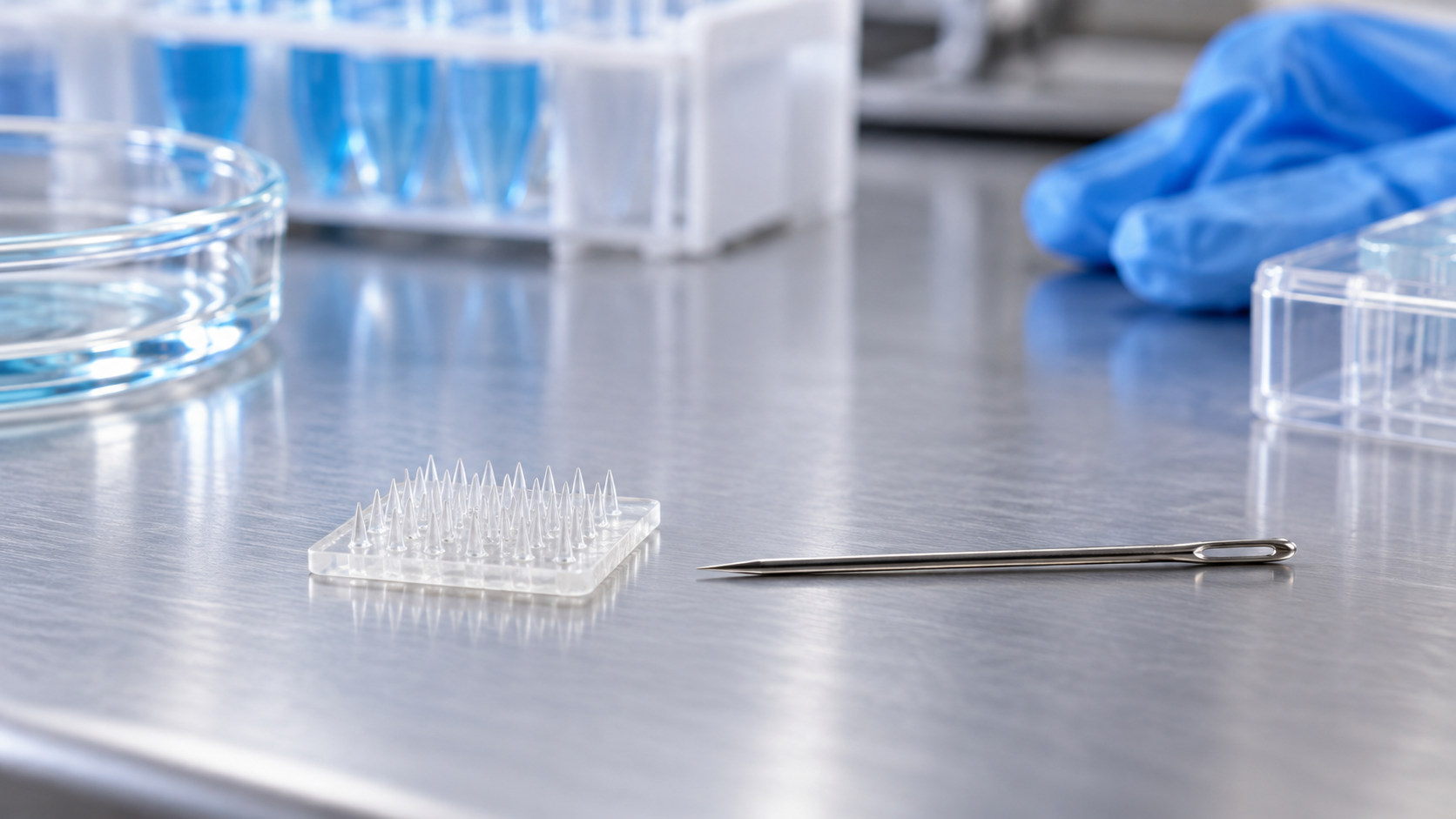
A microneedle is a patch carrying an array of needles a few hundred micrometers long. It crosses the stratum corneum, the skin's barrier layer, to deliver a drug, to draw out interstitial fluid, or to measure with electrodes on the needles. Yet the single word "microneedle" covers solid, coated, dissolving, hollow and hydrogel-forming types, each with different materials and uses. This article sorts out the types, materials and manufacturing, how to make needles that do not break, interstitial fluid sampling and sensor integration, sterilization and safety, and where the technology has reached practical use and where it is still at the research stage, drawing on regulators' documents and peer-reviewed papers.
- What a microneedle is (the short version)
- The types: solid, coated, dissolving, hollow and hydrogel-forming
- A materials engineer's view 1: a needle must both insert and not break
- Materials and manufacturing: etching, casting into molds, coating metal needles
- Sampling interstitial fluid, and integrating sensors
- Our calculation: orders of magnitude for length, dose, survival rate and sampling rate
- A materials engineer's view 2: with dissolving needles, drug, material and shape are decided together
- Sterilization and safety
- Where things stand: authorized or approved examples, and research-stage examples
- A materials engineer's view 3: regulation draws its line at the claim, not at the needle material
- Open issues and what could not be confirmed / Glossary / References / Claim-to-source audit
Sourced = a statement in published material (link given)
Our calculation = a figure this article derived, with the assumptions spelled out
Not yet confirmed = a plan or target with no confirmed record
Structural readings and materials-design interpretations are marked separately as Commentary.
It explains materials, manufacturing, evaluation and regulation. It does not recommend any product or treatment and gives no advice on an individual reader's health. For medical decisions, such as how a drug acts or how to read a glucose value, follow each product's labeling and the explanation of a healthcare professional. Performance numbers are given together with what was tested, on what, and under what conditions (subjects, conditions, source), and figures from different studies are never lined up to rank products.
1. What a microneedle is (the short version)
A microneedle is a small patch or chip carrying an array of tiny needles that are pressed into the skin. FDA's guidance on microneedling products uses "needles" to mean any configuration of needles, "micro-protrusion" tips or pins, and says they can be blunt or sharp and of varying lengths Sourced.
- Why microscopic needles: Henry and colleagues (1998) noted that the skin's main barrier lies in the upper 10-15 µm and that nerves are found only in deeper tissue, so they made needles long enough to cross the barrier but not long enough to stimulate nerves. Needles made by a microfabrication technique called reactive ion etching raised the permeability of excised human skin (in vitro) to a model drug by up to 4 orders of magnitude, and were reported as painless in limited tests on human subjects Sourced
- What they are used for: three uses dominate: delivering drugs or vaccines into the skin, sampling interstitial fluid, and measuring glucose and other analytes with electrodes on the needles. Cosmetic "needling," which injures skin to prompt a response, is treated separately in regulation (chapters 9 and 10)
- How far along they are depends on the use: the regulators' documents we could confirm cover aesthetic devices, an intradermal vaccine and an intradermal glucose sensor. Among drug-delivery patches there are cases that did not reach approval. Being a "microneedle" does not put two products at the same stage (chapter 9)
"Microneedle" is a collective name for several technologies with different materials, manufacturing and uses. A titanium needle coated with a drug, a polymer needle that dissolves in the skin, and a sensor with electrodes on its needles differ in how they are evaluated, how they are regulated, and what is hard about them. Performance and safety numbers must always be read with the type and the subject they were measured on.
2. The types: solid, coated, dissolving, hollow and hydrogel-forming
The review by Kim and colleagues (2012) arranges microneedles for drug delivery into four types Sourced: (i) solid microneedles used to pretreat skin and raise its permeability; (ii) needles coated with drug that dissolves off in the skin; (iii) polymer needles that encapsulate drug and dissolve completely in the skin; and (iv) hollow needles for infusing drug into the skin. To these we add the hydrogel-forming type reported by Donnelly and colleagues in 2012, in which needles of cross-linked polymer swell with interstitial fluid and become conduits from a patch-type drug reservoir to the dermal microcirculation Sourced, and the porous needles that Samant and Prausnitz compared for interstitial fluid sampling Sourced. Fig. 1 summarizes them.
| Type | Role of the needle | Example confirmed (source) | Main use |
|---|---|---|---|
| Solid | Makes holes in the skin; the drug is applied separately | Needles made by reactive ion etching raised permeability of excised human skin by up to 4 orders of magnitude (Henry et al. 1998, in vitro) | Enhancing skin permeation of drugs |
| Coated | Drug on the needle surface dissolves in the skin | Titanium microneedles about 200-350 µm long, coated with drug and mounted on an adhesive patch (Zosano annual report) | Drug delivery |
| Dissolving | The needle dissolves in the skin and releases the drug | Needles of carboxymethylcellulose (CMC) or amylopectin (Lee et al. 2008) | Drug and vaccine delivery |
| Hollow | A channel in the needle infuses or samples fluid | Intradermal influenza vaccine (Kim et al. 2012); infusion in pigs and rats (McAllister et al. 2003); interstitial fluid sampling (Miller et al. 2018) | Infusion, sampling |
| Hydrogel-forming | Absorbs interstitial fluid, swells and becomes a conduit | Cross-linked polymer (Donnelly et al. 2012). Needles of methacrylated hyaluronic acid extract interstitial fluid (Chang et al. 2017) | Drug delivery, interstitial fluid sampling |
All Sourced (see each reference). The assignment to types is this article's arrangement, and one product may combine features of several types.
Where a drug goes in the body and how it is targeted was covered in this series' article on drug delivery systems and targeting. This article concentrates on the materials, manufacture and safety of the needle structure itself.
3. A materials engineer's view 1: a needle must both insert and not break
Getting a needle into skin takes a certain force. If that force exceeds the strength of the needle, the needle breaks. Davis and colleagues (2004) measured and modeled both Sourced.
- Insertion force: the force needed to insert a needle into living skin. Over the needle geometries studied it increased linearly with the interfacial area of the needle tip and ranged from about 0.1 to 3 N, low enough to insert by hand
- Fracture force: the force a needle withstands before it breaks. It increased with wall thickness, wall angle and possibly tip radius, in agreement with finite element simulations and a thin-shell analytical model
- Margin of safety: the ratio of fracture force to insertion force. For almost all geometries it was much greater than one, and it increased with greater wall thickness and smaller tip radius
The design direction is therefore a fine tip (lower insertion force) and a thick wall (higher fracture force). Thinness and strength pull in opposite directions, and the problem is to balance them through both material and shape (Commentary). Because Davis et al. use "wall thickness" as a variable, these results read as being for hollow needles Inference. The abstract does not state the needle material.
Skin is not a uniform solid but a layered, viscoelastic tissue that varies widely between people and stretches readily. Ranamukhaarachchi and colleagues (2019) note that, because of this variability and elasticity, dynamic insertion at speed is being sought, and used a validated artificial skin model to find the factors that govern insertion. They report that the needle's velocity at impact was the most crucial factor, across different tip areas and array sizes (numbers of projections) Sourced. This is a result from an artificial skin model and does not directly show success of insertion in human skin.
4. Materials and manufacturing: etching, casting into molds, coating metal needles
McAllister and colleagues (2003) reported microfabrication techniques for silicon, metal and biodegradable polymer microneedle arrays. The needles could be solid or hollow, with tapered or beveled tips, and feature sizes from 1 to 1,000 µm Sourced. In that work solid needles raised skin permeability by orders of magnitude in vitro, and hollow needles allowed microliter quantities to flow into skin (insulin microinjection lowered blood glucose in diabetic rats) Sourced. There are many combinations of material and process. Below we arrange the ones we could confirm from primary sources into four routes.
Route 2: casting into a mold, how dissolving needles are made
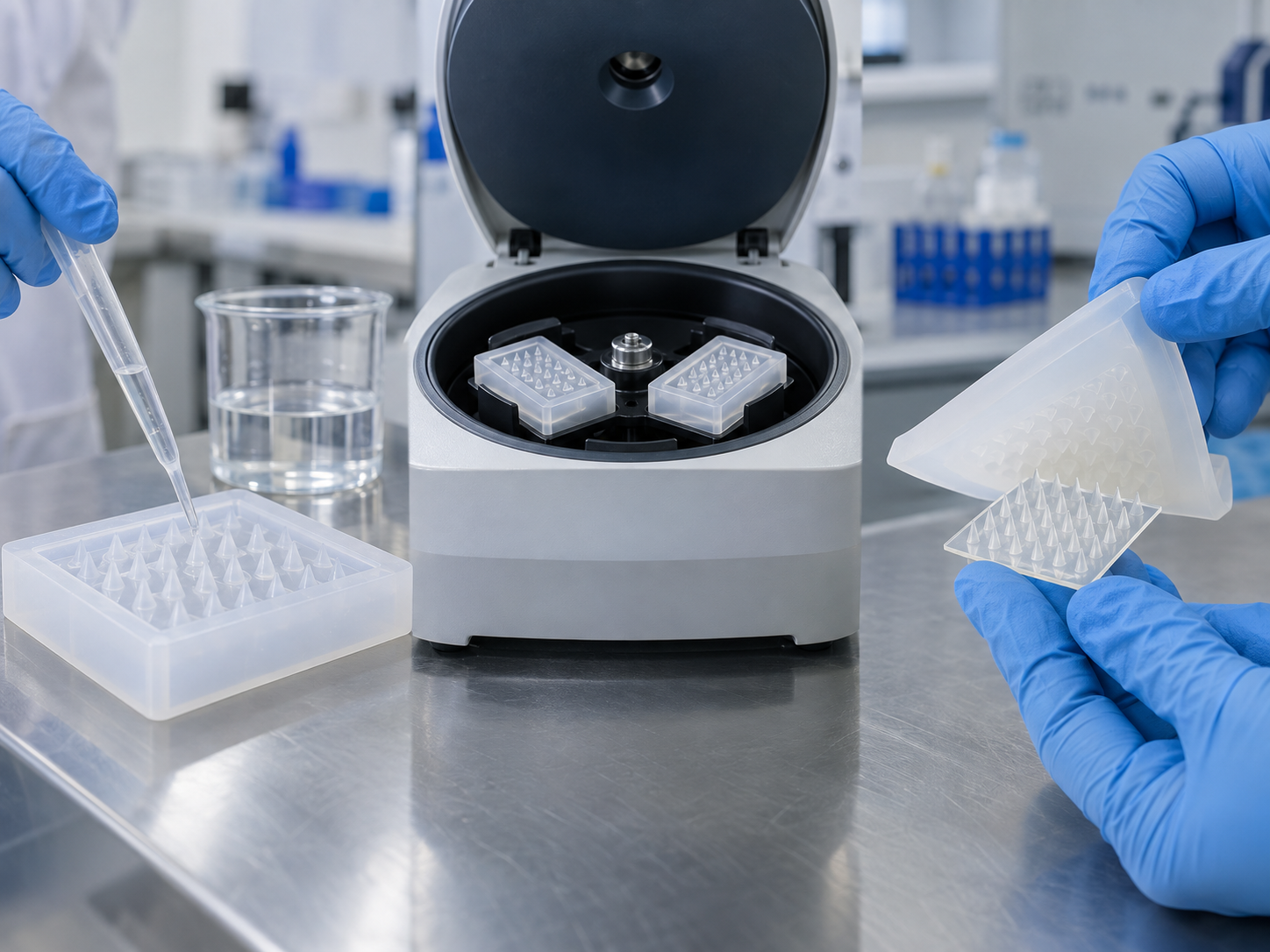
Lee and colleagues (2008) developed a process that casts a viscous aqueous solution of biocompatible carboxymethylcellulose (CMC) or amylopectin into a microfabricated mold during centrifugation, to fill it Sourced. The process encapsulated sulforhodamine B, bovine serum albumin and lysozyme. Lysozyme kept its full enzymatic activity after encapsulation and remained 96% active after two months at room temperature. The needles were strong enough to insert into cadaver skin and dissolved within minutes. When molecules were placed in the needle backing, the backing swelled with interstitial fluid drawn from the skin and the molecules diffused through the channels left by the dissolved needles, giving sustained delivery over hours to days Sourced.
Chu and colleagues (2010) addressed a weakness of dissolving needles, control of the amount and location of drug, with two designs. Localizing drug in the needle tip delivered about 80% within 10 minutes, against about 20% for needles with drug throughout. Adding a pedestal under each needle gave more complete insertion and raised the drug loading per needle from 0.018 µL to 0.053 µL, about threefold Sourced Our calculation (0.053 ÷ 0.018 = 2.9; the paper says "threefold"). Both were experiments with a model molecule (sulforhodamine B) and not results with a real drug.
Route 3: coating a metal needle
Zosano Pharma's annual report (fiscal 2020) describes its drug delivery system as an array of titanium microneedles about 200-350 µm long over 3-6 cm², coated with a hydrophilic drug formulation and mounted on an adhesive patch Sourced. A reusable hand-held applicator presses the needles into the skin to a uniform depth, close to the capillary bed but not deep enough to contact nerve endings. The company also says the maximum amount of drug that can be coated depends on the drug, the weight of the excipients and the coatable surface area of the array Sourced. A coated needle is thus a type in which surface area sets the ceiling on drug loading (Commentary). This contrasts with the dissolving needles of Chu et al., where needle volume and a pedestal raised loading.
Route 4: injection molding and others
Examples include research on making polymer needles by injection molding (Sammoura et al.; the paper's title is "Polymeric microneedle fabrication using a microinjection molding technique"), and Tehrani and colleagues' statement of "cost-effective fabrication via advanced micromachining" for an electrochemical sensor needle array Sourced. We did not check the detailed conditions in these papers (materials, needle dimensions, yield, scalability), so we say nothing about the difficulty of mass production.
5. Sampling interstitial fluid, and integrating sensors
Interstitial fluid (ISF) is the fluid in the spaces between skin cells and it carries biomarkers. Microneedles are being studied as a way to obtain it without drawing blood. There are two broad routes: 1. sample it and measure outside, and 2. measure on the needle.
1. Sample it and measure outside
- Samant and Prausnitz (2018): ISF sampling had had limited impact because no simple, clinically useful technique could collect more than nanoliter volumes. The paper analyzed ISF transport experimentally and theoretically, using solid, porous or hollow needles that penetrate the superficial skin and access ISF through micropores formed on insertion. In pig skin, collection followed the rank order diffusion < capillary action < osmosis < pressure-driven convection, and the rate-limiting step was transport through the dermis. With an optimized system they collected more than 1 µL within 20 minutes in pig cadaver skin and living human subjects, and identified biomarkers in the collected ISF Sourced
- Chang et al. (2017): a swellable needle patch made of methacrylated hyaluronic acid (MeHA), cross-linked by UV, extracts ISF quickly without extra devices. The covalent network keeps its integrity when swollen and leaves no residue in skin. The extracted metabolites were recovered by centrifugation and used for measuring glucose and cholesterol Sourced
- Miller et al. (2018): with hollow needles, ISF was drawn from humans and rats with minimal pain and no blistering. The volumes sufficed for transcriptome and proteome analysis, similar profiles were seen in ISF, serum and plasma, and the authors say ISF can be a proxy for direct blood sampling. Exosome concentration was 12-13 times that in plasma and serum Sourced
2. Measure on the needle
- Tehrani et al. (2022): a fully integrated wearable needle array that senses two metabolites (lactate and glucose, or alcohol and glucose) in the ISF of volunteers during daily activities, wirelessly, continuously and in real time. It combines reusable electronics with a disposable needle array and is described as optimized for system integration, cost-effective fabrication via advanced micromachining, easier assembly, biocompatibility, pain-free skin penetration and enhanced sensitivity. Single- and dual-analyte measurements correlated well with gold-standard blood or breath measurements. The authors say validation in large populations is needed to judge robustness and utility Sourced
- The FDA De Novo decision summary (Biolinq Shine): seven microelectrodes ("microsensors") reside in the papillary dermis and measure ISF glucose continuously. The method is amperometry, in which the enzyme glucose oxidase passes electrons to the electrode to give a current. The summary records factory calibration (user calibration is not accepted), fixation by an adhesive patch, and storage of up to five days of data Sourced. Note that the decision summary does not say "microneedle"; it uses the term "microsensor" (microelectrode)
Interstitial glucose lags blood glucose slightly. Basu and colleagues (2013) inserted microdialysis catheters into the abdominal subcutaneous space of eight healthy adults (overnight fasted), gave intravenous glucose tracers, and sampled plasma and ISF in sequence. The mean time lag of tracer appearance in the interstitial space was 5.3-6.2 minutes Sourced. This is a value for the subcutaneous space in healthy, fasting subjects. Whether the ISF around a needle in the very shallow dermis shows the same lag, and how it changes after meals or exercise, could not be confirmed in the sources we consulted.
The sensing principle (enzyme-electrode interfaces, electrochemical measurement) is covered in this series' articles on enzymes and electrode interfaces and on electrochemical sensors, and fixing a patch to skin in the article on patch sensors and skin adhesion. Here we restrict ourselves to the needle as a structure that obtains ISF or places an electrode at a defined depth in the skin.
6. Our calculation: orders of magnitude for length, dose, survival rate and sampling rate
These conversions, from published numbers, are meant to give a sense of scale Our calculation.
- Needle length versus barrier thickness: Zosano's titanium needles are about 200-350 µm. Henry et al. say the skin barrier lies in the upper 10-15 µm. 200 ÷ 15 = about 13 times; 350 ÷ 10 = 35 times. The needle is about 13-35 times as long as the barrier layer it must cross.
- Dose and effect of the abaloparatide sMTS: in the phase 3 trial (NCT04064411) the sMTS dose was 300 µg a day against 80 µg by subcutaneous injection: 300 ÷ 80 = 3.75 times the dose. The 12-month change in lumbar spine bone mineral density was 7.14% for the sMTS and 10.86% for injection: 7.14 ÷ 10.86 = about 0.66 (a difference of -3.72 points).
- Biolinq Shine survival at five days: of 190 primary wearables, 161 remained at day 5: 161 ÷ 190 = 84.7%. 29 (15.3%) did not "survive."
- ISF sampling rate: Samant et al.'s "more than 1 µL in 20 minutes" corresponds to more than 3 µL per hour (1 ÷ 20 × 60 = 3).
Assumptions and limits: the needle length and the barrier thickness come from different sources and are placed side by side. They are not the depth to which a real needle enters (because skin is elastic, the whole length may not enter). The sMTS and the injection differ in both dose and route, so 300 µg and 80 µg cannot simply be compared to argue about "delivery efficiency." The decision summary does not define "survival." The survival rate is a value under the design of a five-day clinical study (five days of wear, no user calibration) and should not be compared with values for other products or conditions. The sampling rate is for an optimized system in pig cadaver skin and human subjects and is not the sampling volume needed for continuous measurement.
7. A materials engineer's view 2: with dissolving needles, drug, material and shape are decided together
In a dissolving needle the material is at once the structural member that gives strength, the container that holds the drug, and the material that dissolves and disappears in the skin. All three roles are fixed together by the choice of a single polymer (Commentary).
- Strength: the needle must be strong enough to insert into excised skin (Lee et al.). A material that dissolves weakens on contact with water, so both the dry strength before insertion and the dissolution behavior after it are design targets
- Stability of the drug: lysozyme kept full activity after encapsulation and 96% after two months at room temperature (Lee et al.). By contrast, radiation sterilization (gamma, 25 kGy) lowered the recovery of ovalbumin (a model protein) in dissolving needles from about 101% to about 58% (McCrudden et al.). The manufacturing process and sterilization can change the amount of drug Sourced
- Where the drug is and how much: localizing drug in the tip delivered about 80% in 10 minutes, against about 20% without localizing (Chu et al.). A pedestal raised loading per needle about threefold. Shape governs delivery efficiency and loading
- The role of the backing: when drug is placed in the base, the base swells with interstitial fluid and the drug diffuses through the channels of the dissolved needles (Lee et al.). The needles and the backing are separate materials-design problems
A hydrogel-forming needle holds no drug itself, so this constraint is looser. Donnelly and colleagues say the needles of cross-linked polymer absorb ISF to become unblockable conduits, can be easily sterilized, resist hole closure and are removed intact from the skin Sourced (the authors' claims). McCrudden and colleagues also report that hydrogel-forming needles were unchanged in physical properties and delivery capability after 25 kGy of gamma irradiation Sourced. The price is that the drug enters from a separate reservoir, so the amount delivered is governed not by the needle but by the cross-link density (Donnelly et al.).
8. Sterilization and safety
A product that pierces the skin temporarily breaches the skin's outer barrier. Sterilization, skin recovery, needle breakage and biocompatibility are therefore the pillars of evaluation.
Sterilization
McCrudden and colleagues (2015) prepared polymeric dissolving and hydrogel-forming microneedles either aseptically or sterilized them with commonly used techniques, and compared them Sourced.
- Moist and dry heat sterilization damaged all devices, leaving aseptic production and gamma sterilization as the only viable options
- No measurable bioburden was detected, and endotoxin levels were below the FDA limit (20 endotoxin units per device)
- Hydrogel-forming needles were unaffected in physical properties and delivery by 25 kGy of gamma radiation. In dissolving needles, by contrast, recovery of the encapsulated ovalbumin fell from about 101% to about 58%, that in lyophilized wafer-type reservoirs from about 99.7% to about 60.1%, and the skin permeation profile of ibuprofen sodium changed
The authors say microneedles pose a very low risk to human health when used appropriately, and that if regulators require guarantees of absolute sterility, the effect of lower gamma doses will need investigating.
For an authorized product, the FDA decision summary for Biolinq Shine records that e-beam sterilization was validated to a sterility assurance level (SAL) of 10-6 at a dose range of 20-35 kGy, a storage shelf life of six months (2-30 °C, 10-90% humidity), and seal integrity testing Sourced. The standards the summary lists as referenced for sterilization include ISO 11137-1 and -2 (radiation), ISO 11737-1 and -2 (microbiological methods), ISO 13004 (substantiating the dose) and ISO 11607-1 (packaging for terminally sterilized devices) Sourced (the text of the standards was not checked).
Skin recovery and pain
Gupta and colleagues (2011) used electrical impedance spectroscopy in human subjects, for the first time, to study how the skin barrier reseals after microneedle insertion. Without occlusion, all sites recovered barrier properties within 2 hours. Occluded sites resealed more slowly, over 3 to 40 hours depending on needle geometry, and resealed rapidly once occlusion was removed. Longer needles, more needles and a larger cross-sectional area slowed resealing Sourced. On pain, Henry and colleagues reported "painless" in limited human tests, and Miller and colleagues describe ISF sampling with minimal pain and no blistering Sourced. These are small studies and do not give the pain or skin-reaction frequency of any product.
Biocompatibility, and FDA's special controls
According to the Biolinq Shine decision summary, the patient-contacting components were tested following ISO 10993-1, covering cytotoxicity, sensitization, irritation, acute systemic toxicity and pyrogenicity, subacute/subchronic toxicity, genotoxicity, implantation, and chronic toxicity and carcinogenicity, and a toxicological risk assessment was done under ISO 10993-17:2023 Sourced. A device with electrodes entering the shallow skin evidently has more endpoints than one that is merely stuck on the skin (Commentary). Biocompatibility of the adhesive patch is covered in this series' article on patch sensors and skin adhesion.
For aesthetic microneedling devices, the special controls of 21 CFR 878.4430 call for identification of needle length, geometry, maximum penetration depth and puncture rate, accuracy of penetration depth, safety features against cross-contamination, sterility of patient-contacting components, sterility and package integrity over the shelf life, and biocompatibility, among others Sourced. FDA's guidance lists as risks these controls address adverse tissue reaction, cross-contamination and infection, and damage to underlying tissue including nerves and blood vessels, scarring and hyper- or hypopigmentation, which can result from exceeding safe penetration depth, mechanical failure or software malfunction Sourced.
9. Where things stand: authorized or approved examples, and research-stage examples
We line up examples by stage, limited to what regulators' documents and official records let us confirm. Stages differ greatly even under the one word "microneedle."
| Example | Type, use | What regulators' documents and official records confirm | Stage |
|---|---|---|---|
| Aesthetic microneedling devices | Needles puncture and injure skin for aesthetic purposes | 21 CFR 878.4430. Class II (special controls). Explicitly "does not include devices intended for transdermal delivery." Federal Register 8 June 2018 (83 FR 26577). FDA guidance (November 2020) | Marketed (510(k)) |
| Fluzone Intradermal Quadrivalent | Intradermal vaccine; 0.1 mL prefilled "microinjection system" | FDA label (2016-2017 formula): "Initial US Approval: 2014," ages 18 through 64, intradermal use only. Listed in the CDC recommendations for 2017-18; the word "intradermal" does not appear in the 2018-19 recommendations | Approved (2014); later supply not confirmed |
| Qtrypta (M207, zolmitriptan) | Coated titanium needles, 3-6 cm², about 200-350 µm long | NDA filed 20 December 2019; Complete Response Letter received 20 October 2020. The CRL cited inconsistent zolmitriptan exposure across clinical pharmacology studies, differences between lots, and inadequate PK bridging, among other points (company annual report) | Not approved (as confirmed) |
| Abaloparatide sMTS | Solid microstructured transdermal system | ClinicalTrials.gov NCT04064411 (Radius Health). Phase 3, postmenopausal women, 12 months. Change in lumbar spine BMD: sMTS 7.14%, subcutaneous injection 10.86%, difference -3.72 (95% CI -5.01 to -2.43). The non-inferiority criterion (lower bound -2.0%) was not met | Phase 3, target missed |
| Biolinq Shine | Glucose range display with seven intradermal microelectrodes | De Novo DEN240080. Received 27 December 2024, decided 23 September 2025. Class II, 21 CFR 862.1359 (as FDA states). Prescription, age 22 and over, not using insulin. Not for hypoglycemia awareness | Authorized (De Novo) |
| ISF sampling, continuous monitoring | Hollow and swelling needles, multi-analyte electrode arrays | Miller et al. 2018, Chang et al. 2017, Samant et al. 2018, Tehrani et al. 2022 | Research |
All Sourced (see each reference). The "Stage" column is this article's arrangement based on regulators' documents and official records as far as we could confirm them, and does not show current sales or development status.
The Biolinq Shine pivotal study ran at six US centers from 29 February to 10 June 2024. It enrolled 247 adults with diabetes, 202 of whom wore the device (66.8% type 1, 20.3% type 2 on intensive insulin therapy, 5.9% type 2 on basal insulin only, 6.9% type 2 not on insulin). Wear was for up to five days on the volar forearm, with a laboratory glucose analyzer (YSI) as the comparator Sourced. The FDA decision summary lists as conditions of use that the output is color-coded ranges and not a quantitative value, that it is not for hypoglycemia awareness, that the sensor must be removed before MRI, CT and X-ray, and that acetaminophen and hydroxyurea can interfere with readings Sourced.
The above reports what a regulator's document says. It is neither an evaluation of the product's accuracy or use nor advice to any individual. The authorization is for the specific intended use set out in the decision summary (understanding glucose ranges in adults not using insulin) and does not mean the device is safe and accurate for any other use. A De Novo is also a pathway that grants Class I or II classification to a device for which general controls, or general and special controls, give reasonable assurance of safety and effectiveness but for which there is no legally marketed predicate device (FDA's description). It is a different category from PMA approval and 510(k) clearance.
10. A materials engineer's view 3: regulation draws its line at the claim, not at the needle material
FDA's guidance on microneedling products decides whether a product is a medical device mainly on two things Sourced.
- The firm's claims: claims of penetration into living layers of skin (epidermis, dermis) are treated as evidence of intent to affect the body's structure or function. Claims such as treating scars or wrinkles or stimulating collagen production make a product a device. A product that does not penetrate living skin and claims only exfoliation or a smoother look is generally not a device
- Design and technological features: needle length and arrangement, needle sharpness, and how far the product controls the movement of the needles and the depth of penetration. FDA looks at whether these facilitate penetration into living layers
Specifications that a materials engineer sets, such as needle length, tip radius and arrangement, thus become regulatory evidence (Commentary). Furthermore, the aesthetic classification (21 CFR 878.4430) does not include devices for transdermal delivery, and products combined with drugs or biologics are "combination products" handled by CDER, CBER and/or CDRH, and are outside the scope of that guidance. The same kind of needle can go down different roads: an aesthetic device, a drug patch (filed as a drug), a vaccine injection system (a biologic), or a glucose sensor (a device De Novo).
11. Open issues and what could not be confirmed
(1) The situation in Japan and the EU was not checked
The regulatory statements here rest on US FDA documents. We did not check the classification or approval of microneedle products in Japan (PMDA, the Pharmaceuticals and Medical Devices Act) or the EU (MDR, IVDR).
(2) No approved dissolving or hydrogel-forming product was confirmed
What we could confirm in regulators' documents were an aesthetic device, an intradermal vaccine (hollow type) and an intradermal glucose sensor. We could not find, in the official records we confirmed, an approved drug in dissolving or hydrogel-forming form. We did not check the present state of clinical development of microarray patches (MAPs) for vaccines and drugs.
(3) Difficulty of mass production, needle fragments and infection frequency could not be confirmed
The primary sources we confirmed give no concrete figures on needle dimension uniformity, coating uniformity, yield or manufacturing challenges (the lot-to-lot difference in exposure cited in Zosano's CRL is one hint). We also found no source for how often needle tips are left in the skin or how often skin infection occurs.
(4) The lag in dermal ISF, and long-term sensor stability
For the glucose lag from blood to ISF we could confirm only values for healthy subjects in the subcutaneous space (chapter 5). How protein adsorption on intradermal microelectrodes, enzyme deactivation and the foreign-body response affect measurement is not something we confirmed. The Biolinq clinical study covered five days of wear, and we did not confirm results for longer wear.
(5) The text of statutes and standards was not checked
The text of standards such as ISO 10993 and ISO 11137 is paid and was not checked. 21 CFR 862.1359 appears in FDA's classification database and decision summary, but in the eCFR as of 30 September 2026 we found no such section.
(6) The main reviews were not read in full
For the reviews by Larrañeta et al. (Mater Sci Eng R, 2016), Donnelly et al. (Drug Deliv, 2010) and Ventrelli et al. (Adv Healthc Mater, 2015) we checked only the abstract or title and do not quote details of their accounts of manufacturing and commercialization.
- "Microneedle" is not one technology. Solid, coated, dissolving, hollow and hydrogel-forming types differ in both materials and use Sourced
- A needle is designed on the ratio of "inserts" to "does not break." A thicker wall and a finer tip raise the margin of safety Sourced
- In some types, sterilization is tied to the drug. Gamma irradiation at 25 kGy lowered ovalbumin recovery in dissolving needles from about 101% to about 58% Sourced
- The degree of practical use differs by application. An aesthetic device, an intradermal vaccine and an intradermal glucose sensor are authorized or approved by regulators. Among drug patches there are cases that were not approved and cases that missed their target Sourced
- Regulation draws its line at the claim and the design, not the needle material.
12. Glossary
- Microneedle
- A patch or chip carrying an array of needles a few hundred micrometers long, pressed into the skin.
- Stratum corneum
- The outermost skin layer, made of dead cells. The main skin barrier; about 10-15 µm thick per Henry et al.
- Interstitial fluid (ISF)
- The fluid in the spaces between skin cells. It carries biomarkers.
- Solid type
- Needles that make holes in the skin and are removed; drug is applied afterwards.
- Coated type
- Needles with drug on the surface, which dissolves off in the skin.
- Dissolving type
- Needles of water-soluble polymer that encapsulate drug and dissolve in the skin.
- Hollow type
- Needles with a channel for infusing fluid into, or sampling fluid from, the skin.
- Hydrogel-forming type
- Needles of cross-linked polymer that absorb ISF, swell and become conduits.
- Insertion force / fracture force
- The force to insert a needle into skin, and the force it withstands before breaking. Their ratio is the margin of safety.
- Backing
- The base of a needle array. In dissolving types it can be designed as a drug reservoir.
- SAL (sterility assurance level)
- The probability that a viable microorganism remains after sterilization. 10-6 means one in a million or fewer.
- De Novo
- An FDA pathway that classifies a device into Class I or II when there is no legally marketed predicate device.
- CRL (Complete Response Letter)
- A letter in which FDA says it cannot approve an application in its present form.
- Combination product
- A product that combines a drug or biologic with a device; several FDA centers can be involved.
13. References
- Kim YC, Park JH, Prausnitz MR Microneedles for drug and vaccine delivery. Adv Drug Deliv Rev 64(14): 1547-1568 (2012). DOI 10.1016/j.addr.2012.04.005 (abstract checked on PubMed, PMID 22575858) — doi.org
- Henry S, McAllister DV, Allen MG, Prausnitz MR Microfabricated microneedles: a novel approach to transdermal drug delivery. J Pharm Sci 87(8): 922-925 (1998). DOI 10.1021/js980042+ (abstract checked on PubMed, PMID 9687334) — doi.org
- McAllister DV et al. Microfabricated needles for transdermal delivery of macromolecules and nanoparticles: fabrication methods and transport studies. Proc Natl Acad Sci U S A 100(24): 13755-13760 (2003). DOI 10.1073/pnas.2331316100 (PMC283494) — doi.org
- Davis SP et al. Insertion of microneedles into skin: measurement and prediction of insertion force and needle fracture force. J Biomech 37(8): 1155-1163 (2004). DOI 10.1016/j.jbiomech.2003.12.010 (abstract checked on PubMed, PMID 15212920) — doi.org
- Lee JW, Park JH, Prausnitz MR Dissolving microneedles for transdermal drug delivery. Biomaterials 29(13): 2113-2124 (2008). DOI 10.1016/j.biomaterials.2007.12.048 (PMC2293299) — doi.org
- Chu LY, Choi SO, Prausnitz MR Fabrication of dissolving polymer microneedles for controlled drug encapsulation and delivery: bubble and pedestal microneedle designs. J Pharm Sci 99(10): 4228-4238 (2010). DOI 10.1002/jps.22140 (abstract checked on PubMed, PMID 20737630) — doi.org
- Donnelly RF et al. Hydrogel-forming microneedle arrays for enhanced transdermal drug delivery. Adv Funct Mater 22(23): 4879-4890 (2012). DOI 10.1002/adfm.201200864 (PMC3627464) — doi.org
- McCrudden MTC et al. Considerations in the sterile manufacture of polymeric microneedle arrays. Drug Deliv Transl Res 5(1): 3-14 (2015). DOI 10.1007/s13346-014-0211-1 (abstract checked on PubMed, PMID 25787335) — doi.org
- Gupta J et al. Kinetics of skin resealing after insertion of microneedles in human subjects. J Control Release 154(2): 148-155 (2011). DOI 10.1016/j.jconrel.2011.05.021 (PMC3164267) — doi.org
- Samant PP, Prausnitz MR Mechanisms of sampling interstitial fluid from skin using a microneedle patch. Proc Natl Acad Sci U S A 115(18): 4583-4588 (2018). DOI 10.1073/pnas.1716772115 (PMC5939066) — doi.org
- Chang H et al. A swellable microneedle patch to rapidly extract skin interstitial fluid for timely metabolic analysis. Adv Mater 29(37): 1702243 (2017). DOI 10.1002/adma.201702243 (abstract checked on PubMed, PMID 28714117) — doi.org
- Miller PR et al. Extraction and biomolecular analysis of dermal interstitial fluid collected with hollow microneedles. Commun Biol 1: 173 (2018). DOI 10.1038/s42003-018-0170-z (PMC6197253) — doi.org
- Tehrani F et al. An integrated wearable microneedle array for the continuous monitoring of multiple biomarkers in interstitial fluid. Nat Biomed Eng 6(11): 1214-1224 (2022). DOI 10.1038/s41551-022-00887-1 (abstract checked on PubMed, PMID 35534575) — doi.org
- Basu A et al. Time lag of glucose from intravascular to interstitial compartment in humans. Diabetes 62(12): 4083-4087 (2013). DOI 10.2337/db13-1132 (PMC3837059) — doi.org
- Ventrelli L et al. Microneedles for transdermal biosensing: current picture and future direction. Adv Healthc Mater 4(17): 2606-2640 (2015). DOI 10.1002/adhm.201500450 (abstract checked on PubMed, PMID 26439100) — doi.org
- Sammoura F et al. Polymeric microneedle fabrication using a microinjection molding technique. Microsyst Technol 13(5-6): 517-522 (2007). DOI 10.1007/s00542-006-0204-1 (title only checked) — doi.org
- Ranamukhaarachchi SA et al. Determining the factors affecting dynamic insertion of microneedles into skin. Biomed Microdevices 21(4): 100 (2019). DOI 10.1007/s10544-019-0449-y (abstract checked on PubMed, PMID 31745652) — doi.org
- Donnelly RF, Singh TRR, Woolfson AD Microneedle-based drug delivery systems: microfabrication, drug delivery, and safety. Drug Deliv 17(4): 187-207 (2010). DOI 10.3109/10717541003667798 (abstract checked on PubMed, PMID 20297904) — doi.org
- U.S. Food and Drug Administration (FDA) De Novo decision summary DEN240080, "Biolinq Shine Autonomous Time-in-Range Microsensor" (PDF) — accessdata.fda.gov
- FDA De Novo database record DEN240080 (received 27 December 2024; decision 23 September 2025) — accessdata.fda.gov
- FDA (openFDA) Product classification SFU "Glucose Range Monitoring System" (definition, Class II) — api.fda.gov
- FDA "Regulatory Considerations for Microneedling Products: Guidance for Industry and Food and Drug Administration Staff" (November 2020; nonbinding recommendations) — fda.gov
- eCFR 21 CFR 878.4430 Microneedling device for aesthetic use — ecfr.gov
- FDA Fluzone Intradermal Quadrivalent prescribing information (2016-2017 formula, Sanofi Pasteur) — fda.gov
- CDC (MMWR) Prevention and Control of Seasonal Influenza with Vaccines: Recommendations of the ACIP, United States, 2017-18 Influenza Season. MMWR Recomm Rep 66(2) — cdc.gov
- Zosano Pharma Form 10-K for fiscal 2020, filed with the SEC — sec.gov
- CDC (MMWR) Prevention and Control of Seasonal Influenza with Vaccines: Recommendations of the ACIP, United States, 2018-19 Influenza Season. MMWR Recomm Rep 67(3) — cdc.gov
- ClinicalTrials.gov NCT04064411 "Efficacy & Safety of Abaloparatide-Solid Microstructured Transdermal System in Postmenopausal Women With Osteoporosis" (Radius Health; results posted) — clinicaltrials.gov
- eCFR 21 CFR Part 862 (clinical chemistry and clinical toxicology devices; no text for 862.1359 as of 30 September 2026) — ecfr.gov
- Larrañeta E et al. Microneedle arrays as transdermal and intradermal drug delivery systems: Materials science, manufacture and commercial development. Mater Sci Eng R 104: 1-32 (2016). DOI 10.1016/j.mser.2016.03.001 (title only checked) — doi.org
14. Claim-to-source audit
| Claim in the text | Basis | Type |
|---|---|---|
| Four types of microneedle for drug delivery (solid for pretreatment, coated, dissolving, hollow). Hollow-needle influenza vaccination in widespread clinical use. Solid microneedle products sold for cosmetic purposes | Abstract of Kim et al. (2012) Reference 1 https://doi.org/10.1016/j.addr.2012.04.005 | Sourced |
| Needles made by reactive ion etching that cross the skin barrier (upper 10-15 µm) without stimulating nerves; up to 4 orders of magnitude increase in permeability of excised human skin to a model drug; painless in limited human tests | Abstract of Henry et al. (1998) Reference 2 https://doi.org/10.1021/js980042+ | Sourced |
| Microfabrication of silicon, metal and biodegradable polymer arrays (solid and hollow; feature sizes 1-1,000 µm). Orders-of-magnitude increase in permeability with solid needles; microliter infusion with hollow needles (insulin lowered blood glucose in diabetic rats) | Abstract of McAllister et al. (2003) Reference 3 https://doi.org/10.1073/pnas.2331316100 | Sourced |
| Insertion force rises linearly with tip area, about 0.1-3 N. Fracture force rises with wall thickness, wall angle and possibly tip radius. The margin of safety (fracture ÷ insertion) is much greater than one for almost all geometries and rises with thicker walls and smaller tip radius | Abstract of Davis et al. (2004) Reference 4 https://doi.org/10.1016/j.jbiomech.2003.12.010 | Sourced |
| A viscous aqueous solution of CMC or amylopectin cast into a mold under centrifugation. Lysozyme fully active after encapsulation and 96% after two months at room temperature. Needles insert into excised skin and dissolve within minutes. Drug in the backing gives sustained delivery | Abstract of Lee et al. (2008) Reference 5 https://doi.org/10.1016/j.biomaterials.2007.12.048 | Sourced |
| Drug localized in the tip delivered about 80% in 10 minutes (about 20% otherwise). A pedestal raised loading from 0.018 to 0.053 µL (about threefold). Model molecule sulforhodamine B | Abstract of Chu et al. (2010) Reference 6 https://doi.org/10.1002/jps.22140 | Sourced |
| Hydrogel-forming needles of cross-linked polymer absorb ISF and become conduits, can be easily sterilized, resist hole closure and are removed intact (the authors' claims). Delivery is controlled by cross-link density | Abstract of Donnelly et al. (2012) Reference 7 https://doi.org/10.1002/adfm.201200864 | Sourced |
| Moist and dry heat damaged all devices; aseptic production and gamma radiation were the only options. No bioburden detected; endotoxin below the limit. Hydrogel-forming needles unchanged at 25 kGy. Ovalbumin recovery in dissolving needles and wafer reservoirs fell from about 101% to 58% and from 99.7% to 60.1%. Ibuprofen sodium permeation changed. The authors' conclusion of very low risk to human health | Abstract of McCrudden et al. (2015) Reference 8 https://doi.org/10.1007/s13346-014-0211-1 | Sourced |
| In human subjects, without occlusion all sites recovered barrier properties within 2 hours, occluded sites in 3-40 hours; longer needles, more needles and larger cross-sections reseal more slowly; resealing is rapid once occlusion is removed | Abstract of Gupta et al. (2011) Reference 9 https://doi.org/10.1016/j.jconrel.2011.05.021 | Sourced |
| ISF sampling with solid, porous and hollow needles; rank of transport mechanisms (diffusion < capillary action < osmosis < pressure-driven convection); the rate-limiting step is dermal transport; more than 1 µL in 20 minutes in pig cadaver skin and human subjects; a suction prototype | Abstract of Samant and Prausnitz (2018) Reference 10 https://doi.org/10.1073/pnas.1716772115 | Sourced |
| A swellable needle patch of UV-cross-linked methacrylated hyaluronic acid extracts ISF without extra devices and leaves no residue; metabolites recovered by centrifugation and used to measure glucose and cholesterol | Abstract of Chang et al. (2017) Reference 11 https://doi.org/10.1002/adma.201702243 | Sourced |
| Hollow needles extract ISF from humans and rats with minimal pain and no blistering; volumes sufficient for transcriptome and proteome analysis; similar profiles in ISF, serum and plasma; exosomes 12-13 times more concentrated than in plasma and serum | Abstract of Miller et al. (2018) Reference 12 https://doi.org/10.1038/s42003-018-0170-z | Sourced |
| A fully integrated wearable needle array that senses lactate and glucose, or alcohol and glucose, wirelessly and continuously in volunteers; reusable electronics and disposable array; good correlation with blood or breath gold standards; validation in large populations needed | Abstract of Tehrani et al. (2022) Reference 13 https://doi.org/10.1038/s41551-022-00887-1 | Sourced |
| In eight healthy overnight-fasted adults, the mean lag of glucose tracer from plasma to subcutaneous ISF was 5.3-6.2 minutes | Abstract of Basu et al. (2013) Reference 14 https://doi.org/10.2337/db13-1132 | Sourced |
| In an artificial skin model, needle velocity at impact was the most crucial factor for insertion across different tip areas and array sizes | Abstract of Ranamukhaarachchi et al. (2019) Reference 17 https://doi.org/10.1007/s10544-019-0449-y | Sourced |
| The title of Sammoura et al. (injection molding of polymer microneedles), the titles of the reviews by Ventrelli et al. and Larrañeta et al., and the title and abstract scope of the review by Donnelly et al. | Titles and abstracts of each paper (checked on Crossref and PubMed) Reference 16 https://doi.org/10.1007/s00542-006-0204-1, Reference 15 https://doi.org/10.1002/adhm.201500450, Reference 18 https://doi.org/10.3109/10717541003667798, Reference 30 https://doi.org/10.1016/j.mser.2016.03.001 | Sourced |
| The Biolinq Shine De Novo decision summary: seven microelectrodes in the papillary dermis, glucose-oxidase amperometry, factory calibration, adhesive patch, up to five days of data, intended use (age 22 and over, not on insulin, not for hypoglycemia awareness), conditions of use, the clinical study (six centers, 247 enrolled, 202 wore), five-day survival of 84.7% (161/190), e-beam sterilization at 20-35 kGy and SAL 10-6, six-month shelf life, biocompatibility endpoints, referenced standards, Class II, 21 CFR 862.1359, product code SFU | FDA De Novo decision summary DEN240080 Reference 19 https://www.accessdata.fda.gov/cdrh_docs/reviews/DEN240080.pdf | Sourced |
| DEN240080 received 27 December 2024, decision 23 September 2025, granted | FDA De Novo database Reference 20 https://www.accessdata.fda.gov/scripts/cdrh/cfdocs/cfpmn/denovo.cfm?id=DEN240080 | Sourced |
| Definition of product code SFU (glucose range monitoring system; qualitative or semi-quantitative; not for acute medical decision-making), Class II | openFDA product classification Reference 21 https://api.fda.gov/device/classification.json?search=product_code:SFU&limit=1 | Sourced |
| The scope of the term "needles," how a microneedling product is judged a device (claims and design), examples of non-penetrating products that are not devices, combination products outside the guidance, Class II 21 CFR 878.4430, the risks the special controls address, November 2020 | FDA guidance (nonbinding recommendations) Reference 22 https://www.fda.gov/media/107708/download | Sourced |
| The definition in 21 CFR 878.4430 (not including devices for transdermal delivery) and its special controls (needle specifications, depth accuracy, cross-contamination protection, sterility, shelf life, biocompatibility and others); Federal Register 8 June 2018 (83 FR 26577) | eCFR Reference 23 https://www.ecfr.gov/current/title-21/section-878.4430 | Sourced |
| Fluzone Intradermal Quadrivalent: Initial US Approval 2014, ages 18 through 64, intradermal use only, 0.1 mL prefilled microinjection system | FDA prescribing information (2016-2017 formula) Reference 24 https://www.fda.gov/media/90251/download | Sourced |
| Fluzone Intradermal Quadrivalent is listed in the 2017-18 ACIP recommendations | CDC MMWR 2017-18 Reference 25 https://www.cdc.gov/mmwr/volumes/66/rr/rr6602a1.htm | Sourced |
| The word "intradermal" does not appear in the 2018-19 ACIP recommendations (confirmed by searching the text of the document) | CDC MMWR 2018-19 Reference 27 https://www.cdc.gov/mmwr/volumes/67/rr/rr6703a1.htm | Sourced |
| Zosano's system (titanium needles about 200-350 µm, 3-6 cm², hydrophilic drug coating, adhesive patch, reusable applicator, depth short of nerve endings, coating capacity set by surface area and others). NDA 20 December 2019, CRL 20 October 2020 and the points it cited | Zosano Pharma annual report (Form 10-K, fiscal 2020) Reference 26 https://www.sec.gov/Archives/edgar/data/1587221/000158722121000007/zsan-20201231.htm | Sourced |
| Abaloparatide sMTS: phase 3, 511 participants, 12 months, change in lumbar spine BMD (sMTS 7.14%, injection 10.86%), difference -3.72 (95% CI -5.01 to -2.43), non-inferiority bound -2.0%, doses 300 µg and 80 µg | ClinicalTrials.gov record (results posted) Reference 28 https://clinicaltrials.gov/study/NCT04064411 | Sourced |
| The eCFR (30 September 2026) text of 21 CFR Part 862 does not include a section 862.1359 | Check of the eCFR structure Reference 29 https://www.ecfr.gov/current/title-21/part-862 | Sourced |
| Needle length versus barrier thickness (about 13-35 times), dose ratio 3.75 and BMD-change ratio about 0.66, five-day survival 161 ÷ 190 = 84.7% (29 non-surviving, 15.3%), sampling rate of 3 µL per hour or more, pedestal loading 0.053 ÷ 0.018 = about 2.9 | Our calculation. Assumptions: Zosano's needle length (200-350 µm) and Henry et al.'s barrier layer (10-15 µm) are placed side by side; dose and BMD are ClinicalTrials.gov values; survival is the decision-summary value; sampling volume is Samant et al.'s value | Our calculation |
| The reading that the needle material of a dissolving needle serves as structure, container and dissolving material at once. The reading that surface area caps drug loading in a coated needle. The reading that needle specifications (length, tip radius, arrangement) become regulatory evidence. The inference that the Davis et al. results are for hollow needles. The reading that intradermal electrodes have more endpoints than a device merely stuck on the skin. The grouping of types in the figures | Commentary by this article, built on the sourced facts above (the needle form in Davis et al. is an inference) | Commentary |
| Regulation in Japan and the EU; approved drugs in dissolving or hydrogel-forming form; the present state of clinical development of vaccine and drug patches; manufacturing difficulty; the frequency of retained needle tips and infection; the lag of dermal ISF glucose; long-term sensor stability; the text of standards; the text of 21 CFR 862.1359; the full text of the reviews; later development of Zosano's and Radius's products | Could not be confirmed in the public official sources consulted for this article, and so are not stated | Commentary |
| That Figs. 1 to 3 and 5 to 8 are explanatory drawings, and that the hero image and Fig. 4 are AI-generated images that do not show real products, production equipment or dimensions | Note by this article | Commentary |
Last updated 4 October 2026. Sources are limited to primary material (FDA decision summaries, guidance and labeling, the eCFR, CDC recommendations, an SEC annual report, ClinicalTrials.gov records and peer-reviewed papers). Because the article includes structural readings and materials-design interpretations, those are marked as Commentary and kept separate from sourced fact. Regulation in Japan and the EU, approved dissolving or hydrogel-forming drugs, the present state of clinical development of vaccine and drug patches, manufacturing challenges, the frequency of retained needle tips and infection, the lag of dermal interstitial glucose, and long-term sensor stability are not stated, because no published primary source could be confirmed. All figures are explanatory concept graphics. Figs. 1 to 3 and 5 to 8 are vector drawings; the hero image and Fig. 4 are AI-generated images, and none of them shows a real product, production equipment or cross-section. This article explains materials, evaluation and regulation and is not medical advice.