TECHNOLOGY EXPLAINER
Genome Editing
— the part that decides “where to cut” has moved from protein to RNA
Genome editing is a technology that cuts DNA at a chosen site and lets the cell's own repair machinery rewrite it. In 2012, CRISPR-Cas9, which specifies the cutting site with a guide RNA, was demonstrated, and design suddenly became far easier. Among the products on the FDA's list of approved cellular and gene therapy products, CASGEVY is the only genome-editing product this article could identifySourced. What it consists of is two raw materials: a synthetic, chemically modified oligonucleotide (the guide RNA) and a recombinant protein (Cas9).
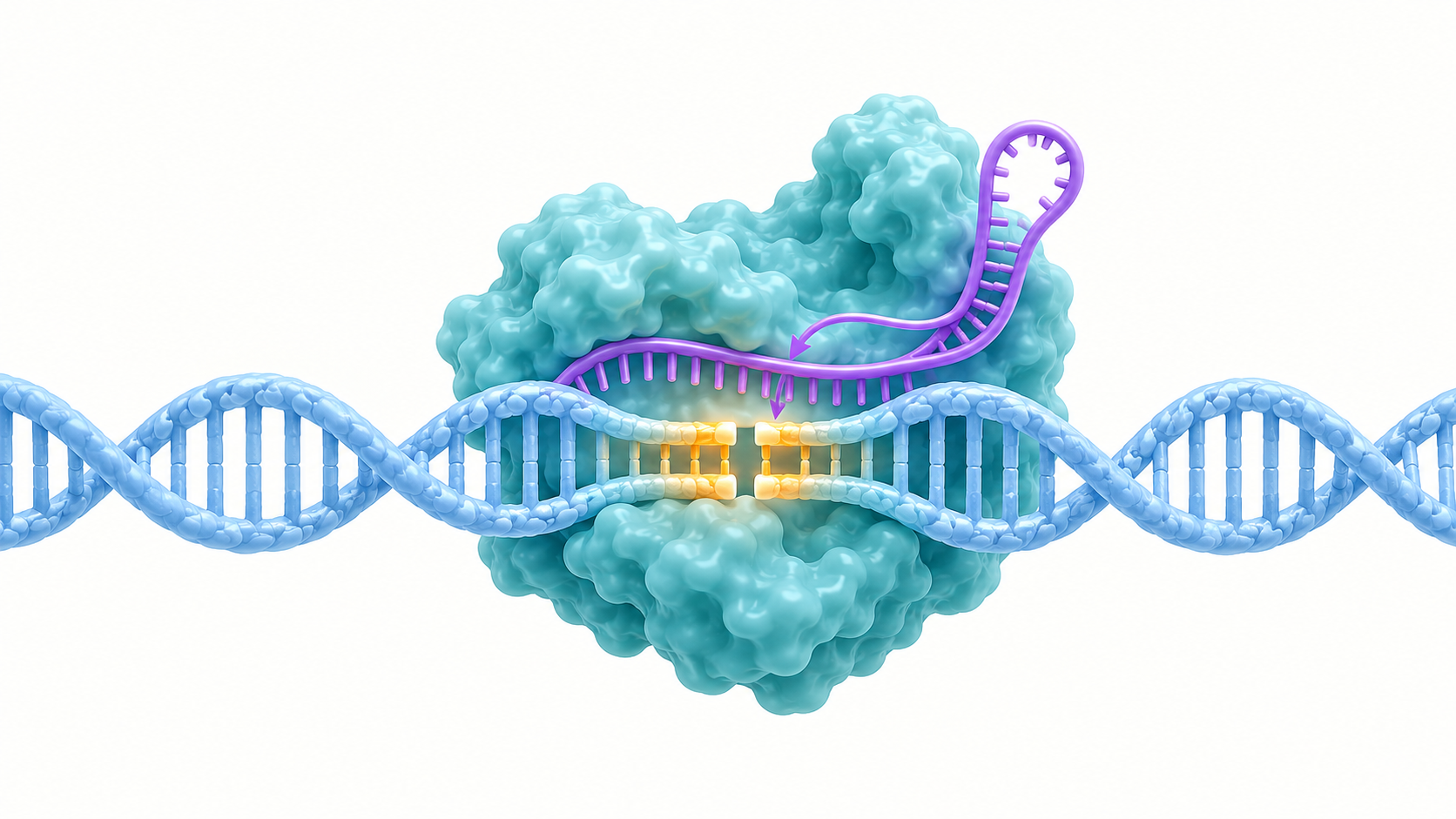
- What genome editing is, in three points
- What happens after the cut: two repair pathways
- Three cutting tools: ZFN, TALEN and CRISPR-Cas9
- The parts of CRISPR-Cas9: Cas9, guide RNA and PAM
- A materials engineer's view (1): the two raw materials are a chemically synthesised product and a recombinant protein
- Editing without cutting: base editing and prime editing
- How to deliver it: electroporation, lipid nanoparticles and AAV
- A materials engineer's view (2): “how long it persists” becomes a design variable
- Approval status and where clinical use stands
- Open problems, and what is not yet settled
- Glossary / References / Claim-to-source audit
Sourced = stated in published material (link given)
Our calculation = a value this article derived from assumptions it states
Not yet confirmed = a plan or target with no confirmed track record
Structural readings and interpretations from a materials or process-design standpoint are marked separately as Commentary.
This article is an explanation of materials and manufacturing technology. It does not recommend any treatment, does not rank treatments against one another, and is not medical advice. Statements on approval status, indications and figures are quoted only where they appear in published material from regulators (FDA, EMA, the UK MHRA) and in peer-reviewed papers. The assessment of off-target effects (unintended edits at other sites) is covered in the companion article in this series, “Off-target effects and safety assessment”.
1. What genome editing is, in three points
Genome editing is a technology that cuts DNA at a chosen site with an engineered enzyme and uses the cell's repair of that cut to rewrite the sequence.
- What it does: FDA guidance defines human genome editing as “a process by which DNA sequences are added, removed, altered or replaced at specific locations in the genome of somatic cells”, and the definition covers both approaches that use a nuclease (a DNA-cutting enzyme) and approaches that do notSourced
- Why it was a breakthrough: the 2020 Nobel Prize in Chemistry went to Charpentier and Doudna “for the development of a method for genome editing”. The Nobel Foundation says that with these CRISPR/Cas9 “genetic scissors” researchers can change the code of life “within a few weeks”Sourced
- How far it has reached products: on the FDA's list of “Approved Cellular and Gene Therapy Products” (checked on 1 October 2026; the list gives only product names and nonproprietary names and has no “genome editing” category), the only product that can be identified as using CRISPR/Cas9 from those names is CASGEVY (nonproprietary name exagamglogene autotemcel) (our check). In its approval announcement the FDA called it “the first FDA-approved treatment to utilize CRISPR/Cas9”Sourced
The history of genome editing is the history of swapping the part that recognises the cutting site. At first a protein (ZFN, TALEN) recognised the site; then RNA (the guide RNA) took over recognition, which made design much easier. Methods that rewrite DNA without cutting it (base editing, prime editing) have since been added (this article's framing). And seen as a product, the editing components are “a synthetic, modified RNA” and “a recombinant protein”: raw materials that can be described in the language of materials and manufacturing.
2. What happens after the cut: two repair pathways
The basic idea of genome editing is to introduce a double-strand break (DSB) at a specific site in the DNA and exploit the cell's own repair machinery. The PMDA Science Board report names two pathways for repairing a DSB: non-homologous end joining (NHEJ) and repair that uses homologous recombination (HDR)Sourced.
The FDA guidance explains that HDR uses a homologous DNA sequence to mend the break, while NHEJ rejoins the two broken ends without a template. It then states that “NHEJ is relatively independent of the cell cycle, whereas HDR is most active in S and G2 phase”, and that although either pathway can be accurate, both can produce insertions and deletions (indels)Sourced.
The PMDA Science Board report likewise says that NHEJ is an emergency-repair reaction that operates throughout the cell cycle and can add or lose a few bases at the junction, so that it can be used to disrupt a gene, and that HDR, when a template DNA carrying the normal sequence is introduced, can also be used to correct a genetic defectSourced.
The example of this “break it by NHEJ” approach in practice is CASGEVY, discussed later. A 2019 paper by Wu and colleagues reported that when a Cas9–guide RNA ribonucleoprotein (RNP) cuts the GATA1 binding site within the BCL11A enhancer, that motif is disrupted at a high frequency, BCL11A expression falls, and fetal γ-globin is inducedSourced. The paper notes that haematopoietic stem cells favour non-homologous end joining over homologous recombination, and concludes that “NHEJ-based editing of the BCL11A enhancer is a feasible therapeutic strategy for durable HbF (fetal haemoglobin) induction”Sourced.
3. Three cutting tools: ZFN, TALEN and CRISPR-Cas9
Three families of engineered enzymes have been used to cut DNA at one chosen site. What they share is that each combines a “part that finds the site” with a “part that cuts”; what differs is what the site-finding part is made of.
(1) ZFN: recognises with protein, cuts with FokI
A 1996 paper by Kim and colleagues reported an engineered enzyme that cuts DNA in a sequence-specific manner by linking two zinc-finger proteins to the cleavage domain of the restriction enzyme FokISourced. Another paper showed that FokI cannot cut DNA unless it forms a dimerSourced. The PMDA report describes ZFN as “an artificial nuclease that links a domain binding the target sequence, carrying 3 to 6 zinc-finger motifs that each recognise a specific three-base sequence, to the DNA-cleaving enzyme FokI”, and states that “designing this DNA-binding protein requires advanced technology”Sourced. As a clinical use of ZFN in humans, there is a report of editing the CCR5 gene in the CD4 T cells of people with HIVSourced.
(2) TALEN: recognises with repeats, cuts with FokI
TAL effectors are DNA-binding proteins used by plant pathogenic bacteria. Two papers in 2009 revealed a simple code: two residues that vary from repeat to repeat, within a repeat of about 34 amino acids, determine one baseSourced. Joining these repeats to the FokI cleavage domain gave TALEN, reported in 2010Sourced. The PMDA report states that four types of module, each recognising A, G, C or T, are linked together to recognise the target sequence, and that in many cases 15 to 20 modules are used to recognise 15 to 20 basesSourced.
Because FokI cuts only one side of the double strand, both ZFN and TALEN require two engineered enzymes, designed to recognise the sequences upstream and downstream of the cutting site. According to the PMDA report, the recognition sequence needed for one DSB comes to roughly 18 to 40 bases, so recognition specificity is high and off-target activity is less frequent than with CRISPR/Cas9. The same report also states, however, that information on off-target activity of ZFN and TALEN is not yet sufficient and careful evaluation is neededSourced.
(3) CRISPR-Cas9: recognises with RNA, cuts with Cas9
A 2012 paper by Jinek and colleagues showed that Cas9, part of the bacterial adaptive immune system, cuts double-stranded DNA when guided by a pair of RNAs (crRNA and tracrRNA), and that fusing the two into a single chimeric RNA works just as well, pointing to the possibility of RNA-programmable genome editingSourced. The HNH domain of Cas9 cuts the strand complementary to the target, and the RuvC-like domain cuts the other strandSourced. In 2013, Cong and colleagues and Mali and colleagues made the system work in human cellsSourced. Mali and colleagues also reported a targeting rate of 2 to 4% in human iPS cellsSourced.
| Item | ZFN | TALEN | CRISPR-Cas9 |
|---|---|---|---|
| Part that recognises the site | Protein (3 to 6 zinc fingers) | Protein (TAL modules) | RNA (guide RNA) + PAM |
| Part that cuts | FokI (dimer) | FokI (dimer) | Cas9 (a single enzyme molecule) |
| Components needed | Two engineered enzymes | Two engineered enzymes | Cas9 + guide RNA |
| To change the target | Redesign the protein | Rearrange the order of modules | Change the guide RNA sequence |
| PMDA report's assessment | Design needs advanced skill, time and cost | Same as ZFN | Easy to design, low cost |
The recognition and cutting mechanisms follow References 2 and 5 to 8, and the PMDA report's statements follow Reference 22 (Sourced). Arranging them in a table is this article's own work.
4. The parts of CRISPR-Cas9: Cas9, guide RNA and PAM
CRISPR-Cas9 consists of three elements.
- Cas9: the enzyme that cuts the DNA. The Cas9 used to manufacture CASGEVY is described as derived from Streptococcus pyogenesSourced
- Guide RNA: the RNA that specifies where to cut. The PMDA report explains that it carries a guide sequence that binds complementarily to 20 bases of the target DNASourced
- PAM (protospacer adjacent motif): a short sequence that must sit next to the target. Sternberg and colleagues showed that both binding and cleavage of DNA by Cas9–RNA require recognition of a short three-base PAM, and a sequence that is fully complementary to the guide RNA is ignored if no PAM is nearbySourced. The EMA public assessment report gives NGG and NAG as the most active PAMs for SpCas9Sourced
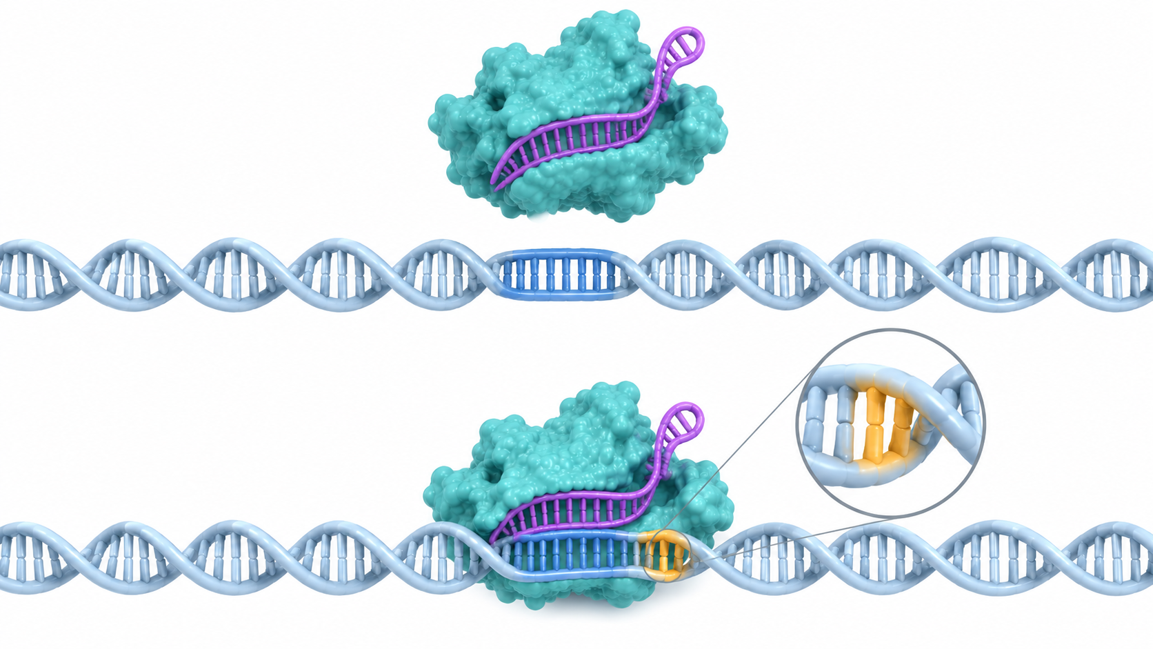 Top: no PAM → not recognized as a target
Bottom: PAM present → binding possible
5. A materials engineer's view (1): the two raw materials are a chemically synthesised product and a recombinant protein
As a product, CRISPR-Cas9 rests on two raw materials: the guide RNA and Cas9. For CASGEVY, the EMA public assessment report treats these two as “two critical starting materials” and evaluates the manufacture, specification and stability of each independentlySourced.
| Item | Cas9 (cutting enzyme) | Guide RNA (SPY101) |
|---|---|---|
| What kind of substance | A protein derived from S. pyogenes. The host-cell DNA is assessed as of prokaryotic origin | A synthetic oligonucleotide (100-mer), supplied as a freeze-dried powder. The FDA review summary writes “100 base pairs” |
| Starting material and how it is made | Protein production and purification (details are in the undisclosed part) | The starting materials are phosphoramidites, made by solid-phase chemical synthesis |
| Chemical modification | — | 2′-O-methylation and phosphorothioate linkages at both ends to resist degradation by nucleases. The phosphorothioate is a mixture of stereoisomers (diastereomers), because coupling and oxidation are not stereospecific |
| Specification tests | Identity, content, purity, host-cell protein (HCP), monomer, functionality (activity), endotoxin, bioburden / sterility | A functionality test (can it guide Cas9 to the target and cut?) and control of organic impurities. Purity was improved by optimising synthesis and purification conditions |
| Storage (proposed shelf life) | 24 months at −75°C for both bulk and aseptically filled vials | Bulk (freeze-dried) 18 months at −20°C; aseptically filled vials 18 months at −70°C |
| Container | Bottles for bulk and vials for aseptically filled product. Extractables and leachables data have been submitted for the vials and caps | Bulk in HDPE bottles with PP caps; aseptically filled product in vials (stopper and flip-off cap) |
| Reference standards | A two-tier system is planned: a primary reference standard (PRS) plus a working reference standard (WRS). A new WRS is verified against the PRS to avoid drift in activity | — |
All Sourced (EMA public assessment report [Ref. 27], FDA review summary SBRA [Ref. 25]). Storage periods are those “proposed” at the time of the assessment report. The manufacturer's name and detailed process steps are in the undisclosed (redacted) parts.
The mRNA described in this series' article on mRNA is a long RNA made by an enzymatic reaction from a template DNA (in vitro transcription). Guide RNA is different. Its starting materials are phosphoramidites, and it is a modified oligonucleotide assembled by solid-phase synthesisSourced. Even with the same word “RNA”, manufacture splits into an enzymatic process and an organic-synthesis process (our commentary).
- End protection as a materials-design choice: methylation at the 2′ position and phosphorothioate linkages are placed at both ends to protect against nucleasesSourced. Hendel and colleagues reported that chemically modified synthetic sgRNA raises editing efficiency in primary human T cells and CD34-positive haematopoietic stem and progenitor cellsSourced. FDA guidance also says that guide RNA can be optimised to resist degradationSourced
- Writing a specification around a mixture of stereoisomers: because phosphorothioate synthesis does not control stereochemistry, the product is explicitly described as a mixture of diastereomersSourced. One reading is that the specification is built on the premise of controlling the distribution of the mixture rather than making a single molecular species (our commentary)
- Cas9 is both an “enzyme” and a “raw material”: host-cell protein (HCP), endotoxin and bioburden are controlled, and a commercial HCP-ELISA kit is used to measure HCPSourced. That activity (functionality) is a specification item, and drift in it is monitored with reference standards is the same thinking as drug-substance control for biologics (our commentary)
- Not meant to remain in the final product: the FDA review summary states that process validation demonstrated removal of process-related impurities including Cas9 and SPY101Sourced. So these raw materials are held to purity control in two directions: “put in at high purity, then remove cleanly” (our commentary)
FDA guidance frames this positioning as follows. When genome editing is done ex vivo, the editing components are “critical components” of the final cell product, whereas when they are administered directly in vivo, the editing components themselves are the drug substance (or drug product)Sourced. Each component is recommended to be tested for sterility, identity, purity and activity, and its stability evaluatedSourced. For later-stage trials and marketing applications, manufacture of the components must follow CGMPSourced.
6. Editing without cutting: base editing and prime editing
Methods that cut the double strand have a weakness: the outcome of the repair cannot be fully controlled (Section 2). That led to the development of methods that rewrite DNA without making a cut. The PMDA report also says that, to reduce genome instability, genome editing that does not cut the genome (single-base editing with a deaminase) and similar methods are being developedSourced.
| Method | What the paper abstract says (results in cultured cells and similar systems) |
|---|---|
| Cytosine base editing (Komor et al. 2016) | A fusion of Cas9 and a cytidine deaminase. Needs neither a double-strand break nor a template DNA; it converts cytidine to uridine, producing C to T (G to A). It converts cytosines within a window of about 5 nucleotides. With the second- and third-generation editors, about 15 to 75% of total DNA was permanently corrected in four types of cultured cells, with indels usually at 1% or below |
| Adenine base editing (Gaudelli et al. 2017) | Converts A•T to G•C. A tRNA adenosine deaminase, evolved to act on DNA, is fused to an impaired Cas9. The seventh-generation editor reached an efficiency of about 50% in human cells, with product purity usually 99.9% or higher and indels usually 0.1% or below |
| Prime editing (Anzalone et al. 2019) | A fusion of an impaired Cas9 and an engineered reverse transcriptase, driven by a pegRNA that encodes both the target and the content of the new sequence. In human cells it carried out more than 175 edits (insertions, deletions and all 12 types of point mutation) without double-strand breaks or template DNA. The authors state that, “in principle, prime editing could correct up to 89% of known genetic variants associated with human disease” |
All Sourced (abstracts of References 12, 13 and 14). Figures are for the experimental conditions each paper reported. “Up to 89%” is the share the paper described as correctable in principle, not a rate achieved in actual treatment.
Examples of use in humans without cutting have also begun to appear. A case reported in NEJM in 2025 involved one infant with a severe inherited metabolic disease (CPS1 deficiency), who received two doses of a base-editing therapy made to match the patient and delivered in lipid nanoparticles. The authors say that no serious adverse events occurred in the 7 weeks after the first dose, but that “longer follow-up is needed to assess safety and efficacy”Sourced. This is a single-patient report and does not show general efficacy or safety. A phase 1/2 trial of base editing (BEAM-101) in sickle cell disease (NCT05456880) is also registered on ClinicalTrials.gov as “Active, not recruiting”Sourced (checked on 1 October 2026).
7. How to deliver it: electroporation, lipid nanoparticles and AAV
The editing components work only if they get inside the cell. FDA guidance says that ex vivo, they can be introduced by transfection, transduction, electroporation and similar methods as DNA, RNA, protein, or a ribonucleoprotein (RNP). In the body, they are delivered by viral vectors or nanoparticlesSourced.
- 1. Ex vivo, RNP by electroporation: the CASGEVY prescribing information states that the patient's CD34-positive cells are edited by introducing a CRISPR/Cas9 ribonucleoprotein (RNP) complex by electroporationSourced. Kim and colleagues introduced purified recombinant Cas9 protein and guide RNA into cultured human cells (including hard-to-transfect fibroblasts and pluripotent stem cells) and reported mutation rates of up to 79% and fewer off-target mutations than with plasmid deliverySourced. Hendel and colleagues also state that chemically modified synthetic sgRNA combined with Cas9 mRNA or protein is an efficient method without the toxicity that accompanies DNA deliverySourced
- 2. In vivo, lipid nanoparticles: the 2021 report by Gillmore and colleagues (NTLA-2001) was a phase 1 trial in six people with hereditary transthyretin amyloidosis, who received a single intravenous dose of a formulation in which Cas9 mRNA and sgRNA are encapsulated in lipid nanoparticles. Adverse events were mild, and serum TTR on day 28 fell by a mean of 52% (0.1 mg/kg group) and 87% (0.3 mg/kg group)Sourced. This is a result from a small early-stage study; details are in this series' article on transthyretin amyloidosis. For the design of the LNP itself, see this series' article on lipid nanoparticles
- 3. In vivo, AAV: because of the AAV payload limit, Ran and colleagues put SaCas9 (more than 1 kb shorter than SpCas9) into AAV and obtained more than 40% modification of the Pcsk9 gene in mouse liverSourced. AAV capacity is also a point discussed in this series' article on viral vectors
8. A materials engineer's view (2): “how long it persists” becomes a design variable
FDA guidance states that the longer the genome-editing components remain functional, the greater the risk of unintended genomic changes (off-target editing and chromosomal rearrangements), and that “the duration of the components should be minimised, as far as possible, to the time needed for the intended edit”Sourced. The same guidance sets out that viral vectors can support sustained expression, whereas nanoparticles can deliver the components transientlySourced. The PMDA report likewise says that sustained expression of a genome-editing enzyme raises the risk of off-target activity, so when a viral vector is used the persistence of expression must be analysedSourced.
In other words, “delivered as DNA, as RNA or as protein (RNP)” is a choice among three material forms, with different lifetimes, in which to wrap the same sequence information (our commentary). Kim and colleagues state that RNP “cleaves chromosomal DNA almost immediately after introduction and is rapidly degraded in cells”, which leads to fewer off-target effectsSourced.
This shows the conditions that materials design has to satisfy at the same time. The ends of the guide RNA are made resistant to degradation (Section 5), yet once inside the cell the complex is wanted to disappear quickly. The balance between storage stability and lifetime in the body is thus built in on the materials side: “do not break before delivery, do not linger after it” (our commentary).
From the round numbers in the paper by Ran and colleagues, we calculate how much room is left in the payloadOur calculation.
- Assumptions: the AAV payload is about 4.5 kb and SpCas9 is about 4.2 kb (Ran et al. 2015). SaCas9 is more than 1 kb shorter than SpCas9
- With SpCas9: 4.5 − 4.2 = about 0.3 kb. The promoter, polyA and the guide RNA expression cassette and so on must all fit in this
- With SaCas9: about 4.2 − 1 = about 3.2 kb or less, so the remainder is 4.5 − 3.2 = about 1.3 kb or more
Assumptions and limits: all the figures are “round numbers” from the text of the paper, and the real payload varies with vector design. Ran and colleagues state that putting SpCas9 and an sgRNA into a single AAV is “technically feasible, but leaves little room for expression and regulatory elements”Sourced. The subtraction above is meant to show the order of magnitude of that lack of room.
9. Approval status and where clinical use stands
Among genome-editing products, the one approved by regulators, within the scope this article checked, is CASGEVY (exagamglogene autotemcel, Vertex Pharmaceuticals). The details of the diseases, clinical trial results and the issues around pre-treatment conditioning are covered in this series' article on genome-editing therapy for blood disorders. Here we set out only the facts of approval.
| Regulator | Date | What was approved |
|---|---|---|
| UK MHRA | 16 November 2023 | Approved for sickle cell disease and transfusion-dependent β-thalassaemia in patients aged 12 and over. The MHRA described it as the first medicine to use CRISPR |
| US FDA | 8 December 2023 | Sickle cell disease (with recurrent vaso-occlusive crises), aged 12 and over (BLA STN 125787). The FDA described it as the first FDA-approved treatment to use CRISPR/Cas9 |
| US FDA | 16 January 2024 | Transfusion-dependent β-thalassaemia, aged 12 and over (BLA STN 125785) |
| EU | 9 February 2024 | Conditional marketing authorisation (the CHMP positive opinion was on 14 December 2023). Aged 12 and over. As of the version updated on 19 February 2026, the EMA's public page says nothing about conversion to a standard authorisation or an extension of the age range |
| US FDA | 1 July 2026 | Indication extended to include ages 2 to under 12 (approval letter STN BL 125787/457). The current prescribing information says “aged 2 and over” |
| Japan | — | No approved product using genome editing could be confirmed within the scope of this article's search of official PMDA and Ministry of Health, Labour and Welfare information |
For the FDA, see its official pages, approval letters and review summary [Refs. 23, 24, 25, 29 and 30]; for the MHRA, the GOV.UK announcement [Ref. 31]; for the EU, the EMA public page and public assessment report [Refs. 26 and 27] (Sourced). “Could not be confirmed” for Japan is a limit of the search scope and does not mean that none exists.
For Cas9 and the guide RNA (SPY101), the EMA public assessment report separately evaluates the manufacturing process, specification, analytical method validation, stability and container compatibility, and lists minor unresolved quality points as post-approval commitmentsSourced. The FDA review summary likewise records that the manufacturing processes of Cas9 and SPY101 themselves were subject to verificationSourced. So approval extends beyond the “edited cells” to the quality systems of the two raw materials used for the editing (our commentary). From the raw-material supplier's side, having a complete set of GMP-compliant manufacture, the basis for specifications, reference standards and stability data is a condition of entry.
Main clinical examples other than approvals (as far as could be confirmed on 1 October 2026)
| Example | Editing method and delivery | What the published material says |
|---|---|---|
| CCR5 editing with ZFN (Tebas et al. 2014, NCT00842634) | ZFN. CD4 T cells from people with HIV are edited ex vivo and returned | 12 people enrolled. Concluded that “infusion of CCR5-modified autologous CD4 T cells is safe within the scope of this study” |
| NTLA-2001 (Gillmore et al. 2021, NCT04601051) | CRISPR-Cas9. Cas9 mRNA and sgRNA delivered into the body in LNP | 6 people. Adverse events were mild. Serum TTR on day 28 fell in a dose-dependent way. The phase 1 status on ClinicalTrials.gov is “COMPLETED” |
| Patient-tailored base editing (Musunuru et al. 2025) | Base editing. Delivered into the body in LNP | One infant with CPS1 deficiency. “Longer follow-up is needed” |
| BEAM-101 (NCT05456880) | Base-edited autologous CD34-positive cells | A phase 1/2 trial in sickle cell disease. Status on ClinicalTrials.gov: “Active, not recruiting” |
All Sourced (References 18, 19, 32 and 33). Products in development are Not yet confirmed and nothing here indicates a prospect of approval.
10. Open problems, and what is not yet settled
(1) Methods that cut cannot fully control the outcome of the repair
FDA guidance lists as risks specific to genome editing off-target editing, unintended consequences of on-target editing, and the fact that the long-term effects of editing are unknown, and notes that repair of a DSB can also cause chromosomal rearrangements (including translocations)Sourced. The PMDA report states that, with methods that involve a DSB, there are reports of large chromosomal deletions that conventional assessment methods cannot detect, and insertion of unintended sequences at the cut siteSourced. Specific detection methods, mitigation measures and the regulators' thinking are covered in the next article, “Off-target effects and safety assessment”.
(2) Immunogenicity of the editing enzyme itself
The PMDA report says that, because Cas proteins and the like are bacterial proteins, then even for cells edited ex vivo, if the cells express the editing enzyme they may be recognised in the body as a foreign antigen. Because animal studies are poor at predicting human immunogenicity, clinical trials need to be planned with weakening of the clinical effect through an immune reaction, and anaphylaxis, in mindSourced.
(3) With in vivo editing, cells other than the target are hard to check or remove
The PMDA report states that with in vivo genome editing it is difficult to confirm or remove edits in cells other than the target cells, or changes to genes other than the intended one, and that effects on germ cells are a particular concern for children and for patients of reproductive ageSourced.
(4) Long-term follow-up
Because the long-term effects of editing are unknown at the time of treatment, FDA guidance recommends long-term follow-up for up to 15 years after administrationSourced. In the approval of CASGEVY too, the FDA made a 15-year observational study a mandatory post-marketing requirement [Ref. 25]Sourced.
(5) What could not be confirmed
Whether public standards common to genome-editing components (Cas9 and guide RNA), such as pharmacopoeial monographs, have been established could not be confirmed within the scope of this article. Regulators' documents are worded so as to require that a specification be set product by product, with its basis shown (FDA guidance). Also, whether any product using genome editing has been approved in Japan could not be confirmed from official information.
- Genome editing is a technology that cuts DNA and uses repair to rewrite it. The repair pathways after a cut are NHEJ and HDR; NHEJ is used to disrupt a gene and HDR to replace a sequenceSourced
- The axis of progress is the swapping of the part that recognises the site. ZFN and TALEN recognise with protein; CRISPR-Cas9 recognises with a guide RNA. The PMDA notes that CRISPR is easy to design and low in costSourced
- “Editing without cutting” has been added. Base editing makes single-base conversions of C to T or A to G, and prime editing makes insertions, deletions and all 12 point mutations, both without a double-strand breakSourced
- The editing components of a product are two raw materials. The guide RNA is a 100-mer synthetic oligonucleotide (modified at both ends) and Cas9 is a recombinant protein, and the specification, stability and reference standards of each are evaluated separatelySourced
- The “form” of the component is a choice of lifetime. RNP, RNA and DNA are said to persist in the cell for progressively longer, and the FDA asks that the duration be minimisedSourced
- Putting SpCas9 in a single AAV leaves only about 0.3 kb. Our calculation
- CASGEVY is the only approved product (within what was checked on the FDA list)Sourced. No approved product in Japan could be confirmed
11. Glossary
- Genome editing
- Technology that adds, removes, alters or replaces DNA sequences at specific locations in the genome.
- Double-strand break (DSB)
- A break in both strands of DNA at the same place. The starting point of genome editing.
- NHEJ
- Non-homologous end joining. Repair that rejoins broken ends without a template; it often adds or loses a few bases.
- HDR
- Homology-directed repair, which uses homologous recombination. A template DNA can be used to replace a sequence. Most active in S and G2 phase.
- Indel
- Collective term for insertions and deletions.
- ZFN
- Zinc finger nuclease. An engineered enzyme linking zinc-finger proteins to FokI.
- TALEN
- TAL effector nuclease. An engineered enzyme linking the repeat sequences of a TAL effector to FokI.
- CRISPR-Cas9
- An RNA-guided DNA-cutting system derived from bacterial adaptive immunity.
- Guide RNA (sgRNA)
- The RNA that leads Cas9 to its target. It carries a sequence of about 20 bases complementary to the target.
- PAM
- Protospacer adjacent motif. A short sequence required next to the target; Cas9 recognises it before cutting.
- RNP
- Ribonucleoprotein complex. Here, Cas9 protein pre-assembled with its guide RNA.
- Electroporation
- A method of introducing substances by opening temporary pores in the cell membrane with electric pulses.
- Base editing
- A method that uses a deaminase to convert one base into another without a double-strand break.
- Prime editing
- A method that writes a new sequence using a fusion of an impaired Cas9 and a reverse transcriptase, together with a pegRNA.
- Ex vivo / in vivo
- Editing cells outside the body and returning them / administering the editing components directly into the body.
- Off-target
- Unintended editing at a site other than the intended one, where a similar sequence exists.
12. References (primary sources)
- Nobel Foundation “The Nobel Prize in Chemistry 2020 — Press release”, 7 October 2020 — nobelprize.org
- Jinek M et al. “A programmable dual-RNA-guided DNA endonuclease in adaptive bacterial immunity”, Science 337(6096):816-821 (2012). doi:10.1126/science.1225829 — pubmed.ncbi.nlm.nih.gov
- Cong L et al. “Multiplex genome engineering using CRISPR/Cas systems”, Science 339(6121):819-823 (2013). doi:10.1126/science.1231143 — pubmed.ncbi.nlm.nih.gov
- Mali P et al. “RNA-guided human genome engineering via Cas9”, Science 339(6121):823-826 (2013). doi:10.1126/science.1232033 — pubmed.ncbi.nlm.nih.gov
- Kim YG et al. “Hybrid restriction enzymes: zinc finger fusions to Fok I cleavage domain”, PNAS 93(3):1156-1160 (1996). doi:10.1073/pnas.93.3.1156 — pubmed.ncbi.nlm.nih.gov
- Bitinaite J et al. “FokI dimerization is required for DNA cleavage”, PNAS 95(18):10570-10575 (1998). doi:10.1073/pnas.95.18.10570 — pubmed.ncbi.nlm.nih.gov
- Boch J et al. “Breaking the code of DNA binding specificity of TAL-type III effectors”, Science 326(5959):1509-1512 (2009). doi:10.1126/science.1178811 — pubmed.ncbi.nlm.nih.gov
- Moscou MJ, Bogdanove AJ “A simple cipher governs DNA recognition by TAL effectors”, Science 326(5959):1501 (2009). doi:10.1126/science.1178817 — pubmed.ncbi.nlm.nih.gov
- Christian M et al. “Targeting DNA double-strand breaks with TAL effector nucleases”, Genetics 186(2):757-761 (2010). doi:10.1534/genetics.110.120717 — pubmed.ncbi.nlm.nih.gov
- Miller JC et al. “A TALE nuclease architecture for efficient genome editing”, Nat Biotechnol 29(2):143-148 (2011). doi:10.1038/nbt.1755 — pubmed.ncbi.nlm.nih.gov
- Sternberg SH et al. “DNA interrogation by the CRISPR RNA-guided endonuclease Cas9”, Nature 507(7490):62-67 (2014). doi:10.1038/nature13011 — pubmed.ncbi.nlm.nih.gov
- Komor AC et al. “Programmable editing of a target base in genomic DNA without double-stranded DNA cleavage”, Nature 533(7603):420-424 (2016). doi:10.1038/nature17946 — pubmed.ncbi.nlm.nih.gov
- Gaudelli NM et al. “Programmable base editing of A•T to G•C in genomic DNA without DNA cleavage”, Nature 551(7681):464-471 (2017). doi:10.1038/nature24644 — pubmed.ncbi.nlm.nih.gov
- Anzalone AV et al. “Search-and-replace genome editing without double-strand breaks or donor DNA”, Nature 576(7785):149-157 (2019). doi:10.1038/s41586-019-1711-4 — pubmed.ncbi.nlm.nih.gov
- Hendel A et al. “Chemically modified guide RNAs enhance CRISPR-Cas genome editing in human primary cells”, Nat Biotechnol 33(9):985-989 (2015). doi:10.1038/nbt.3290 — pubmed.ncbi.nlm.nih.gov
- Kim S et al. “Highly efficient RNA-guided genome editing in human cells via delivery of purified Cas9 ribonucleoproteins”, Genome Res 24(6):1012-1019 (2014). doi:10.1101/gr.171322.113 — pubmed.ncbi.nlm.nih.gov
- Ran FA et al. “In vivo genome editing using Staphylococcus aureus Cas9”, Nature 520(7546):186-191 (2015). doi:10.1038/nature14299 — pubmed.ncbi.nlm.nih.gov
- Gillmore JD et al. “CRISPR-Cas9 in vivo gene editing for transthyretin amyloidosis”, N Engl J Med 385(6):493-502 (2021). doi:10.1056/NEJMoa2107454 — pubmed.ncbi.nlm.nih.gov
- Musunuru K et al. “Patient-specific in vivo gene editing to treat a rare genetic disease”, N Engl J Med 392(22):2235-2243 (2025). doi:10.1056/NEJMoa2504747 — pubmed.ncbi.nlm.nih.gov
- Wu Y et al. “Highly efficient therapeutic gene editing of human hematopoietic stem cells”, Nat Med 25(5):776-783 (2019). doi:10.1038/s41591-019-0401-y — pubmed.ncbi.nlm.nih.gov
- US Food and Drug Administration (FDA) “Human Gene Therapy Products Incorporating Human Genome Editing — Guidance for Industry”, January 2024 (PDF) — fda.gov
- PMDA Science Board “Report on considerations for the quality and safety of gene therapy products using genome editing technology” (in Japanese), 7 February 2020 (PDF; the table of contents is at the start of the file) — pmda.go.jp
- US FDA “CASGEVY” product page (approval dates, indications, list of approval documents) — fda.gov
- US FDA “CASGEVY Prescribing Information”, revised July 2026 (PDF) — fda.gov
- US FDA CBER “Summary Basis for Regulatory Action — CASGEVY (BLA STN 125787)”, 8 December 2023 (PDF) — fda.gov
- EMA “Casgevy — European public assessment report (EPAR)” product page — ema.europa.eu
- EMA “Casgevy — EPAR Public assessment report”, EMA/6332/2024 (PDF) — ema.europa.eu
- US FDA “Approved Cellular and Gene Therapy Products” (checked 1 October 2026) — fda.gov
- US FDA “FDA Approves First Gene Therapies to Treat Patients with Sickle Cell Disease”, 8 December 2023 — fda.gov
- US FDA “CASGEVY approval letter (STN BL 125787/457)”, 1 July 2026 (PDF) — fda.gov
- UK MHRA “MHRA authorises world-first gene therapy that aims to cure sickle-cell disease and transfusion-dependent thalassemia”, 16 November 2023 — gov.uk
- Tebas P et al. “Gene editing of CCR5 in autologous CD4 T cells of persons infected with HIV”, N Engl J Med 370(10):901-910 (2014). doi:10.1056/NEJMoa1300662 — pubmed.ncbi.nlm.nih.gov
- ClinicalTrials.gov “BEACON: BEAM-101 in patients with severe sickle cell disease (NCT05456880)” — clinicaltrials.gov
13. Claim-to-source audit
| Claim in the text | Basis | Label |
|---|---|---|
| That human genome editing is “a process by which DNA sequences are added, removed, altered or replaced at specific locations in the genome of somatic cells”, covering both nuclease-dependent and nuclease-independent approaches. The explanation of HDR and NHEJ (NHEJ is relatively independent of the cell cycle, HDR is most active in S and G2 phase, and either can produce indels). That a DSB can cause chromosomal rearrangements (including translocations). The risks specific to genome editing (off-target editing, unintended consequences of on-target editing, unknown long-term effects). That ex vivo, components can be introduced as DNA, RNA, protein or RNP by electroporation and similar methods; in vivo, by viral vectors or nanoparticles. That the duration of the components should be minimised, and that viral vectors can support sustained expression whereas nanoparticles allow transient delivery. That guide RNA can be optimised to resist degradation. That the components are critical components in ex vivo editing and the drug substance or product in direct in vivo administration. That the components are tested for sterility, identity, purity and activity and their stability evaluated, and that CGMP is required in later-stage trials and at approval. That long-term follow-up is set at up to 15 years | FDA guidance “Human Gene Therapy Products Incorporating Human Genome Editing” (January 2024). Reference 21 https://www.fda.gov/media/156894/download | Sourced |
| That the repair pathways of a DSB are NHEJ and HDR, and that NHEJ can be used to disrupt genes and HDR for correction. That ZFN carries 3 to 6 zinc fingers, each recognising 3 bases, linked to FokI, and that its design needs advanced technology. That TALEN recognises one base per 34-amino-acid module and links 15 to 20 of them. That ZFN and TALEN recognise 18 to 40 bases as a pair of engineered enzymes, are said to have a lower off-target frequency than CRISPR/Cas9, but that information is insufficient and careful evaluation is needed. That CRISPR/Cas recognises with an sgRNA, is easy to design and low in cost, has a guide sequence complementary to 20 target bases plus a PAM, and can bind even with up to 5 mismatches. That genome editing without cutting the genome is being developed, and that even then the mechanism of reduction has to be explained. That in viral vectors, sustained enzyme expression increases the off-target risk. That Cas proteins are of bacterial origin and may be recognised as foreign antigens. That in vivo, edits outside the target are hard to confirm or remove and effects on germ cells are a concern. That large chromosomal deletions that conventional assessment cannot detect, and insertion of unintended sequences, have been reported with DSBs | PMDA Science Board “Report on considerations for the quality and safety of gene therapy products using genome editing technology” (7 February 2020; in Japanese). Reference 22 https://www.pmda.go.jp/files/000265952.pdf | Sourced |
| That the 2020 Nobel Prize in Chemistry was awarded to Charpentier and Doudna “for the development of a method for genome editing”, and that CRISPR/Cas9 is called “genetic scissors” and can change the code within a few weeks | Nobel Foundation press release (7 October 2020). Reference 1 https://www.nobelprize.org/prizes/chemistry/2020/press-release/ | Sourced |
| That Cas9 cuts the double strand guided by a pair of crRNA and tracrRNA (or a single chimeric RNA), with the HNH domain cutting the complementary strand and the RuvC-like domain the non-complementary strand | Abstract of Jinek et al. (2012). Reference 2 https://pubmed.ncbi.nlm.nih.gov/22745249/ | Sourced |
| That the CRISPR/Cas system works in human and mouse cells (2013), and that Mali et al. reported targeting rates in human genome editing including iPS cells, 2 to 4% in iPS cells | Abstract of Cong et al. (2013). Reference 3 https://pubmed.ncbi.nlm.nih.gov/23287718/; abstract of Mali et al. (2013). Reference 4 https://pubmed.ncbi.nlm.nih.gov/23287722/ | Sourced |
| That an engineered nuclease was made by linking two kinds of zinc-finger protein to the FokI cleavage domain. That FokI needs dimerisation to cut. The clinical trial of ZFN in humans (CCR5, 12 people, concluded safe) | Abstract of Kim et al. (1996). Reference 5 https://pubmed.ncbi.nlm.nih.gov/8577732/; abstract of Bitinaite et al. (1998). Reference 6 https://pubmed.ncbi.nlm.nih.gov/9724744/; abstract of Tebas et al. (2014). Reference 32 https://pubmed.ncbi.nlm.nih.gov/24597865/ | Sourced |
| That the TAL effector repeat is about 34 amino acids and two residues that vary from repeat to repeat determine one base. That the TALE–FokI fusion nuclease was reported in 2010 | Boch et al. (2009). Reference 7 https://pubmed.ncbi.nlm.nih.gov/19933107/; Moscou et al. (2009). Reference 8 https://pubmed.ncbi.nlm.nih.gov/19933106/; Christian et al. (2010). Reference 9 https://pubmed.ncbi.nlm.nih.gov/20660643/; Miller et al. (2011). Reference 10 https://pubmed.ncbi.nlm.nih.gov/21179091/ | Sourced |
| That binding and cleavage by Cas9–RNA need recognition of a three-base PAM, and that without a PAM even a fully complementary sequence is ignored | Abstract of Sternberg et al. (2014). Reference 11 https://pubmed.ncbi.nlm.nih.gov/24476820/ | Sourced |
| Base editing (C to T, window of about 5 nucleotides, about 15 to 75%, indels usually 1% or below), adenine base editing (A•T to G•C, about 50%, purity 99.9% or higher, indels 0.1% or below), prime editing (more than 175 edits, 12 types of point mutation, no double-strand break or template DNA needed, up to 89%) | Komor et al. (2016). Reference 12 https://pubmed.ncbi.nlm.nih.gov/27096365/; Gaudelli et al. (2017). Reference 13 https://pubmed.ncbi.nlm.nih.gov/29160308/; Anzalone et al. (2019). Reference 14 https://pubmed.ncbi.nlm.nih.gov/31634902/ | Sourced |
| That chemically modified synthetic sgRNA raises editing efficiency in primary human T cells and CD34-positive haematopoietic stem and progenitor cells, and that there is no toxicity from DNA delivery. That introducing purified Cas9 and guide RNA gave mutation rates of up to 79% and fewer off-target mutations, and that RNP cleaves almost immediately after introduction and is rapidly degraded | Abstract of Hendel et al. (2015). Reference 15 https://pubmed.ncbi.nlm.nih.gov/26121415/; abstract of Kim et al. (2014). Reference 16 https://pubmed.ncbi.nlm.nih.gov/24696461/ | Sourced |
| That the AAV payload is about 4.5 kb, SpCas9 about 4.2 kb, and SaCas9 more than 1 kb shorter than SpCas9. That SaCas9 and an sgRNA were put into a single AAV and gave more than 40% gene modification at Pcsk9 in mouse liver. The statement that putting SpCas9 in a single AAV is technically feasible but leaves little room | Abstract and text of Ran et al. (2015). Reference 17 https://pubmed.ncbi.nlm.nih.gov/25830891/ | Sourced |
| The phase 1 results of NTLA-2001 (Cas9 mRNA and sgRNA in LNP) given to six people with hereditary ATTR amyloidosis (mild adverse events, serum TTR down 52% and 87% on day 28) | Abstract of Gillmore et al. (2021). Reference 18 https://pubmed.ncbi.nlm.nih.gov/34215024/ | Sourced |
| The report of two doses of LNP-delivered base-editing therapy to one infant with CPS1 deficiency: no serious adverse events in 7 weeks, and longer follow-up needed | Abstract of Musunuru et al. (2025). Reference 19 https://pubmed.ncbi.nlm.nih.gov/40373211/ | Sourced |
| That cutting the GATA1 binding site of the BCL11A enhancer with a Cas–guide RNA RNP disrupts the motif, lowers BCL11A expression and induces fetal γ-globin. That haematopoietic stem cells favour NHEJ | Abstract of Wu et al. (2019). Reference 20 https://pubmed.ncbi.nlm.nih.gov/30911135/ | Sourced |
| That the Cas9 used for CASGEVY is from S. pyogenes. That the SPY101 guide RNA is a synthetic oligonucleotide of “100 base pairs”. That process validation demonstrated removal of process-related impurities including Cas9 and SPY101. That a 15-year post-marketing observational study was made mandatory for CASGEVY | FDA review summary (SBRA, 8 December 2023). Reference 25 https://www.fda.gov/media/175179/download?attachment | Sourced |
| That Cas9 and SPY101 are two critical starting materials. That the host DNA of Cas9 is of prokaryotic origin, the specification tests (identity, content, purity, HCP, monomer, functionality, endotoxin, bioburden / sterility), the HCP-ELISA kit, the 24-month shelf life at −75°C, bulk in bottles and aseptically filled product in vials, and the two-tier reference standards. That SPY101 is a 100-mer synthetic oligonucleotide (freeze-dried powder) whose starting materials are phosphoramidites, that the phosphorothioate is a mixture of diastereomers, the 2′-O-methylation and phosphorothioate linkages at both ends, the optimisation for purity, 18 months at −20°C for bulk and −70°C for aseptically filled product, and HDPE bottles with PP caps. That the most active PAMs for SpCas9 are NGG and NAG. The EU conditional marketing authorisation and the CHMP positive opinion (14 December 2023) | EMA public assessment report EMA/6332/2024. Reference 27 https://www.ema.europa.eu/en/documents/assessment-report/casgevy-epar-public-assessment-report_en.pdf | Sourced |
| The approval dates of CASGEVY (FDA: 8 December 2023 and 16 January 2024, STN 125787 and 125785), the indications, and the list of approval documents | FDA CASGEVY product page. Reference 23 https://www.fda.gov/vaccines-blood-biologics/casgevy | Sourced |
| That CASGEVY is a product in which autologous CD34-positive haematopoietic stem cells are edited with CRISPR/Cas9, that editing is done by introducing a CRISPR/Cas9 RNP complex by electroporation, and that the indication in the current prescribing information (revised July 2026) is aged 2 and over | FDA prescribing information. Reference 24 https://www.fda.gov/media/174615/download?attachment | Sourced |
| That the FDA announced it as the first FDA-approved treatment to use CRISPR/Cas9 (8 December 2023) | FDA announcement. Reference 29 https://www.fda.gov/news-events/press-announcements/fda-approves-first-gene-therapies-treat-patients-sickle-cell-disease | Sourced |
| That the indication was extended to ages 2 to under 12 on 1 July 2026 (STN BL 125787/457) | FDA approval letter. Reference 30 https://www.fda.gov/media/193444/download?attachment | Sourced |
| That, within what this article checked from the product names and nonproprietary names on the FDA list of approved cellular and gene therapy products, CASGEVY is the only product using CRISPR/Cas9 (checked on 1 October 2026; the list itself has no genome-editing category) | FDA list of approved products. Reference 28 https://www.fda.gov/vaccines-blood-biologics/cellular-gene-therapy-products/approved-cellular-and-gene-therapy-products; FDA announcement (Reference 29). This is this article's own check, and because the list has no such category it does not guarantee that there are no other genome-editing products | Commentary |
| That the UK MHRA approved CASGEVY on 16 November 2023 for ages 12 and over and described it as the first medicine to use CRISPR | MHRA announcement. Reference 31 https://www.gov.uk/government/news/mhra-authorises-world-first-gene-therapy-that-aims-to-cure-sickle-cell-disease-and-transfusion-dependent-thalassemia | Sourced |
| The EU marketing authorisation date (9 February 2024), that it is a conditional authorisation, and that as of the version updated on 19 February 2026 there is no mention of conversion to a standard authorisation or an extension of the age range | EMA CASGEVY product page. Reference 26 https://www.ema.europa.eu/en/medicines/human/EPAR/casgevy | Sourced |
| That BEAM-101 is a phase 1/2 trial in sickle cell disease using base-edited autologous CD34-positive cells, with a status of “Active, not recruiting” on ClinicalTrials.gov (checked on 1 October 2026) | ClinicalTrials.gov NCT05456880. Reference 33 https://clinicaltrials.gov/study/NCT05456880 | Sourced |
| Whether products in development (BEAM-101 and others) will be approved in future | The status of the trials could be confirmed, but the prospect of approval cannot be judged from published material and is not discussed in this article | Not yet confirmed |
| That the remainder when SpCas9 is put into AAV is 4.5 − 4.2 = about 0.3 kb, and for SaCas9 about 1.3 kb or more | Our calculation. The AAV figure of about 4.5 kb, SpCas9 at about 4.2 kb and SaCas9 being more than 1 kb shorter are round numbers from Ran et al. used as assumptions for a subtraction; it does not show the real margin in any vector design | Our calculation |
| Whether any product using genome editing has been approved in Japan | It could not be confirmed in official PMDA and Ministry of Health, Labour and Welfare information, so the article says “could not be confirmed”. This does not mean that none exists | Commentary |
| Whether public standards common to genome-editing components (pharmacopoeial monographs and so on) have been established | Not stated in this article because it could not be confirmed within its scope | Commentary |
| Sorting the three families of tool by what carries out recognition. The contrast that guide RNA is a solid-phase synthetic product unlike enzymatically made mRNA. The reading about a specification for a mixture of diastereomers. The reading that Cas9 control follows the same thinking as drug-substance control for biologics. The framing of DNA, RNA and RNP forms as a choice of lifetime. The reading about the balance between storage stability and lifetime inside the cell. The reading that approval extends to the quality systems of the raw materials | This article's own framing and commentary based on published content. Not views expressed by the institutions | Commentary |
| That Figs. 1, 2, 3, 5, 6 and 7 are explanatory drawings, and that the hero image and Fig. 4 are AI-generated images | A note by this article | Commentary |
Last updated 1 October 2026. Sources are limited to primary material (published documents from regulators, public assessment reports, peer-reviewed papers, and the Nobel Foundation's announcement). Because the article includes structural readings and materials- and process-design interpretations, those are marked as Commentary and kept separate from sourced fact. Whether any product is approved in Japan, public standards common to genome-editing components, and the prospects of approval for products in development are not asserted, because they could not be confirmed in published primary sources. Facts on approval are limited to what could be confirmed on the official FDA, EMA and MHRA pages on 1 October 2026. All figures are concept diagrams for explanation. Figs. 1, 2, 3, 5, 6 and 7 are vector drawings; the hero image and Fig. 4 are AI-generated images, and none of them shows a real molecular structure, a microscope image or an actual product.