TECHNOLOGY EXPLAINER
Theranostics and Nano Contrast Agents
— How Far Has “Diagnosis and Therapy in One” Become a Product?
Theranostics is the idea of combining diagnostics and therapy. The best-established examples are in fact not nanoparticles but pairs of radiopharmaceuticals (NETSPOT, approved by the FDA in 2016, and Lutathera, approved in 2018). Among nanoparticles, we could confirm iron oxide liver MRI contrast (Resovist, approved in Japan in 2002) and a magnetic particle tracer for locating lymph nodes (Magtrace, approved by the FDA in 2018), but we could not confirm any approval of quantum dots or gold nanoparticles as in vivo contrast agents within the scope of our research.

- What theranostics is (the short version)
- The practical core is a pair that is “not nano” — radiopharmaceuticals
- The idea of combining them in a nanoparticle, and kinds of contrast agent
- How iron oxide darkens the liver on MRI
- Examples with documented approval or authorisation
- A materials engineer’s view (1): there are three kinds of particle size — core, coated, colloid
- Biodistribution and safety — excreted, or taken up by the reticuloendothelial system
- Our calculation: contrast and therapy differ by orders of magnitude in particle “amount”
- Examples at the trial and research stage
- A materials engineer’s view (2): why combining is hard
- What is still open, and what we could not confirm
- Glossary / Primary sources / Claim-to-source audit
Sourced = content stated in published material (link given)
Our calculation = a figure this article derived, with the assumptions spelled out
Not yet confirmed = a plan, outlook or research-stage item with no confirmed record
Readings and interpretations are marked separately as Commentary.
This article is an explanation of materials technology. It does not evaluate or compare the efficacy or safety of individual products and is not diagnostic or therapeutic advice. Statements from labels and approval documents are quoted only to confirm what a material is made of and the fact of approval; they do not indicate whether or how a product should be used. Use of any product is a matter for a physician. Results from animal studies or small trials do not necessarily apply to people.
1. What theranostics is (the short version)
- The idea: Lammers and colleagues call nanomedicines that contain both a drug and an imaging agent in a single formulation “(nano) theranostics”, and summarise their advantages as being able to follow the drug’s destination, release and effect by imaging, and to help pre-screen patientsSourced.
- What is actually a product: radiopharmaceuticals, in which a diagnostic and a therapeutic agent are used as a pair against the same target (the somatostatin receptor). They are small molecules or peptides carrying a radionuclide, not nanoparticlesSourced.
- For nanoparticles: what we could confirm as approved or authorised is limited to a few iron oxide particles (liver MRI contrast, lymph-node localisation) and similar; a product that does diagnosis and therapy in one particle could not be confirmed within the scope of our research.
“An iron oxide nanoparticle approved as a contrast agent” and “an iron oxide nanoparticle approved as a drug” are different products. The iron replacement product Feraheme is a superparamagnetic iron oxide with a particle size of 17 to 31 nm, but the FDA label’s indication is iron deficiency anemia and no imaging indication is written. If anything, the label cautions that it “can alter” MRI test resultsSourced. Even within one family of materials, they should be treated as separate things according to the use for which each was approved (Commentary).
2. The practical core is a pair that is “not nano” — radiopharmaceuticals
Two products already use the idea of pairing diagnosis and therapySourced.
- NETSPOT (a kit for preparation of gallium Ga 68 dotatate injection): approved by the FDA on 1 June 2016. After radiolabelling, it is a diagnostic used with positron emission tomography (PET) to localise somatostatin-receptor-positive neuroendocrine tumours.
- Lutathera (lutetium Lu 177 dotatate): approved by the FDA on 26 January 2018 (the date of the electronic signature on the approval letter). It is a radiolabelled somatostatin analog used to treat somatostatin-receptor-positive gastroenteropancreatic neuroendocrine tumours.
Both are molecules that bind the same receptor, carrying a diagnostic or a therapeutic radionuclide. This is a working example of the theranostic idea: “for a patient whose receptor was visible on the diagnostic, use the therapeutic version of the same molecule” (Commentary). Nanoparticle theranostics tries to do the same with a particle as the carrier, but as Section 9 shows, most of it remains at the trial or research stage.
3. The idea of combining them in a nanoparticle, and kinds of contrast agent
The advantages of nanoparticles as contrast agents are that surface modification can change where they go in the body, and that one particle can carry both an imaging signal source and a drug. The main materials are as follows.
| Material | Imaging signal source | Status we could confirm |
|---|---|---|
| Superparamagnetic iron oxide | Lowers the signal on MRI (T2 shortening). Also detectable with a magnetic probe | Documented approval as contrast agent and tracer (Section 5) |
| Ultrasmall silica particles (C dots) | Fluorescence (Cy5 dye) and radioiodine 124I for PET | First-in-human trial reported in 2014 (Section 9) |
| Quantum dots | Fluorescence | Phillips et al. state that clinical translation of nanoparticle probes, including quantum dots, has not kept pace with minimally invasive surgical toolsSourced. No approval as an in vivo contrast agent confirmed |
| Gold nanoparticles | CT, photoacoustic, SERS and others (research) | No approval as an in vivo contrast agent confirmed. For in vitro diagnostic labels, see the explainer on nanoparticles and nanozymes |
The arrangement of materials and signal sources is ours (Commentary). The toxicity of quantum dots is covered in this series’ explainer on nanoparticles and nanozymes.
4. How iron oxide darkens the liver on MRI
Superparamagnetic iron oxide liver contrast agents make use of the fact that particles are taken up by the liver’s phagocytes (Kupffer cells). The PMDA label for Resovist (ferucarbotran) explains that superparamagnetic iron oxide microparticles coated with carboxydextran are taken up promptly, mainly by Kupffer cells of the liver, shorten the transverse relaxation time (T2) of protons in tissue and so lower the MR signal, which gives the contrast effectSourced.
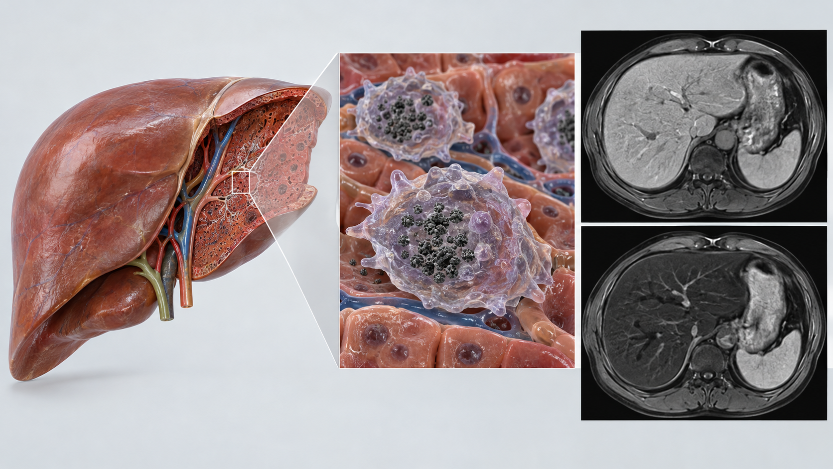
Shapes, colors and dimensions are illustrative. Consult the original schematic below for exact values and label correspondence.
View the original SVG schematic and exact annotations
Reading guide and original schematic labels: How iron oxide nanoparticles “darken” the liver on T2-weighted MRI (conceptual) / lesion / Blue dots = Kupffer cells (liver phagocytes) that took up particles / The lesion has few Kupffer cells and takes up few particles / lesion (relatively bright) / normal liver (darkens) / T2-weighted image: normal liver with particles darkens / Lesions with Kupffer cells may appear indistinct / * Explanatory schematic, not an actual image or particle size. Mechanism per the PMDA label and a review (Wang 2011). / * Lesion signal has exceptions, and diagnosis is for physicians. This article gives no diagnostic advice.
The review says these particles produce strong T2/T2* relaxation effects. The label also states that the contrast effect on T2-weighted images is seen from 10 minutes after administration and lasts up to 8 hours, while the effect on liver signal in T1-weighted images is not seen at 48 hoursSourced.
5. Examples with documented approval or authorisation
The table below separates what we could confirm in official documents (labels, approval documents) from what we could confirm only from company statements.
| Product | Particle material | Use | Documented approval or authorisation | Published particle values |
|---|---|---|---|---|
| Resovist (ferucarbotran) | Superparamagnetic iron oxide coated with carboxydextran | Liver contrast for MRI localisation of liver tumours | Japan: approved 8 October 2002, marketing began December 2002. Label revised March 2022 (PMDA) | Hydrodynamic diameter about 45 to 60 nm (review). Dose 0.45 mg iron/kg |
| Feridex (ferumoxides) | Superparamagnetic iron oxide coated with dextran | Liver MRI | The review says it was approved in the US, Europe and Japan (we did not check a primary approval document) | 120 to 180 nm (review) |
| Magtrace (used with the Sentimag magnetic probe) | Superparamagnetic iron oxide coated with carboxydextran | Locating lymph nodes in sentinel lymph node biopsy in breast cancer patients | FDA premarket approval (PMA) P160053, 24 July 2018 | Iron oxide core 3.5 to 10 nm; whole particle after coating 45 to 65 nm (FDA approval documents) |
| Feraheme (ferumoxytol) | Non-stoichiometric magnetite coated with polyglucose sorbitol carboxymethylether | Treatment of iron deficiency anemia (no imaging indication) | FDA initial approval 2009; label revised May 2026 | Whole colloidal particle 17 to 31 nm (FDA label) |
| NanoTherm (MagForce Nano-Cancer therapy) | Magnetic iron oxide nanoparticles (injected into the tumour and heated in an alternating magnetic field) | Treatment of brain tumours (thermotherapy) | The company announced EU regulatory approval on 28 June 2010 (no regulator document checked) | Not found |
| Hensify (NBTXR3) | Hafnium oxide nanoparticles | Radioenhancer in locally advanced soft tissue sarcoma (therapy) | The company announced a CE mark on 4 April 2019 (no regulator document checked) | Not found |
Resovist, Magtrace and Feraheme: Sourced (PMDA and FDA documents, Sources 1 to 4). Feridex and some particle sizes: from a review (Source 5). NanoTherm and Hensify: company statements (Sources 15 and 16). The arrangement of the table is ours.
- No approved product in the table does diagnosis and therapy in one particle. NanoTherm and Hensify are therapies, Resovist and Magtrace are diagnostic or localisation, and Feraheme is iron replacement.
- We confirmed that the Resovist label is posted by PMDA, but we have not confirmed whether it is still supplied. Likewise we could not confirm the current supply status of Feridex.
- Particle-size values come from sources whose methods and conditions differ. They should not be ranked against each other.
6. A materials engineer’s view (1): there are three kinds of particle size — core, coated, colloid
The Magtrace approval document describes the particle in two steps. The iron oxide core is 3.5 to 10 nm, which is what makes it detectable. A carboxydextran coating then brings the overall particle diameter to 45 to 65 nm, and the coating keeps the particles in solution and prevents the iron oxide from aggregatingSourced. For one product, the core diameter and the diameter including the coating differ by nearly an order of magnitude. Measuring by DLS should give a value closer to the latter, and looking at the core alone in an electron microscope a value closer to the former (Commentary; how values vary with the method is covered in this series’ explainer on nanometrology).
The Feraheme label is more specific about the role of the coating. The carbohydrate shell isolates the bioactive iron from plasma components until the particle enters reticuloendothelial macrophages of the liver, spleen and bone marrow, and then the iron is released from the shell within vesicles and either enters the storage pool (ferritin) or is transferred to transferrinSourced.
So the coating has three jobs: preventing aggregation during manufacture and storage, deciding where the particle goes in the body, and letting the iron be processed once its job is done. Meeting dispersion stabilisation and in vivo degradability at the same time is the heart of materials design here (Commentary).
7. Biodistribution and safety — excreted, or taken up by the reticuloendothelial system
Choi and colleagues studied intravenously administered quantum dots in rats and reported that renal excretion requires a final hydrodynamic diameter below 5.5 nm. They also showed that serum-protein adsorption increases the hydrodynamic diameter by more than 15 nm and prevents renal excretion, and that zwitterionic or neutral coatings prevent the adsorptionSourced. This result is also touched on in this series’ explainer on drug delivery systems and targeting.
- Every approved iron oxide particle is larger than 5.5 nm: the values in Fig. 3 (17 to 31 nm, 45 to 65 nm, 120 to 180 nm and so on) all exceed this guide. Labels and the review say these particles are taken up by Kupffer cells of the liver and macrophages of the reticuloendothelial systemSourced. We read this as a design in which they are taken into cells and processed, not excreted whole through the kidney (Commentary; the rat quantum-dot result is not applied directly to the behaviour of iron oxide).
- An example of an excreted ultrasmall particle: the first-in-human trial of Phillips et al. used C dots (silica, labelled with radioiodine 124I, modified with cRGDY peptide and containing the dye Cy5). It reports that the particles are renally excreted and that no toxic or adverse events attributable to the particles were observed (a small trial)Sourced.
- Hypersensitivity: the Feraheme label carries a boxed warning on serious hypersensitivity reactions including anaphylaxis, and the Resovist label lists a history of hypersensitivity to the product’s components or to iron injections as a contraindicationSourced. The safety of a particle is assessed separately for each coating, size, dose and route, and cannot be generalised as “because it is nano” (Commentary).
- Quantum-dot toxicity: Derfus et al. report that CdSe-core quantum dots can be toxic to cultured liver cells depending on conditions, that toxicity varies with synthesis conditions, UV exposure and surface coating, and that it correlates with free Cd2+Sourced (details in the explainer on nanoparticles and nanozymes).
- Regulation: the FDA finalised its guidance on human drug products, including biological products, that contain nanomaterials in April 2022Sourced. For definitions and national frameworks, see this series’ explainer on nanomaterial definitions and regulation.
8. Our calculation: contrast and therapy differ by orders of magnitude in particle “amount”
From the approval-document values (core 3.5 to 10 nm, whole particle 45 to 65 nm), and assuming one core per particle, we calculate the fraction of the particle’s volume taken by the coreOur calculation.
- Minimum: (3.5 ÷ 65)3 = 1.56 × 10−4 = about 0.016%
- Maximum: (10 ÷ 45)3 = 0.011 = about 1.1%
Assumptions and limits: if the real particle is a cluster with several cores in one coating, the fraction would be larger. We could not confirm the number of cores per particle from the approval documents. It is a simplified calculation, but it shows that the iron oxide that provides the signal does not occupy most of the particle’s volume.
The Resovist dose is 0.45 mg/kg as iron (8 µmol/kg) (PMDA label). For a 60 kg body weight this is 27 mg of iron. One vial of Feraheme holds 510 mg of iron, and the regimen is 510 mg twice, 3 to 8 days apart (FDA label). Comparing one dose, 510 ÷ 27 = about 19 timesOur calculation.
Assumptions and limits: the two products serve different purposes (contrast and iron replacement), and this is not a ranking. The 60 kg body weight is our assumption, and this is not a statement of any individual dose. The point is a materials-design problem: a small amount is enough to give an imaging signal, but making a drug act may need orders of magnitude more (Commentary).
9. Examples at the trial and research stage
- Ultrasmall silica particles (C dots): in 2014 Phillips et al. reported a first-in-human trial in patients with metastatic melanoma. The particles are PET imaging particles labelled with radioiodine 124I, targeted with a cRGDY peptide and fluorescent through the dye Cy5. They report renal excretion and no toxic or adverse events attributable to the particlesSourced.
- Hafnium oxide particles (NBTXR3): Bonvalot et al. report a randomised phase 2-3 trial (Act.In.Sarc, NCT02379845) in locally advanced soft-tissue sarcoma, comparing a single intratumoural dose of NBTXR3 plus preoperative radiotherapy with radiotherapy alone (recruitment complete, long-term follow-up ongoing)Sourced. The particle is used not as a contrast agent but as a “radioenhancer” that boosts the effect of radiation.
- Thermotherapy with magnetic iron oxide particles: Maier-Hauff et al. report a single-arm study of 66 patients, including recurrent glioblastoma, in which an aqueous dispersion of iron oxide (magnetite) nanoparticles was injected into the tumour, heated in an alternating magnetic field and combined with radiotherapySourced.
- Quantum dots and gold nanoparticles: we could not confirm approval as in vivo contrast agents within the scope of our research. Much research exists, but it is not covered hereNot yet confirmed.
10. A materials engineer’s view (2): why combining is hard
Diagnostic and therapeutic requirements diverge in materials design as follows (the following is Commentary).
- Amount: as Section 8 shows, the amount that gives an imaging signal and the amount that makes a drug act can differ by orders of magnitude. Loading both into one particle tends to produce a design where one is short or the other is excessive.
- Size: in the rat quantum-dot result, renal excretion needed below 5.5 nm. Approved iron oxide particles, by contrast, are in the 17 to 180 nm range taken up by the reticuloendothelial system. “A particle that is excreted safely” and “a particle that stays in the body and works” point in opposite design directions.
- Coating: what decides where a particle goes in the body is not the core but the coating and the proteins that adsorb onto it (see the explainer on drug delivery systems and targeting). The coating must at once prevent aggregation in storage, set the destination in the body and allow processing afterwards (Section 6).
- Manufacture: the Feraheme label describes the product as a sterile aqueous colloid formulated with mannitol, with a pH of 6 to 8 and an osmolality of 270 to 330 mOsm/kg, containing no preservatives. The Resovist packaging is a 1.6 mL vial with one filter suppliedSourced. The challenge behind this is likely to be making a colloid sterile while keeping it dispersed, and controlling its quality (Commentary).
- Approval is per use: even within one family of iron oxide, contrast, localisation, iron replacement and thermotherapy have each been approved or authorised as separate products (Section 5). A single particle that does both diagnosis and therapy would widen what has to be assessed to both.
11. What is still open, and what we could not confirm
- Any approval of quantum dots or gold nanoparticles as in vivo contrast agents (none found in the FDA and PMDA public information we checked; not an exhaustive survey).
- The current supply status of Resovist, Feridex and similar products.
- Regulator documents for the EU authorisation of NanoTherm (Nano-Cancer therapy) and Hensify (NBTXR3) (confirmed only from company statements). The 2026 status of the NBTXR3 late-stage trials.
- How many cores a Magtrace particle contains, and the methods used to measure core and whole-particle diameters.
- The approval document for Feridex (the review’s statement only).
- The later clinical development of C dots.
- The efficacy and safety data of individual products (not covered here; this is a materials explainer).
- The practical core of theranostics is a pair of radiopharmaceuticals, not nanoparticles. NETSPOT (2016) and Lutathera (2018)Sourced
- Nanoparticles with documented approval are mainly iron oxide. Resovist (Japan, 2002) and Magtrace (FDA, 2018)Sourced
- We could not confirm an approved product that does diagnosis and therapy in one particle. Feraheme is an iron replacement drug, not a contrast agentSourced
- Particle size has three meanings. Magtrace: core 3.5 to 10 nm, whole particle 45 to 65 nm; Feraheme: colloid 17 to 31 nmSourced
- Approved particles are designed to be taken up by the reticuloendothelial system. The 5.5 nm renal guide is a rat quantum-dot resultSourced
- The order of magnitude of dose depends on use. Iron per dose is about 27 mg for contrast and 510 mg for iron replacement (about 19 times)Our calculation
- Quantum-dot and gold in vivo contrast agents are at a research stage with no approval confirmed.Not yet confirmed
12. Glossary
- Theranostics
- The idea of combining therapy and diagnostics.
- Contrast agent
- A substance given in imaging examinations to make differences between tissues easier to see.
- Superparamagnetism
- A property of being strongly magnetised only in an external field, with no remanence once the field is removed. It appears in iron oxide of a few nanometres.
- T2-weighted image
- An MRI image emphasising differences in T2 (transverse) relaxation. Iron oxide particles lower the signal.
- Kupffer cell
- A phagocyte in the liver that takes up particles from the blood.
- Reticuloendothelial system (RES)
- The system of macrophages in the liver, spleen, bone marrow and elsewhere that takes up particles from the blood.
- Hydrodynamic diameter
- A particle diameter converted from diffusion, including solvation layer and adsorbates.
- PET
- Positron emission tomography. It images the distribution of a drug carrying a radionuclide.
- Radioenhancer
- A substance that locally boosts the effect of radiotherapy. NBTXR3 is a hafnium oxide particle.
- Magnetic thermotherapy
- Treatment by heating magnetic particles in a tumour with an alternating magnetic field.
- PMA
- FDA premarket approval, required for higher-risk medical devices.
- CE mark
- A marking showing that a product placed on the EU market meets EU requirements.
- Protein corona
- The layer of proteins that adsorbs onto a particle surface in blood. It influences where the particle goes in the body.
- Renal filtration
- Filtration of blood in the kidney’s glomeruli. The route by which small particles are excreted in urine.
13. Primary sources
- U.S. FDA FERAHEME (ferumoxytol injection) prescribing information, revised May 2026 (PDF) — accessdata.fda.gov
- PMDA Resovist injection (ferucarbotran) package insert, revised March 2022 (PDF, in Japanese) — pmda.go.jp
- U.S. FDA Magtrace and Sentimag Magnetic Localization System, PMA P160053, Summary of Safety and Effectiveness Data (PDF) — accessdata.fda.gov
- U.S. FDA "Magtrace and Sentimag Magnetic Localization System – P160053/S002" (recently approved devices page) — fda.gov
- Wang YX "Superparamagnetic iron oxide based MRI contrast agents: Current status of clinical application", Quant Imaging Med Surg 2011;1:35-40 — pmc.ncbi.nlm.nih.gov
- U.S. FDA NETSPOT approval letter (NDA 208547), 1 June 2016 (PDF) — accessdata.fda.gov
- U.S. FDA Lutathera approval letter (NDA 208700), 26 January 2018 (PDF) — accessdata.fda.gov
- U.S. FDA NETSPOT prescribing information, 2016 (PDF) — accessdata.fda.gov
- U.S. FDA LUTATHERA prescribing information, 2018 (PDF) — accessdata.fda.gov
- Lammers T et al. "Theranostic Nanomedicine", Acc Chem Res 2011;44:1029-1038 — doi.org/10.1021/ar200019c
- Choi HS et al. "Renal clearance of quantum dots", Nat Biotechnol 2007;25:1165-1170 — doi.org/10.1038/nbt1340
- Phillips E et al. "Clinical translation of an ultrasmall inorganic optical-PET imaging nanoparticle probe", Sci Transl Med 2014;6(260):260ra149 — doi.org/10.1126/scitranslmed.3009524
- Bonvalot S et al. "NBTXR3, a first-in-class radioenhancer hafnium oxide nanoparticle, plus radiotherapy versus radiotherapy alone in patients with locally advanced soft-tissue sarcoma (Act.In.Sarc)", Lancet Oncol 2019;20:1148-1159 — doi.org/10.1016/S1470-2045(19)30326-2
- Maier-Hauff K et al. "Efficacy and safety of intratumoral thermotherapy using magnetic iron-oxide nanoparticles combined with external beam radiotherapy on patients with recurrent glioblastoma multiforme", J Neurooncol 2011;103:317-324 — doi.org/10.1007/s11060-010-0389-0
- Nanobiotix (company) "Nanobiotix announces first ever radioenhancer to receive European market approval", 4 April 2019 (press release) — biospace.com
- MagForce (company) "MagForce Nanotechnologies receives European regulatory approval for its Nano-Cancer therapy", 28 June 2010 (press release) — biospace.com
- Derfus AM, Chan WCW, Bhatia SN "Probing the Cytotoxicity of Semiconductor Quantum Dots", Nano Lett 2004;4:11-18 — doi.org/10.1021/nl0347334
- U.S. FDA "Drug Products, Including Biological Products, that Contain Nanomaterials", Guidance for Industry, April 2022 — fda.gov
14. Claim-to-source audit
| Claim in the text | Basis | Label |
|---|---|---|
| NETSPOT (kit for Ga 68 dotatate) was approved by the FDA on 1 June 2016 and Lutathera (Lu 177 dotatate) on 26 January 2018. NETSPOT is a diagnostic used with PET to localise somatostatin-receptor-positive neuroendocrine tumours; Lutathera is a radiolabelled somatostatin analog used to treat them | FDA approval letters and labels[Source 6] https://www.accessdata.fda.gov/drugsatfda_docs/appletter/2016/208547Orig1s000ltr.pdf[Source 7] https://www.accessdata.fda.gov/drugsatfda_docs/appletter/2018/208700Orig1s000ltr.pdf[Source 8] https://www.accessdata.fda.gov/drugsatfda_docs/label/2016/208547s000lbl.pdf[Source 9] https://www.accessdata.fda.gov/drugsatfda_docs/label/2018/208700s000lbl.pdf | Sourced |
| Lammers et al.’s review: (nano) theranostics that contain a drug and an imaging agent in one formulation suit monitoring of drug delivery, release and effect, and patient selection | Lammers et al. 2011[Source 10] https://doi.org/10.1021/ar200019c | Sourced |
| Feraheme: superparamagnetic iron oxide (non-stoichiometric magnetite) coated with polyglucose sorbitol carboxymethylether; overall colloidal particle size 17 to 31 nm. Indication iron deficiency anemia; initial approval 2009; label revised May 2026. The caution on MRI test interference. The boxed warning on hypersensitivity. The carbohydrate shell isolating iron until entry into reticuloendothelial macrophages, and iron released in vesicles. One vial 510 mg; 510 mg twice, 3 to 8 days apart; sterile aqueous colloid with mannitol, pH 6 to 8, osmolality 270 to 330 mOsm/kg, no preservatives | FDA label[Source 1] https://www.accessdata.fda.gov/drugsatfda_docs/label/2026/022180s026lbl.pdf | Sourced |
| Resovist: hydrophilic colloidal solution of superparamagnetic iron oxide coated with carboxydextran. Liver contrast for MRI localisation of liver tumours. Approved 8 October 2002, marketing began December 2002, label revised March 2022. Uptake by Kupffer cells and T2 shortening; contrast effect on T2-weighted images from 10 minutes to 8 hours; some hepatocellular carcinomas contain Kupffer cells and become indistinct; 0.45 mg iron/kg; contraindications (including a history of hypersensitivity); packaging (1.6 mL vial with one filter) | PMDA package insert[Source 2] https://www.pmda.go.jp/PmdaSearch/iyakuDetail/ResultDataSetPDF/270173_7290413A1029_3_09 | Sourced |
| Magtrace: PMA P160053 approved 24 July 2018. Superparamagnetic iron oxide coated with carboxydextran. Core 3.5 to 10 nm; 45 to 65 nm after coating; the coating keeps particles in solution and prevents aggregation. Locating lymph nodes in sentinel lymph node biopsy in breast cancer patients. The FDA approved-devices page (supplement S002) | FDA approval documents (SSED)[Source 3] https://www.accessdata.fda.gov/cdrh_docs/pdf16/P160053B.pdf. FDA page[Source 4] https://www.fda.gov/medical-devices/recently-approved-devices/magtrace-and-sentimag-magnetic-localization-system-p160053s002 | Sourced |
| Feridex: dextran-coated, 120 to 180 nm, liver MRI, approved in the US, Europe and Japan. Resovist: hydrodynamic diameter about 45 to 60 nm. Uptake by Kupffer cells darkens the liver parenchyma | Wang 2011 (review)[Source 5] https://pmc.ncbi.nlm.nih.gov/articles/PMC3496483 | Sourced |
| Choi et al.: renal excretion needs a final hydrodynamic diameter below 5.5 nm; protein adsorption adds more than 15 nm; zwitterionic or neutral coatings prevent it (quantum dots in rats) | Choi et al. 2007[Source 11] https://doi.org/10.1038/nbt1340 | Sourced |
| First-in-human trial of C dots (124I-labelled, cRGDY, Cy5, renal excretion, no adverse events attributable to the particles). The statement that clinical translation of nanoparticle probes, including quantum dots, has lagged | Phillips et al. 2014[Source 12] https://doi.org/10.1126/scitranslmed.3009524 | Sourced |
| The NBTXR3 randomised phase 2-3 trial (Act.In.Sarc, NCT02379845). The design of the single-arm study of thermotherapy with magnetic iron oxide particles in 66 patients | Bonvalot et al. 2019[Source 13] https://doi.org/10.1016/S1470-2045(19)30326-2. Maier-Hauff et al. 2011[Source 14] https://doi.org/10.1007/s11060-010-0389-0 | Sourced |
| The company statement that Hensify (NBTXR3) obtained a CE mark on 4 April 2019, and the statement that MagForce obtained EU regulatory approval on 28 June 2010 | Nanobiotix press release[Source 15] https://www.biospace.com/nanobiotix-announces-first-ever-radioenhancer-to-receive-european-market-approval. MagForce press release[Source 16] https://www.biospace.com/magforce-nanotechnologies-receives-european-regulatory-approval-for-its-nano-cancer-r-therapy. These are company statements, and we did not check regulator documents | Not yet confirmed |
| That toxicity of CdSe-core quantum dots varies with synthesis conditions, UV exposure and coating, and correlates with free Cd2+ | Derfus et al. 2004[Source 17] https://doi.org/10.1021/nl0347334 | Sourced |
| That the FDA finalised its guidance on human drug products, including biological products, that contain nanomaterials in April 2022 | FDA guidance[Source 18] https://www.fda.gov/regulatory-information/search-fda-guidance-documents/drug-products-including-biological-products-contain-nanomaterials-guidance-industry | Sourced |
| The core takes 0.016 to 1.1% of a Magtrace particle’s volume (one core per particle assumed) | Our calculation: (3.5÷65)3 and (10÷45)3. We could not confirm the number of cores per particle from the approval documents | Our calculation |
| Iron per dose is 27 mg for Resovist (0.45 mg/kg × 60 kg) and 510 mg for Feraheme, about 19 times | Our calculation. The 60 kg body weight is our assumption. The Resovist dose is from the PMDA label[Source 2] https://www.pmda.go.jp/PmdaSearch/iyakuDetail/ResultDataSetPDF/270173_7290413A1029_3_09 and the iron in one Feraheme vial from the FDA label[Source 1] https://www.accessdata.fda.gov/drugsatfda_docs/label/2026/022180s026lbl.pdf | Our calculation |
| Approval of quantum dots or gold nanoparticles as in vivo contrast agents, the current supply status of Resovist and Feridex, regulator documents for NanoTherm and Hensify, the 2026 status of NBTXR3, the number of cores in a Magtrace particle and how it was measured, the Feridex approval document, and later development of C dots | Not stated, because we could not confirm them in public primary sources within the scope of our research | Commentary |
| Our readings and organisation: contrast and therapeutic products are separate; the three jobs of a coating; approved particles are designed to be taken up by the reticuloendothelial system; diagnostic and therapeutic requirements diverge; a PET and radiopharmaceutical pair as the working example of theranostics; the table of materials and signal sources (including uses of gold nanoparticles) | Our reading and commentary on the published content. They are not views stated by any of the bodies | Commentary |
| That Figs. 1 and 6 are AI-generated images and Figs. 2 to 5 are explanatory vector drawings | Our note | Commentary |
Last updated 9 October 2026. Sources are limited to primary material (FDA labels and approval documents, PMDA package inserts and peer-reviewed papers), plus company statements where this is stated. Readings and organisation are marked as Commentary and kept separate from sourced fact. We could not confirm the following in primary sources, so we do not state them as fact: approval of quantum dots or gold nanoparticles as in vivo contrast agents, the current supply status of Resovist and similar products, regulator documents for NanoTherm and Hensify, the 2026 status of NBTXR3, and the number of cores in a Magtrace particle and how it was measured. The calculations in Section 8 are illustrations this article made under stated assumptions and are not statements of any individual dose. This article explains materials technology. It is not diagnostic or therapeutic advice and does not evaluate the efficacy or safety of any product. All figures are explanatory concept graphics. Figs. 1 and 6 are AI-generated images, and Figs. 2 to 5 are vector drawings; none of them shows a real instrument, product or image.