

Key Findings
The handheld microfluidic device successfully collects living cells from precise locations within tissue samples without destroying the surrounding structure. In comparative tests, cells harvested via this fluidic method remained viable and grew in culture more readily than those collected through conventional means. The researchers demonstrated that the device is agnostic to disease type, though adherence levels vary: prostate cancer cells detach at stress levels as low as one pascal, while bone cancer cells require higher stress, with few releasing even at five pascals.
Technical Details
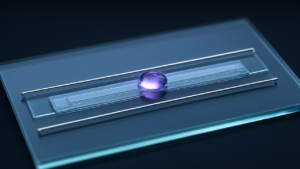
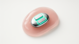
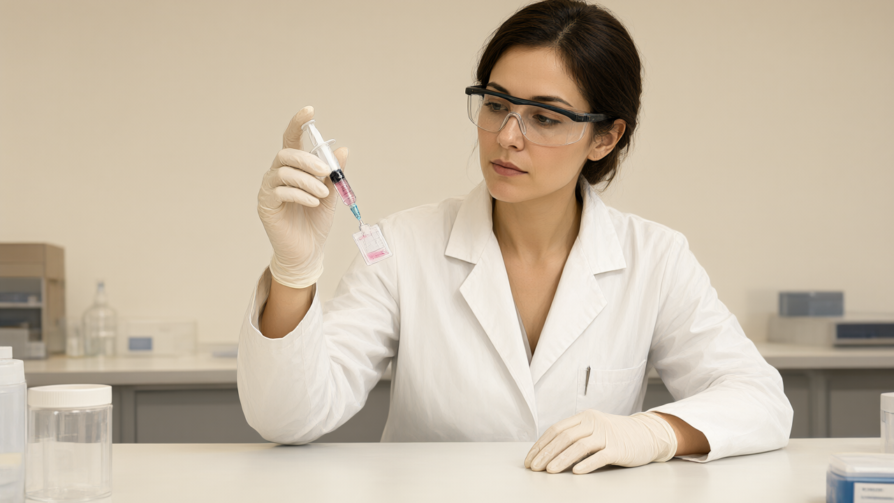
The system utilizes a 3D-printed microfluidic chip and a dual-syringe mechanism:
- Vacuum Seal: One syringe creates a vacuum that seals the device against the surface of freshly excised tissue to prevent leaks.
- Shear Stress Application: A second syringe pushes fluid through a microfluidic channel, creating a flow that applies shear stress parallel to the tissue surface.
- Non-Destructive Sampling: This process gently detaches living cells from targeted sections, leaving the tissue intact for subsequent conventional pathological examination.
Background & Context
The project addresses the difficulty of detecting high-grade serous ovarian cancer, which often begins as microscopic precursor lesions in the fallopian tubes. Traditional diagnostic workflows rely on chemical preservatives that kill cells, making it impossible to cultivate them. This collaboration between Professor Kripa Varanasi’s lab at MIT and gynecologic oncologist Rebecca Stone at Johns Hopkins University was funded by Break Through Cancer to bridge the gap between fluid mechanics engineering and clinical oncology.
Significance & Outlook
Collecting viable cells rather than fixed samples allows researchers to cultivate cells and grow them into organoids. These living models enable the study of how cells from a specific patient behave, providing a foundation for personalized treatment development. The team validated the technology using fresh human fallopian tube samples, ensuring the samples were returned for standard pathology to maintain diagnostic records.
Source: https://bioengineer.org/handheld-microfluidic-device-collects-living-cells-to-catch-cancer-earlier/
Get our weekly technology intelligence — free
Receive an infographic that lets you judge at a glance whether each field’s analysis report is worth reading.
Subscribe Free — Weekly Tech Intelligence
By subscribing, you’ll receive Troy-Technical’s weekly technology intelligence newsletter.
- Your email and selected fields are used only to deliver the newsletter.
- We never share your information with third parties.
- You can unsubscribe anytime via the link in each email.
See our Privacy Policy for details.
Takes about a minute · Unsubscribe anytime