

Key Findings
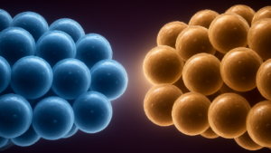
The 3D Cell Painting method successfully revealed striking differences in how cancer cells respond to drugs that appeared nearly identical in 2D screenings. Specifically, among 52 tested compounds, the researchers found that the chemotherapy drug 5-fluorouracil and the PARP inhibitor olaparib produced indistinguishable morphological profiles in 2D cultures but showed clearly distinguishable effects in 3D spheroids when analyzed at the single-cell level.
Technical Details
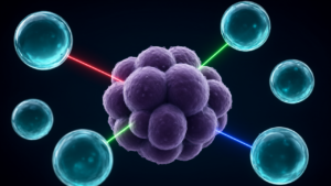
To overcome the limitations of 2D layers, the Uppsala team grew colorectal cancer cells as small three-dimensional spheroids. The process involved several technical steps:
- Transparency: A clearing solution containing sugar and glycerol was used to make spheroids transparent, enabling the imaging of individual cells throughout the structures.
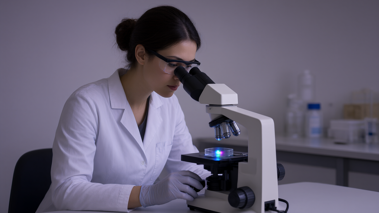
- Imaging: Fluorescent dyes were used to highlight the nucleus, mitochondria, and cytoskeleton to measure changes in cell morphology.
- Analysis: Thousands of morphological features were extracted from individual cells. This avoided the loss of detail that occurs when 3D samples are collapsed into a single picture or when measurements are averaged across entire spheroids.
Further analysis showed that 5-fluorouracil was more effective at preventing cell division and caused less DNA damage than olaparib, a response linked to stronger activation of the p53 pathway.
Background & Context
Cell Painting is a high-content imaging technique used in drug discovery to identify changes in cellular structures. While widely used, it has traditionally relied on 2D cultures, which fail to fully reproduce the organization of cells in actual tissues. The Uppsala team sought to maintain single-cell detail while testing multiple drugs simultaneously.
Significance & Outlook
The method was tested in collaboration with AstraZeneca to verify reproducibility outside the original laboratory. While the study used two laboratory cancer cell lines, the findings suggest 3D Cell Painting can provide critical data on compound activity and mechanisms of action. The researchers intend to extend this technique to more complex models, such as patient-derived organoids.
Get our weekly technology intelligence — free
Receive an infographic that lets you judge at a glance whether each field’s analysis report is worth reading.
Subscribe Free — Weekly Tech Intelligence
By subscribing, you’ll receive Troy-Technical’s weekly technology intelligence newsletter.
- Your email and selected fields are used only to deliver the newsletter.
- We never share your information with third parties.
- You can unsubscribe anytime via the link in each email.
See our Privacy Policy for details.
Takes about a minute · Unsubscribe anytime