Key Findings
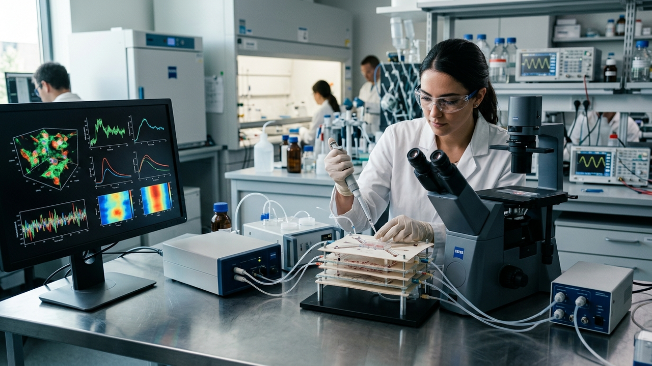
Researchers have developed a scalable 3D biosensing platform for label-free, time-resolved cell analysis by ingeniously repurposing off-the-shelf photo paper. This groundbreaking, low-cost platform effectively overcomes significant limitations associated with conventional, often expensive and complex, 3D cell culture systems. A key innovation is the integration of coplanar aluminum electrodes, which enables highly reproducible electrical impedance spectroscopy (EIS). This technique shows a gradual increase in impedance correlated with increasing cell confluence and barrier formation. Furthermore, the cellulose-rich surface of the photo paper efficiently supports direct cell attachment and proliferation, establishing a robust foundation for future in vitro tissue models and cell-based bioelectronic assays.
Technical / Clinical Details
The core of this novel 3D biosensing platform lies in its ability to transform readily available photo paper into a sophisticated cell analysis device. Specific technical elements include:
- Repurposing Photo Paper: Commercial inkjet photo paper offers an excellent porous cellulose fiber structure, which acts as a scaffold promoting 3D cell proliferation. It is also low-cost and easy to manufacture.
- Integration of Coplanar Aluminum Electrodes: Using techniques like photolithography, minute aluminum electrodes are patterned onto the photo paper, enabling electrical impedance spectroscopy (EIS) measurements. This electrode design allows for highly sensitive detection of changes in cellular electrical properties.
- Label-Free, Time-Resolved Analysis: EIS eliminates the need for cell labeling, allowing for real-time, non-invasive monitoring of physiological changes without disturbing the cells’ intrinsic state. It can track diverse cellular behaviors such as adhesion, proliferation, morphological changes, and barrier function formation over time.
- Reproducible Impedance Changes: As cells attach, proliferate on the electrode surface, and form dense monolayers or 3D structures, the impedance between the electrodes and the solution increases. This impedance change has been confirmed to correlate highly with cell confluence and barrier formation, enabling quantitative cell analysis.
This platform is expected to have broad applications in biomedical fields, including drug screening for toxicity and disease modeling, as well as quality assessment of cell therapies.
Background & Context
Traditional 2D cell culture methods have well-known limitations, leading to an increasing demand for 3D cell culture systems that can more closely mimic in vivo physiological conditions. However, existing 3D cell culture platforms are often expensive to manufacture, require complex operation, and suffer from scalability issues. There is a strong need for systems capable of label-free, real-time functional cell analysis, particularly for high-throughput screening and personalized medicine research. The photo paper-based platform developed in this study offers a low-cost, scalable solution to these challenges, with the potential to significantly accelerate the adoption of cell analysis technology in both academic and industrial settings.
Strategic Significance & Outlook
This photo paper-based 3D biosensing platform is poised to significantly impact the future development of in vitro tissue models. It is particularly expected to contribute to the culture of organoids mimicking specific tissues or organs, evaluation of drug efficacy and toxicity, and elucidation of disease mechanisms. Furthermore, it can be applied as a foundation for cell-based bioelectronic assays, such as monitoring neuronal network activity or assessing cardiac cell contractility. Its low-cost manufacturing characteristic also promises widespread adoption as a diagnostic tool and research platform in developing countries, contributing to global health. Future research will likely focus on improving long-term culture stability, cell-type-specific surface modifications, and integration into automated systems.
Get our weekly technology intelligence — free
Receive an infographic that lets you judge at a glance whether each field’s analysis report is worth reading.
Subscribe Free — Weekly Tech Intelligence
By subscribing, you’ll receive Troy-Technical’s weekly technology intelligence newsletter.
- Your email and selected fields are used only to deliver the newsletter.
- We never share your information with third parties.
- You can unsubscribe anytime via the link in each email.
See our Privacy Policy for details.
Takes about a minute · Unsubscribe anytime







Comments